A
NNAS
ADAKIERSKA−C
HUDY1, D
AGMARAB
ACZYŃSKA2, J
ANS
KÓRA3,
E
LŻBIETAG
ĘBAROWSKA4, A
RTURP
UPKA3, T
ADEUSZD
OBOSZ1Transfection Efficiency and Cytotoxicity of Transfection
Reagents in Human Umbilical Vein Endothelial Cells
Wydajność transfekcji i cytotoksyczność czynników transfekcyjnych
w komórkach śródbłonka ludzkiej żyły pępowinowej
1 Department of Forensic Medicine, Molecular Techniques Unit, Silesian Piasts University of Medicine
in Wrocław, Poland
2 Institute of Biochemistry and Molecular Biology, Department of Cell Pathology, Wrocław University, Poland 3 Department and Clinic of General and Vascular Surgery and Transplantology, Silesian Piasts University
of Medicine in Wrocław, Poland
4 Department of Histology and Embryology, Silesian Piasts University of Medicine in Wrocław, Poland
Adv Clin Exp Med 2008, 17, 6, 625–634 ISSN 1230−025X
ORIGINAL PAPERS
© Copyright by Silesian Piasts University of Medicine in Wrocław
Abstract
Background.Gene therapy is a new approach in the treatment of cardiovascular diseases such as atherosclerosis, restenosis, hypertension, and thrombotic disease. Non−viral delivery systems have low transfection efficiency, especially in primary cells such as vascular endothelial cells. To improve the delivery efficiency of genetic mater− ial into cells by non−viral carriers, further investigations are required.
Objectives.The purpose of this study was to evaluate non−viral transfection reagents with respect to transfection efficiency and cytotoxicity in the HUVEC line. The transcriptional activities of the CMV and SV40 promoters in human endothelial cells were also analyzed and compared.
Material and Methods.Four commercially available transfection reagents (ExGen 500, jetPEITM, FuGENE®HD,
and MetafecteneTMPRO) and EGFP−encoding plasmid were studied. In vitrotransfection efficiency in the HUVEC
line was assayed by the number of EGFP−expressing cells using fluorescence microscope. Cytotoxicity was assessed 24 h later by Hoechst 33258 staining.
Results. The results indicated that transfection efficiency depended on the type of reagent. The linear poly− ethylenimine agents ExGen 500 and jetPEITMwere significantly more cytotoxic than the lipid compounds; they
changed the cells’ morphology and decreased their viability. Moreover, the CMV promoter had a higher ability to express the reporter gene (EGFP) than the SV40 promoter.
Conclusions.Considering the transfection efficiency and lack of cytotoxicity in HUVECs, it was established that FuGENE®HD in the ratio of 5 : 2 was the most effective of the four reagents used for HUVEC line transfection
(Adv Clin Exp Med 2008, 17, 6, 625–634).
Key words: cytotoxicity, linear polyethylenimines, lipid reagents, HUVEC.
Streszczenie
Wprowadzenie.Terapia genowa jest nową metodą leczenia chorób sercowo−naczyniowych, takich jak: miażdżyca, restenoza, nadciśnienie i choroby zakrzepowe. Nośniki niewirusowe charakteryzuje mała wydajność transfekcji, szcze− gólnie w przypadku komórek pierwotnych, do których należą komórki śródbłonka naczyń. Aby poprawić wydajność dostarczania materiału genetycznego do komórek z użyciem niewirusowych nośników, są konieczne dalsze badania.
Cel pracy.Ocena wydajności transfekcji i cytotoksyczności niewirusowych czynników transfekcyjnych w komór− kach śródbłonka ludzkiej żyły pępowinowej. Analiza i porównanie aktywności transkrypcyjnej promotorów CMV i SV40 w linii HUVEC.
Materiał i metody.Badano cztery komercyjnie dostępne czynniki transfekcyjne (ExGen 500, jetPEITM, FuGENE®
HD and MetafecteneTMPRO) i plazmid kodujący białko EGFP. Oceny wydajności transfekcji in vitrodokonywa−
The most common cause of death in industri− alized countries is still cardiovascular disease. Invasive methods such as percutaneous coronary intervention (PCI) or percutaneous transluminal coronary angioplasty (PTCA) have been shown to have limitations. One of these is the development of restenosis, which usually occurs within six months after the procedures [1]. Gene transfer into vascular endothelial cells seems to be a novel approach in the treatment of cardiovascular dis− eases such as atherosclerosis, restenosis, hyperten− sion, and vascular thrombotic diseases [2, 3]. Early clinical trials showed that viral and non−viral gene transfers were safe and well tolerated [4]. However, in vivo adenoviral gene transfer was shown to have a 70− to 240−fold higher expression level of the reporter protein than that produced by lipid−DNA complexes [5]. The major problems associated with viral vectors are immune and toxic response, random integration into the host cell genome, and limits on insert size and large−scale production. Non−viral therapy has advantages over viral carriers (e.g. more safety, non−immunogenic− ity, and ease of producing large quantities), but it is less efficient; therefore the delivery methods of naked DNA should be improved. Consequently the search for new treatment methods for resteno− sis after surgical intervention is of prime and basic importance in vascular and cardiac surgery.
The transfer of exogenous DNA into somatic cells is a complex process. Successful gene trans− fer depends on several factors, including reagent/DNA complex formation, entry into the cell, cytosolic transport, release of DNA from the complex, and DNA uptake into the nucleus [6]. To improve gene delivery and overcome biological barriers, physical and chemical methods are used. Physical approaches such as electroporation, parti− cle gun, ultrasound, hydrodynamic pressure, and jet injection allow DNA to penetrate the cell mem− brane directly and bypass endosomes/lysosomes, thus avoiding enzymatic degradation. The chemi− cal methods for transfection with plasmid DNA use natural or synthetic compounds as carriers for transgene delivery into the cells [7]. Non−viral transfer systems may have a (poly)cationic struc− ture for DNA condensation, lipid compounds for
increasing protection and affinity to cell mem− brane, as well as specific ligands for cell receptors. It is known that some chemical transfection reagents can be cytotoxic to different cell types. The aim of this study was to assess the cytotoxici− ty and transfection efficiency of different types of transfection reagents in human umbilical vein cells (HUVECs). The activities of two viral promoters, CMV and SV40, were also compared in these cells.
Material and Methods
Cell Culture
The HUVEC line was cultured in OPTI−Mem supplemented with 2 mM L−glutamine, 10% fetal bovine serum, 100 U/μl penicillin, 100 μg/ml streptomycin (all reagents from Gibco BRL), and 75 μg/ml endothelial cell growth supplement (ECGS, Becton Dickinson). The cells were cul− tured in collagen−coated flasks (5 μg/ml, Sigma) in a humid incubator with 5% CO2at 37°C. One day before transfection, the cells were plated in a 24− −well collagen−coated cluster dish at a density of 5× 10⁴cells and cultivated in the same medium and conditions for 24 hours.
In vitro
Transfection Assay
Plasmids
Transfection was performed with pEGFP−C1 (enhanced green fluorescent protein, Clontech) plasmid, which utilizes a strong CMV−immediate early promoter, and pEGFP−C1 plasmid with sub− cloned SV40 (the sequence of the CMV promoter was replaced by SV40 sequences). These plasmids were amplified in Escherichia coli DH5α and purified from bacterial cultures using an Endotoxin Free Qiagen Mini Kit (Qiagen).
Non−Viral Carriers
Four commercially available transfection reagents were tested. ExGen 500 (Fermentas) and
Wyniki.Uzyskane dane wskazują, że wydajność transfekcji zależy od rodzaju czynnika transfekcyjnego. Liniowe polietylenoiminy (ExGen 500, jetPEITM) wykazywały wyższą cytotoksyczność niż czynniki lipidowe, zmieniały
morfologię komórek i zmniejszały ich żywotność. Promotor CMV wykazywał ponadto większą zdolność ekspre− sji genu reporterowego w badanych komórkach niż promotor SV40.
Wnioski.Uwzględniając wydajność transfekcji i brak toksycznego wpływu na komórki śródbłonka ustalono, że spośród wszystkich badanych czynników FuGENE®HD użyty w stosunku 5 : 2 był optymalnym czynnikiem trans−
fekcyjnym dla komórek śródbłonka (Adv Clin Exp Med 2008, 17, 6, 625–634).
jetPEITM (Polyplus Transfection) are polyethylen− imines (PEIs) and FuGENE® HD Transfection Reagent (Roche) and MetafecteneTM PRO (Biontex) are lipid compounds. The PEIs used in these experiments are linear cationic polymers (LPEIs) and have a nominal molecular weight of 22 kDa. Their mechanism of action allows them to function as a “proton−sponge” that can prevent DNA degradation [8]. The lipid reagent FuGENE® HD is a proprietary blend of lipids and other com− ponents; further information about its composition and its mechanism of action is unavailable. It is a new generation of non−liposomal multicompo− nent reagent, free of animal− and human−derived components. MetafecteneTMPRO is a novel com− position combining RMA technology (Repulsive Membrane Acidolysis) with a new TOP technolo− gy (Toxicity OPtimization Module). All the com− plexes were prepared as recommended by the manufacturers.
Transfection
The plasmid DNA concentration for all the transfection procedures was 1 μg/μl.
ExGen: 1 μl of DNA was diluted in 99 μl of 150 mM NaCl and mixed with 3.3 μl of ExGen 500. The complex was incubated for 10 min at room temperature before addition to cells. The whole volume of polyplex was added to each well.
jetPEITM: 1 μl of DNA was diluted in 49 μl of 150 mM NaCl and 2 μl of jetPEITM solution was diluted in 48 μl of 150 mM NaCl. The two solu− tions were mixed and incubated for 30 min at room temperature before addition to cells. This amount of mixture was added to each well.
FuGENE® HD: 2 μl of DNA was diluted in 93 μl Opti−MEM without serum and antibiotics, then 5 μl of FuGENE® HD was added to obtain a final ratio of 5:2. The complex was incubated for 15 min at room temperature before addition to cells. This volume of lipoplex was added to each well.
MetafecteneTMPRO: 1 μl of DNA was diluted in 29 μl of Opti−MEM without serum and antibi− otics and 2μl of MetafecteneTMPRO was diluted in 28 μl of 150 mM NaCl. The two solutions were mixed and incubated for 20 min at room tempera− ture before addition to cells. This volume of mix− ture was added to each well.
Cell Viability
Twenty−four hours after transfection, cells in a 24−well dish were fixed in 4% paraformaldehyde for 15 min. The cells were stained in Hoechst 33258 diluted in PBS for 10 min; each well was rinsed three times with PBS buffer before and after
to remove the stain. The cells were counted in six different areas under fluorescence microscopy, using an Olympus IX70 FV500 confocal microscope.
Statistical Analysis
Statistical analysis was performed using ANOVA to compare the mean values among the transfection groups. A statistical probability of P< < 0.01 was considered significant.
Results
HUVEC Morphology
and Viability After Exposure
to the Transfection Reagents
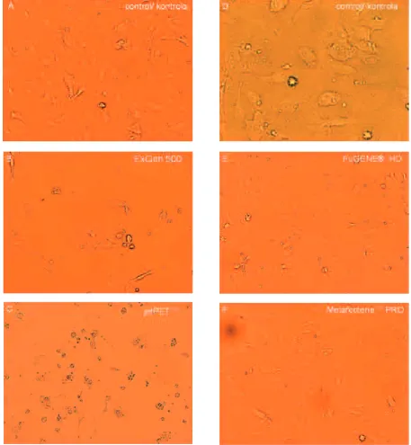
To evaluate HUVEC morphology, the cells were observed 24 hours after transfection under white light using the Olympus microscope. The morphology of the endothelial cells is shown in Figure 1; untreated cells show the typical mor− phology of adherent cells (Figs. 1A and 1D). Parallel cultures of HUVECs exposed to the trans− fection complexes showed changes in morphology (spherical shape). HUVECs treated with the PEI compounds showed a more altered morphology (Figs. 1B and 1C) than those treated with the lipid compounds (Figs. 1E and 1F).
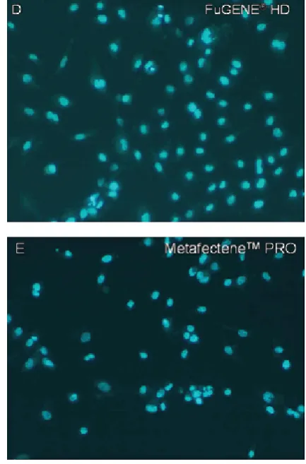
Cytotoxicity was assessed 24 hours after treat− ment with Hoechst 33258 dye staining. The nuclei of the cells are presented in Figure 2. Hoechst can cross intact cell membranes and it binds preferen− tially at A–T base pairs to the DNA molecule. The intact cells were counted and the percentage of liv− ing cells in relation to those of the controls was determined (Fig. 3). ExGen 500 and jetPEITM exerted a more pronounced cytotoxic effect than the lipid compounds FuGENE® HD and Meta− fecteneTMPRO.
Transfection Efficiency
Promoter Activity
The HUVEC line was transfected with two plasmids expressing EGFP protein under CMV or SV40 promoters. Transgene expression was esti−
mated by fluorescence microscopy 24 hours after transfection. As shown in Figures 6B, 6D, 6F, and 6H, the SV40 promoter was unable to drive effi− cient levels of EGFP protein expression in either the PEI or lipid transfection reagents, indicating its
Fig. 1. Morphological changes occurring in HUVECs after exposure to polyethylenimine or lipid transfection reagents. Transfected HUVEC cultures show different degrees of changes in morphology (spherical shape) as a result of the stressful effects exerted by transfection reagents. A, D – control cells (no treatment), typical adherent cell morphology; B – linear polyethylenimine ExGen 500 (altered cell shape); C – linear polyethylenimine jetPEI (altered cell shape); E – lipid reagent FuGENE HD (no cell shape change); F – lipid reagent Metafectene PRO (no cell shape change)
lower activity in this cell line. In contrast, the CMV promoter drove higher expression of the EGFP protein, demonstrating that individual pro− moters could have different activities in HUVECs.
Discussion
In recent years, great progress has been made in gene therapy of cardiovascular diseases. There is great interest in developing carriers for more efficient gene delivery into the vessel wall. In spite
of their relatively low transfection efficiency, viruses are still the most widely used vectors because they can transfect both proliferating and non−proliferating cells [9]. Other viral vectors such as adeno−associated viruses (AAV), retro− viruses, Baculovirus, and herpes simplex virus are used for vascular gene transfer, but these vectors are not ideal. Recently there has been increased interest in non−viral delivery methods due to their safety. The major limitation to their vascular appli− cation is their relatively low transfection efficien− cy. Chemical and physical techniques have been
Fig. 2. Viability of HUVECs following exposure to polyethylenimine or lipid complexes analyzed by fluo− rescence microscopy. A – control cells (no treatment) [147], B – linear polyethylenimine ExGen 500 [37], C – linear polyethylenimine jetPEI [34], D – lipid reagent FuGENE HD [189], E – lipid reagent Metafectene PRO [119]. The numbers in brackets are the average numbers of cells in all counted fields
used to increase the efficiency of DNA uptake and expression. It is known that transfection reagent efficiency depends on the type of cells.
In the present study, an analysis of the trans− fection efficiency and cytotoxicity of four com− mercial transfection reagents was performed. The activities of the CMV and SV40 promoters were also assessed. The results indicate that both the efficiency and the cytotoxicity of gene transfer depended on the type of transfection reagent. The cytotoxicity data showed that linear polyethylen− imines (LPEIs) were more toxic than lipid com− pounds. The percentages of living cells compared with those of the control cells were 25%, 23%, 129%, and 81%, respectively, for ExGen 500, jetTM PEI, FuGENE® HD, and MetafecteneTM PRO. The endothelial cells were rounded and were detached from the well surface 24 hours after transfection. These processes may occur due to the membrane destabilization effects of PEI and suc− cessful transfection, which caused activation and dysfunction of the cells [10]. It has been shown that PEI can be a potent permeabilizer of the outer membrane of Gram−negative bacteria [11]. Because PEI has recently been shown to enter cell nuclei during transfection, its high cytotoxicity can be caused by its binding to host DNA and blocking gene transcriptions [10]. The lipid reagents FuGENE® HD and MetafecteneTM PRO were less toxic and seemed to have no influence on cell morphology. Rughetti et al. also demon− strated that FuGENE 6 showed a less altered mor− phology of dendritic cells compared with
LipofectAMINE PLUS [12]. Comparison of the expression profiles demonstrates that FuGENE® HD affects far fewer genes in two transfected human cell lines, HeLa and MCF7 [13]. This transfection reagent revealed no toxicity in three neuroblastoma cell lines [14]. It is surprising that HUVECs treated with FuGENE® HD had higher viability than controls; FuGENE® HD seemed to be the most efficient gene delivery reagent. FuGENE® HD Transfection Reagent is a new product which is able to transfect a wider range of eukaryotic cells than FuGENE 6 and is designed for application in cell lines that have traditionally been considered difficult to transfect. Previous studies have shown that FuGENE 6 is an efficient reagent for human endothelial cells such as HUVECs [15]. Different results were obtained by Kiefer et al.; FuGENE 6 transfected human aorta endothelial cells (HAECs) only sporadically, but human aorta smooth muscle cells (HASMCs) were transfected very well [16]. Arnold et al. observed that FuGENE 6 was significantly more effective than ExGen 500 for primary human myoblast transfection [17]. These authors suggested that FuGENE 6 formed larger complexes than ExGen 500 and thus produced better efficiency in vitro. However, Haines et al. showed that FuGENE 6 and ExGen 500 had similar transfection activities in HUVECs [18].
It is known that promoter activities depend on the type of cells, which is why viral promoters (CMV, SV40) were evaluated here for their ability to drive the expression of enhanced green fluores−
Fig. 3. The percentage of living HUVECs transfected by polyethylenimine or lipid reagents compared with control cells. Pvalue: ExGen 500 (P= 3.12 × 10⁻¹⁶)*, jetPEITM(P= 3.43 × 10⁻¹⁴)*, FuGENE®HD (P= 0.16), MetafecteneTM
PRO (P= 0.03). Statistical significance: P< 0.01; * statistically significant difference
Ryc. 3. Odsetek przeżywających komórek HUVEC po transfekcji polietyloiminowymi lub lipidowymi czynnikami transfekcyjnymi w porównaniu z grupą kontrolną. Wartość P: ExGen 500 (P = 3,12 × 10⁻¹⁶)*, jetPEITM(P = 3,43 ×
× 10–14)*, FuGENE®HD (P = 0,16), MetafecteneTMPRO (P = 0,03). Statystycznie istotne: P < 0,01; * różnica istotna
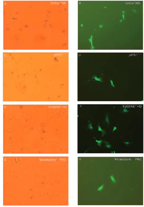
Fig. 4. Efficiency of LPEI and lipid transfection reagents in the HUVEC line assessed under white and fluorescent light. A, B – ExGen 500; C, D – jetPEI; E, F – FuGENE HD; G, H – Metafectene PRO
cent protein (EGFP) in the HUVEC line. The results of this study demonstrated that the CMV promoter was stronger than the SV40. Other inves− tigators demonstrated this in porcine aortic endothelial cells [19]. The strong activity of the CMV promoter in HUVECs has also been con− firmed by other researchers [20], one explanation for the effect being that the CMV promoter con− tains binding elements for transcriptional factors such as NF−κB and cAMP [21]. These transcription factors remain constitutively active in transfected cells. Other observations indicated that CMV pro− moter activity is higher in adherent cells than in suspended cells, in contrast to the SV40 promoter [22]. Chung et al. reported that the CMV promoter was inactive in mouse embryonic stem (ES) cells; one likely explanation is that ES cells cannot express some transcriptional factors required for CMV promoter activity stimulation [23].
In conclusion, the results of the present study show that different promoters exhibit different
transcriptional activities and further suggest that the CMV promoter ensures efficient transgene expression in the HUVEC line. Promoter activity should thus be tested for each cell type. This study showed that the LPEI transfection reagents dis− played high toxicity and decreased cell viability. Further investigations are needed to determine what type of changes (necrosis or apoptosis) are induced by the transfection reagents. Optimal transfection conditions, i.e. a combination of transfection efficiency and a lack of cytotoxicity to HUVECs, were achieved with FuGENE® HD at a ratio of 5:2 (36% transfected cells). We sug− gest that FuGENE®HD can be used as an efficient transfection reagent to deliver foreign DNA into human endothelial cells. In the next stage, trans− fection stability in vitro and in vivo will be assessed. The further development of safe and efficient in vivo gene transfer protocols for the prevention of cardiovascular disease is also nec− essary.
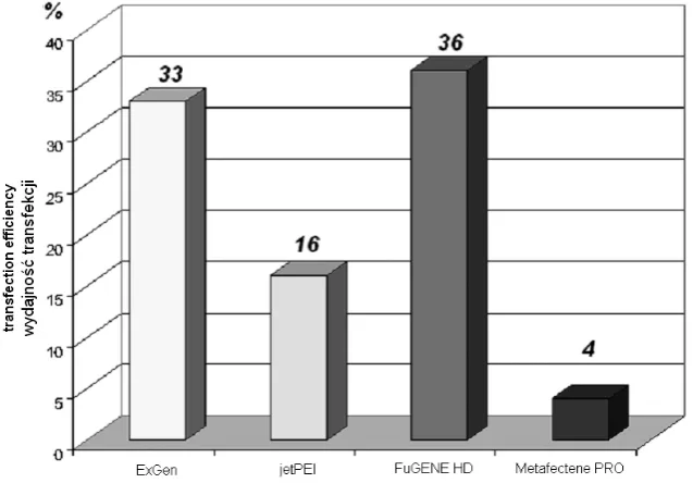
Fig. 5. Percentage of efficiency of the four transfection reagents in the HUVEC line. A statistically significantly dif− ferent efficiency was observed in the cells transfected by Metafectene PRO (P= 0.0023) compared with FuGENE HD. Statistical significance: P< 0.01
Ryc. 5. Procentowa wydajność czterech czynników transfekcyjnych w linii komórek HUVEC. Statystycznie istotną różnicę w wydajności obserwowano w komórkach transfekowanych czynnikiem Metafectene PRO (P = 0,0023) w porównaniu z czynnikiem FuGENE HD. Statystycznie istotne: P < 0,01
Fig. 6. Transcriptional activities of the CMV and SV40 promoters in the HUVEC line. pEGFP−C1 with CMV: A–ExGen 500, C–jetPEI, E–FuGENE HD, G–Metafectene PRO. pEGFP−C1 with SV40: B–ExGen 500, D–jetPEI, F–FuGENE HD, H–Metafectene PRO
References
[1] Serruys PW, Luijten HE, Beatt KJ, Geuskens R, de Feyter PJ, van den Brand M, Reiber JH, ten Katen HJ, van Es GA, Hugenholtz PG:Incidence of restenosis after successful coronary angioplasty: a time−related phenomenon: a quantitative angiographic study in 342 consecutive patients at 1, 2, 3, and 4 months. Circulation 1988, 77, 361–371.
[2] Yla−Herttula S, Martin JF:Cardiovascular gene therapy. Lancet 2000, 355, 213–222.
[3] Ross R:The pathogenesis of atherosclerosis: A perspective for the 1990s. Nature 1993, 362, 801–809.
[4] Rutanen J, Markkanen J, Yla−Herttuala S:Gene therapy for restenosis: current status. Drugs 2002, 62, 1575–1585.
[5] Mazur W, Nadir M, Raizner AE, French BA:Coronary restenosis and gene therapy. Tex Heart Ins J 1994, 21, 104–111.
[6] Lechardeur D, Lukacs G:Nucleocytoplasmic transport of plasmid DNA: A prilous journey from the cytoplasm to the nucleus. Hum Gene Ther 2006, 17, 882–889.
[7] Gao X, Kim KS, Liu D:Nonviral gene delivery: What we know and what is next. AAPS J 2007, 9, E92–E104.
[8] Beher J:The proton sponge – a trick to enter cells the viruses did not exploit. CHMIA 1997, 51, 34–36.
[9] Hiltunen MO, Turunrn MP, Yla−Herttula S:Gene therapy methods in cardiovascular diseases. Meth Enzymol 2002, 346, 311–320.
[10] Godbey WT, Wu KK, Mikos AG:Poly(ethylenimine)−mediated gene delivery affects endothelial cell function and viability. Biomaterials 2001, 22, 471–480.
[11] Helander IM, Alakomi HL, Latva−Kala K, Koski P:Polyethyleneimine is an effective permeabilizer of gram− negative bacteria. Microbiology 1997, 143, 3193–3199.
[12] Rughetti A, Biffoni M, Sabbatucci M, Rahimi H, Pellicciotta I, Fattorossi A, Pierelli L, Scambia G, Lavitrano M, Frati L, Nuti M:Transfected human dendritic cells to induce antitumor immunity. Gene Ther 2000, 7, 1458–1466.
[13] Calvin S, Jeff Emch J, Wang J, Linda Jacobsen L, Pitz S:FuGENE®HD Transfection Reagent: Choice of a transfection reagent with minimal off−target effect as analyzed by microarray transcriptional profiling. Biochemica 2006, 4, 22–25.
[14] Grün M:Efficient transfection of neuroblastoma cells. Biochemica 2007, 1, 12.
[15] Young ATL, Lakey JRT, Murray AG, Moore RB:Gene therapy: A lipofection approach for gene transfer into primary endothelial cells. Cell Transplant 2002, 11, 573–582.
[16] Kiefer K, Clement J, Garidel P, Peschka−Suss R:Transfection efficient and cytotoxicity of nonviral gene trans− fer reagents in human smooth muscle and endothelial cells. Pharm Res 2004, 21, 1009–1017.
[17] Arnold AS, Laporte V, Duont S, Appert−Cillin A, Erbacher P, Coupin G, Levy R, Poindron P, Gies JP:
Comparing reagents for efficient transfection human primary myoblasts: FuGENE 6, Effectene and ExGen 500. Fundam Clin Pharmacol 2006, 20, 81–89.
[18] Haines AMR, Irvine AS, Mountain A, Charlesworth J, Farrow NA, Husain RD, Hyde H, Ketteringham H, McDermott RH, Mulcahy AF, Mustoe TL, Reid SCH, Rouquette M, Shaw JC, Thatcher DR, Welsh JH, Williams DE, Zauner W, Phillips RO:CL−22 a novel cationic peptide for efficient transfection of mammalian cells. Gene Ther 2001, 8, 99–110.
[19] He Z, She R, Sumitran−Holgersson S, Blomberg P, Islam KB, Holgersson J:The in vitroactivity and speci− ficity of human endothelial cell−specific promoters in porcine cells. Xenotransplantation 2001, 8, 201–212.
[20] Kaiser S, Toborek M:Liposome−mediated high−efficiency transfection of human endothelial cells. J Vas Res 2001, 38, 133–143.
[21] Weihl C, Macdonald RL, Stoodley M, Luders J, Li G:Gene therapy for cerebrovascular disease. Neurosurgery 1999, 44, 239–252.
[22] Feng G, Hicks P, Chang PL:Differential expression of mammalian or viral promoter−driven gene in adherent versus suspension cells. In vitro Cell Dev Biol Animal 2003, 39, 420–423.
[23] Chung S, Anderson T, Sonntag KC, Björklund L, Isacson O, Kim KS:Analysis of different promoter systems for efficient transgene expression in mouse embryonic stem cell lines. Stem Cells 2002, 20, 139–145.
Address for correspondence:
Anna Sadakierska−ChudyDepartment of Forensic Medicine Molecular Techniques Unit
Silesian Piasts University of Medicine Curie−Skłodowskiej 52
50−369 Wrocław Poland
Tel.: +48 71 784 15 97
E−mail: [email protected]
Conflict of interest: None declared