Maciej Guziński
1, Tomasz Szczepański
2, Paweł Krukowski
1, Władysław Berny
2,
Włodzimierz Jarmundowicz
2, Marek Sąsiadek
1Comparative Analysis of Endovascular
and Microsurgical Treatment of Intracranial AVMs
Analiza porównawcza wyników leczenia operacyjnego i embolizacyjnego
wewnątrzczaszkowych malformacji tętniczo-żylnych
1 Department of General Radiology, Interventional Radiology and Neuroradiology, Wroclaw Medical University,
Poland
2 Department of Neurosurgery, Wroclaw Medical University, Poland
Abstract
Background. Intracranial arteriovenous malformations (AVMs) are among the most frequent developmental anomalies of brain vessels. The most serious complication of AVM is intracranial hemorrhage. Treatment meth-ods of intracranial AVMs include microsurgical techniques, endovascular embolization, and radiosurgery. As all these methods have their advantages and disadvantages, the choice of treatment depends mostly on the experience and capabilities of the treating hospital.
Objectives. To analyze comparatively two treatment methods of AVMs: endovascular embolization and micro-surgical resection.
Material and Methods. Forty consecutive AVMs in 40 patients aged 4–60 years treated between 2002–2007 were analyzed retrospectively. Twenty of the patients were treated by microsurgical resection (subgroup S) and the remaining 20 (subgroup E) underwent embolization (36 sessions overall). There were 12 emergency cases (8 in subgroup S and 4 in subgroup E). Embolizations were performed with histoacrylic glue. Microsurgery included complete resection of the visible AVM and clipping of the feeding artery, in emergency cases also the removal of hematomas. The results of the treatment method were assessed on the last day of hospitalization according to the Glasgow Outcome Scale (GOS).
Results. In both subgroups E and S, the most common AVMs were those with 3 points in the Spetzler-Martin clas-sification (S-M) (6 and 9 AVMs, respectively). Mean S-M scores were 2.8 and 2.5, respectively. The most frequent feeding artery in both groups was the MCA (14 and 11, respectively). The degree of nidus embolization ranged from 33 to 100% and depended on S-M score (for S-M of 1 an average of 91.7%, of 2 88%, of 3 70.2%, of 4 65.8%, and of 5 40%). In 15 of the 20 surgical cases, AVMs were removed totally, while in 5 cases residual nidi were found in follow-up CT and/or angiography. In group E the mean GOS score at discharge from hospital was 4.35 points and in group S 3.95 points (no significant difference). In the case of elective embolization or surgery, the mean GOS was 4.7 and 4.5, respectively. The mean duration of hospitalization (in the case of multiple embolizations, hospitalizations during all the sessions were summed) was 14 days in subgroup E and substantially longer (23 days) in subgroup S (p < 0.05).
Conclusions. Intravascular and microsurgical methods in many cases do not provide total cure of an AVM. A com-bination of these two methods and close cooperation between interventional neuroradiologists and neurosurgeons seem to be necessary to optimize the treatment results, shorten the hospitalization period, and reduce the costs of AVM therapy (Adv Clin Exp Med 2010, 19, 2, 219–226).
Key words: intracranial arteriovenous malformation, AVM, endovascular embolization.
Streszczenie
Wprowadzenie. Wewnątrzczaszkowe malformacje tętniczo-żylne (AVMs – Intracranial Arteriovenosus Malfor-mations) są wadami rozwojowymi naczyń mózgowych, a ich najpoważniejszym powikłaniem jest krwawienie wewnątrzczaszkowe. Spośród metod leczenia najczęściej stosuje się resekcje mikrochirurgiczne, embolizacje wewnątrznaczyniowe oraz radioterapię celowaną. Wszystkie te metody mają swoje wady i zalety, a ich wybór zale-ży od doświadczenia i możliwości ośrodka.
Adv Clin Exp Med 2010, 19, 2, 219–226 ISSN 1230-025X
ORIGINAl PAPERS
Intracranial arteriovenous malformations (AVMs), also called cerebral angiomas, are con-genital developmental anomalies of brain vessels. Their most serious complication is intracranial hemorrhage; they may additionally cause various symptoms such as epileptic seizures or focal neu-rological disorders. The malformations may be located anywhere within the brain, but they tend to affect areas supplied by the middle cerebral arteries (MCAs). About 90% of them are located supraten-torially [1]. They usually consist of dilated vessels which feed the nidus and of draining veins. In most cases their vascularization includes branches of the internal carotid artery or vertebral artery [2]. The number of supplying arteries and arterioles, and therefore the AVM’s angio-architecture and size, varies with different lesions. The incidence of intracranial AVM is not precisely known. Brown et al. estimated that the incidence of new malfor-mations is 1.84 per 100,000 persons a year [3]. In autopsies the prevalence of malformations is vari-able, ranging from 0.02 to 0.6% [4].
The aim of treating AVMs is to protect the patient from intracranial hemorrhage and the therapy consists of complete exclusion of the pathological vascular bed along with the nidus from the normal circulation, with the least pos-sible interference with the hemodynamics of the intracranial flow. In most cases it is difficult to achieve complete cure even at experienced and skilled institutions. Currently, based on contem-porary knowledge and research results, no
domi-nant treatment protocol has been established to eliminate hemorrhagic complications. In the man-agement of AVMs, three different methods can be applied jointly or separately: surgical microresec-tion, endovascular embolizamicroresec-tion, and radiosur-gery. Selection of the method and procedural risk are strictly related to the malformation’s angio-architecture. In addition, some centers apply con-servative therapy to all angiomas without a history of intracranial hemorrhage and without progres-sion of neurological symptoms [5, 6].
The most frequently used method of assessing the procedure’s risk is the Spetzler and Martin (S-M) classification (Tab. 1). It classifies malformations on the basis of imaging studies and according to surgi-cal risks. It assesses the size of an AVM’s nidus, the type of the malformation’s outflow (to superficial or deep veins) [7], and its location, i.e. whether it affects eloquent or non-eloquent areas of the brain [8].
The aim of the present paper was to compare microsurgery and endovascular embolization as methods applied in the treatment of arteriovenous malformations in two groups with the same num-ber of patients and with similar numnum-bers of points in the Spetzler and Martin risk classification.
Material and Methods
This study is based on the analysis of a group of 40 patients (21 women and 19 men) diagnosed with arteriovenous malformations of the brain. The study
Cel pracy. Analiza porównawcza zabiegów mikrochirurgicznych i embolizacji wewnątrznaczyniowej stosowanych w leczeniu malformacji tętniczo-żylnych.
Materiał i metody. Analizie retrospektywnej poddano 20 pacjentów, u których wykonano zabieg resekcji naczy-niaka (grupa S) i 20 chorych, których leczono metodą embolizacji (grupa E). W sumie analizowano 40 malformacji u 21 kobiet i 19 mężczyzn w wieku 4–60 lat (średnio 39 lat) leczonych w latach 2002–2007. Podczas embolizacji przez system cewników wprowadzonych przez tętnicę udową uzyskiwano dostęp do gniazda naczyniaka i poda-wano porcje kleju histoakrylowego obliterującego naczynia zmienione patologicznie. W rezultacie zabiegu mikro-chirurgicznego po wykonaniu kraniotomii usuwano makroskopowo ewentualnego krwiaka, a następnie malfor-mację i klipsowano naczynia doprowadzające. Wyniki leczenia były oceniane na podstawie wyniku skali Glasgow Outcome Scale (GOS) podczas ostatniego dnia pobytu.
Wyniki. Najliczniej reprezentowane w grupie E i S były naczyniaki z 3 punktami w skali Spetzera-Martina (S-M) (odpowiednio 6 i 9). Średnia liczba punktów dla malformacji wynosiła odpowiednio 2,8 i 2,5. Głównym naczyniem doprowadzającym w obu grupach była MCA (odpowiednio w 14 i w 11 przypadkach). Średni stopień zmniejsze-nia gzmniejsze-niazda naczyzmniejsze-niaka wynosił 33–100% i zależał od liczby punktów w skali S-M (dla 1 – średnio 91,7% i dalej odpowiednio dla 2 – 88%, dla 3 – 70,2% dla 4 – 65,8%, dla 5 – 40%). W 15 z 20 przypadków resekcja mikrochi-rurgiczna okazała się doszczętna, a w 5 przypadkach kontrolne badania pooperacyjne (tomografia komputerowa i/lub angiografia) wykazały obecność części gniazda naczyniaka. Wyniki leczenia w obu grupach oceniano w dniu wypisu zgodnie ze skalą GOS (Glasgow Outcome Scale). Średnia ocena w skali GOS dla malformacji embolizo-wanych wynosiła 4,35, a dla operoembolizo-wanych 3,95 i nie była istotna statystycznie. Biorąc pod uwagę zabiegi planowe średnia GOS wynosiła odpowiednio 4,7 i 4,5. Średni czas pobytu (w zabiegach wieloetapowych suma wszystkich dni pobytów w szpitalu) wynosił dla embolizacji 14 dni oraz 23 w przypadku zabiegu mikrochirurgicznego i był istotnie różny (p < 0,05).
Wnioski. Obie metody lecznicze w wielu przypadkach samodzielnie nie zapewniają pełnego wyleczenia. Dlatego też wydaje się, że skojarzenie obu metod i ścisła współpraca radiologiczno-neurochirurgiczna jest konieczna do optymalizacji wyników i czasu leczenia oraz obniżenia kosztów terapii (Adv Clin Exp Med 2010, 19, 2, 219–226).
group’s age ranged from 4 to 60 years (mean: 39 years). The subjects were treated at the Department of Neurosurgery of Wroclaw Medical University. Microsurgical resection of the AVMs was performed on 20 persons (the S-group) and the remaining 20 patients (the E-group) were treated with one- or multi-stage embolization of the lesions (12 patients underwent one session of embolization, 6 had two sessions, and in 3 persons the embolization was done in five stages); in total, 36 embolization ses-sions were performed. In 8 of the 20 cases an emer-gency procedure was necessary due to intracerebral hematoma and consequential significant mass effect. In 4 patients of the E-group an urgent embolization was performed because of evidence of active bleeding from the malformations and increasing focal neuro-logical symptoms. In the remaining 28 patients the treatment process was elective.
In the first stage of the microsurgical resection procedure the lesion was accessed (craniotomy) and the feeding vessels were prepared and closed. Then the malformation’s nidus was excised and subsequently vein outflow was coagulated. If there were related aneurysms on feeding arteries, the aneurysm was excluded from the circulation first, followed by the malformation. In the case of emer-gency procedures, the intracranial hematoma was evacuated first and then the lesion was excised. After the procedure, a follow-up head CT and/or cerebral angiography was performed.
Endovascular therapy of the malformation (embolization) consisted of the implantation of a portion of the obliterating material (N-butyl cyanoacryl histoacrylic glue, NBCA, Histoacryl®; B. Braun Corp., USA) into the pathological
ves-sels through a catheter system. The vascular sys-tem was accessed by an incision of the common femoral artery and then by introduction through a hemostatic sheath and a microcatheter, by which the embolization material was applied. The micro-catheter’s end was placed inside the angioma’s nidus or close to it, in the pathological feeding vessels. The glue was mixed with a contrast agent (lipiodol®; laboratoire Guerbet, Roissy, France) at 25 to 40% concentration and the mixture’s volume was 0.5 to 1.5 ml. After each procedure, a follow-up cerebral angiography was performed.
Results of the resections and embolizations were assessed based on the follow-up angiogra-phy. If the pathological vessels did not contrast in the post-procedure or follow-up examination, total exclusion of the AVM was confirmed. The patients’ condition was assessed on the day of dis-charge according to the GOS (Glasgow Outcome Scale) [9].
Results
In 17 cases the AVMs were located in the right hemisphere and in 23 cases in the left. The most frequent location (8 cases) was the parietal lobe. In patients treated with neurosurgical pro-cedures, the most frequent location of the AVM was the temporal lobe (5 cases). In 7 cases in the S-group, the lesion involved more than one lobe. In two patients, AVMs were confirmed to affect the brain stem. In the group treated with endo-vascular embolization, the angiomas affected the parietal lobe most frequently (5 cases). The lesion
Table 1. The Spetzler-Martin classification (S-M) [8] Tabela 1. Skala Spetzlera i Martina (S-M) [8]
AVM features (Cechy malformacji) Size (Rozmiar) location (Umiejscowienie) Vein outflow (Spływ żylny) Nidus diameter
(Gniazdo) up to 3 cm 1 point
3 to 6 cm 2 points more than 6 cm 3 points Eloquent area of the brain
(Elokwentny obszar mózgowia) 1 point
Non-eloquent area of the brain
(Nieelokwentny obszar mózgowia) 0 points
Only superficial vein outflow
(Tylko spływ do żył powierzchownych) 0 points
Involvement of deep vein outflow
(Spływ również do żył głębokich) 1 points
Total: 1 (small) to 5 points (marked risk)
involved more than one lobe in 7 cases. A single lesion was found in the basal ganglia (Tab. 2).
In the S-group the vascular supply of the AVMs was most frequently based on the middle cerebral artery (MCA, 11 cases), then the posterior cere-bral artery (PCA, 6 cases) and the anterior cerecere-bral artery (ACA, 3 cases). In the E-group the malfor-mations’ main feeding vessel was also the MCA (14 cases), then the ACA (3 cases) and finally the PCA (3 cases). In the S-group the average Spetzler-Martin assessment result of the AVMs was 2.5 and in the E-group 2.8. There was no statistically sig-nificant difference between these data in the two groups (Mann-Whitney U test, p > 0.05).
In the S-group, the surgery was complete in 15 cases and partial in 5 (a residual nidus of the AVM was revealed). The average reduction of the
malformation’s nidus for the whole 20-person group was 71% (33–100%) and it correlated with the S-M classification result (for an S-M score of 1 the average was 91.7%, of 2 88%, of 3 70.2%, of 4 65.8%, and of 5 40%) (Tab. 3). In the subgroup with one embolization session (9 patients), follow-up angiography confirmed reductions in the mal-formation’s nidus by 40–100% (mean: 88.7%). In the subgroup of 11 patients with more emboliza-tion sessions (in 6 cases two sessions and in 5 cases three), reduction of the nidus’s size ranged from 33–100% (mean: 65.1%). In 4 cases (20%) the angi-oma was embolized entirely (including 2 patients with multiple embolizations). In patients who had multiple embolizations, the average S-M score was markedly higher compared with the patients with a single session (3.36 vs. 2.11, p < 0.05).
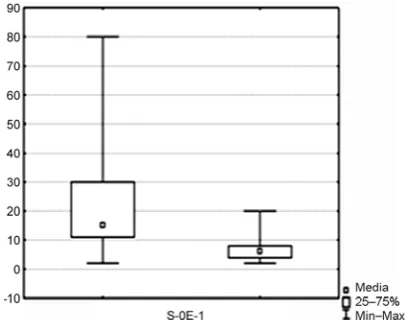
In the S-group, hospitalization lasted 23 days on average (3–80 days). In the E-group the aver-age duration of hospitalization was 7.5 days (3–15) and 13.6 (7–20) days if the total duration of hos-pitalization for the patients who underwent mul-tiple embolizations was considered. A statistically significant difference was observed between hos-pitalization time in the S-group and that in the E-group according to the Mann Whitney U test (p = 0.000023) (Fig. 1).
The average Glasgow Outcome Scale (GOS) score for the patients in the S-group was 3.95 (range: 1–5) and in the E-group 4.35 (3–5). The difference in the GOS result between the S- and E-groups was favorable for embolization and close to statistical significance (p = 0.071, 3.95 vs. 4.35, Tab. 4.). Furthermore, in the embolization group a statistically significant negative correlation was observed between the S-M score and the GOS result (Spearman’s correlation coefficient R = –0.4611, p = 0.04), which means that the patients
Table 2. location of AVMs (N = 40)
Tabela 2. Umiejscowienie malformacji (N = 40) location
(Umiejscowienie) S-group (Grupa S) E-group (Grupa E) ∑ Right hemisphere
(Prawa półkula) 8 9 17
left hemisphere
(lewa półkula) 12 11 23
Region (Okolica) Frontal
(Czołowa) 2 3 5
Parietal
(Ciemieniowa) 3 5 8
Temporal
(Skroniowa) 5 1 6
Occipital
(Potyliczna) 1 3 4
Parieto-occipital (Ciemieniowo-potyliczna)
3 2 5
Temporo-parietal
(Skroniowo-ciemieniowa) 3 2 5 Fronto-temporal
(Czołowo-skroniowa) 1 3 4
Brain stem
(Pień mózgu) 2 0 2
Basal ganglia
(Jądra podstawy) 0 1 1
Total (Suma) 20 20 40
Average score on the S-M scale (Przeciętna liczba punktów w skali S-M)
2.5 2.8 Fig. 1. Duration of hospitalization (days); (0 – S group, 1 – E group)
with a high S-M score had significantly lower GOS scoring.
In the S-group, complications were recorded in four cases: in three patients permanent isch-emia-related focal symptoms confirmed by diag-nostic imaging and in one case an intracranial hemorrhage occurred. One patient died due to massive hemorrhage. In the E-group there were four complications: in two cases permanent isch-emia-related focal symptoms and in two patients acute post-embolization bleeding from the malfor-mation’s nidus.
Discussion
The basic goal in the treatment of arterio-venous malformations of the brain is to exclude the pathological vessels of the malformation from the cerebral circulation. This allows for maximal reduction of the risk of the most serious compli-cation of angiomas, i.e. intracranial hemorrhage. However, as AVMs differ greatly in their
architec-ture, it is impossible to indicate a single method to achieve this goal in all AVMs.
Surgical treatment is the method of choice if bleeding from an AVM leads to a significant life-threatening intracranial hematoma. In the present material, such treatment was applied in 8 patients. In these cases the hematoma was removed and the angioma excised if possible. In this 8-patient group, in 6 cases (75%) the resection was com-plete, which means that follow-up examinations revealed no residual angioma. A similar percentage (75%, 9 of 12 patients) of complete resection was found in the subgroup with an elective procedure. It should be mentioned, however, that in two cases of incompletely excised AVMs, the malforma-tion’s nidus was partially located within the brain-stem structures, which made complete resection of these lesions highly dangerous. No such location was observed in the subgroup of emergency opera-tions. In both subgroups, the S-M score was simi-lar (2.3 and 2.6, respectively), which could suggest that completeness of the procedure depends not only on the malformation’s features (i.e. size and
Table 3. Effectiveness of treatment in relation to the S-M score (N = 40) Tabela 3. Skuteczność leczenia zależna od punktacji w skali S-M (N = 40)
S-M classification points (Punkty w skali
S-M)
E-group (Grupa E) S-group (Grupa S)
Number of patients
(liczba pacjentów) of the nidus’s size (%) Average reduction (Przeciętna wielkość
zmniejsze-nia rozmiaru gzmniejsze-niazda) (%)
Number of patients
(liczba pacjentów) (Całkowita resekcja)Radical resections
1 3 91.7 3 3
2 5 88.0 7 6
3 6 70.2 9 6
4 5 65.8 0 –
5 1 40.0 1 0
Total (Suma) 20 71.1 20 15 (75%)
Table 4. GOS score at discharge from hospital (N = 40)
Tabela 4. Ocena stanu pacjentów przy wypisie wg skali GOS (N = 40)
GOS (Kategorie skali GOS) The patient’s state (Stan chorych) E S E + S
5 patients with no neurological disorders, conscious,
with complete and logical contact 10 7 17
4 patients with neurological deficits, independent 7 8 15
3 conscious, requiring constant care 3 3 6
2 vegetative state 0 1 1
1 death 0 1 1
location), but also on the patient’s general condi-tion and preparacondi-tion for the procedure, although confirmation of this thesis requires a larger group of patients.
In the entire analyzed material the percent-age of radical resections of AVMs was 75% and in the remaining 25% of patients, follow-up angiog-raphy and CT revealed the presence of residual nidi. Compared with data from more experienced institutions, where the percentage of completely excised AVMs is about 97% [10, 11], the results achieved by the present authors’ center are below average. However, in a study by Munshi et al. [12], in which all angioma resection procedures were verified by precise intra-operation and post-operation angiography, the efficiency of resections was lower, ranging from 80 to 92% depending on the time passed after the procedure. According to an assessment by Solomon and Stein [13], the effi-cacy of resection of AVMs located within the brain stem was 89%. Therefore one can assume that the actual efficiency is much lower than the above-mentioned 97%.
Castel and Kantor’s meta-analysis, which included about 2500 patients who had under-gone AVM microsection, revealed that the risk of surgery-related death ranged from 0 to 15%, with a mean of 3.3% (in three thirds of the reports it was ≤ 5%). The percentage of permanent postsurgical complications was 1.5 to 18.7%, 8.6% on average [14]. In the present material, among the patients operated on were 4 cases (20%) of serious compli-cations and 1 death (5%). Among the complica-tions was one hemorrhage which required reop-eration to evaluate the hematoma and the other three cases involved ischemic lesions resulting in permanent neurological deficits. The above-mentioned death was caused by massive bleeding from the angioma in a 4-year-old patient who had undergone emergency surgery because of the rup-ture of a large AVM in the right temporal region (S-M: 5 points) accompanied by an intracerebral hematoma. It should be mentioned that the per-centage of complications and deaths did not vary from reports from other institutions. However, it should be noted that the death and two serious ischemic episodes involved patients of the 8-per-son group with emergency operations. Among the 12 patients with elective operations there was only 1 case of bleeding which required re-surgery and one case of a confirmed (by CT) ischemic stroke. Therefore, the percentage of complications and deaths differed between the emergency and non-emergency patients, as it was 38% (3 patients out of 8) and 17% (2 out of 12), respectively. This would suggest that the percentage of serious postsurgical complications depends not only on the
malforma-tion’s size and features, but also on the neurologic condition before the procedure and especially on the presence of bleeding from the malformations. Such conclusions were statistically confirmed in a study by Pikus et al., who analyzed, among other issues, the surgical treatment of a large group of patients with AVMs [15].
The endovascular treatment of cerebral mal-formations was introduced in the 1980s and now, due to miniaturization and improvement of cath-eter systems, it can be an independent treatment method or part of an integrated therapy. The most frequent procedure is pre-surgery embolization (e.g. preceding the angioma resection procedure), but quite often, especially in small and medium malformations (S-M up to 3 points), embolization is applied also as the only treatment method. In addition, in some institutions it is integrated with radiosurgery because preliminary embolization allows a reduction in the nidus’s size and makes irradiation more efficient. The so-called pallia-tive embolization of giant AVMs (SM = 5 points) is a rare procedure, performed only on angiomas which cause strong neurological symptoms involv-ing the “steal syndrome” because the risk of com-plications is much higher in such cases [16]. In the material of the present comparative analysis there was one similar case of a 44-year-old patient who underwent multiple embolizations and suffered from intensified focal symptoms, probably related to the steal syndrome. There were no complica-tions during the procedures, but the reduction in the AVMs was significantly lower than average (40% vs. 70%).
In the embolization group there were 4 com-plications in total, which consisted of 20% of the patients and 11.1% of the embolization ses-sions (4 out of 36). There were no deaths related to the endovascular procedures. Hemorrhagic complications affected 2 patients and they were related to poor neurological condition (mean GOS: 3) and ischemic complications (hemipa-resis) affected 2 patients, but their effect on the patients’ quality of life was less than in the cases of hemorrhages (mean GOS: 4). It is worth not-ing that in one patient with a giant malforma-tion (S-M = 5 points), GOS at discharge was 3, but the deterioration of her quality of life resulted not from post-procedure complications, but from the natural history of the malformation involving focal symptoms, such as hemiparesis and multiple epileptic fits.
from 4.6 to 12.5%, and the percentage of deaths from 0 to 2.6% [17]. If one compares these results with the data of the group analyzed in the present study, a significant variation in frequency of com-plications is visible. They occurred twice as fre-quently as the average values reported by the lead-ing centers. However, it is worth stresslead-ing that no patient in the E-group died and no patient fell into a vegetative condition as a result of post-emboli-zation complications (GOS 2). To supplement the above data, one should quote reports published by Polish institutions. Kordecki et al. [18] reported complication rates in a group of 34 patients of total permanent and transient complications 15% and deaths 9%, and Tomalski et al. [19] reported, respectively, 9.4% and 4.7% (in a group of 43 pa- tients). Compared with the these Polish reports, the variation in complication rates is not as evi-dent as in the case of comparison with experienced foreign institutions with long-standing expertise in embolization of malformations; the present results were better than the quoted reports as far as the number of deaths is concerned.
The hospitalization period at the neurosurgery ward was significantly shorter in the case of embo-lized patients, both comparing hospitalization for a single session and the combined multi-session hospitalization. Along with the low invasiveness of the procedure, short hospitalization was an advantage for patients. However, considering that the resection procedure led to complete healing (removal of the nidus) three times more frequent-ly than embolization, the mentioned advantage for patients is no longer so obvious. One should men-tion that longer hospitalizamen-tion involved patients with symptoms of acute bleeding from the AVM and there were twice as many of them in the S-group than in the E-group.
Assessing the patients’ condition at discharge according to the GOS, it was shown that in the case of the surgical patients the average score was 3.95 points, while the embolized patients had a slightly better result, i.e. 4.35 points. The difference was not statistically significant at the confidence level of
p < 0.05, but it fell within the range of an extended significance level of p < 0.1. Thus one can state that the patients who had had endovascular treatment were in a slightly better general condition that those who had been operated on. It should also be
stressed that the average surgery risk expressed in S-M points was very similar for the two subgroups (2.5 and 2.8 points, respectively). However, the patients’ condition was probably affected by the facts that there were more emergency patients in the S-group than in the E-group (8 vs. 4) and that the endovascular procedure is by nature not so invasive. The patients’ condition at discharge was assessed as satisfactory (GOS 5 and 4) in 32 patients (80%) and unsatisfactory (GOS 1 to 3) in 8 patients (20%), while ledezma et al. [20], on the basis of embolizations of 168 patients followed by microresections in 124 patients, reported 90.5% and 9.5% respectively. The percentage of patients with unsatisfactory GOS results was two times lower in reports of this experienced institution than the present statistics, which is strictly correlated to the above-described two times higher rate of compli-cations compared with reports from the reference institution. Therefore one can say that the number of direct post-resection or post-embolization com-plications determines the general physical and psy-chological condition of the patients. This is why, in all cases, strict cooperation of neurosurgeons and neuroradiologists is necessary in the selection of the treatment method which brings the least risks to the patient. Such cooperation may contribute to reducing the complication rate and bringing it closer to the rate achieved at more experienced institutions.
The authors concluded that none of the treat-ment methods allows for complete healing of all patients with AVMs. The success rate of surgi-cal management is higher, but it involves higher complication risk and longer hospitalization of the patient. As the majority of malformations can be treated according to an elective schedule, it seems both medically and economically advan-tageous to tailor treatment precisely applying a single method or integrated treatment (partial embolization and then resection). This would also allow stricter cooperation between neurosur-geons and neuroradiologists, which is necessary to optimize treatment results and reduce therapy costs. Serious procedure-related complications of both methods result in deterioration of the patients’ general neurological state in most cases and are more frequent in patients with symptom-atic malformations.
References
Stieg P, Batjer H, Samson D:
[1] Intracranial Arteriovenous Malformations. Infolma Healthcare, New York 2006, 70–79.
Morris P:
[2] Practical neuroangiography. lippincott Williams & Wilkins, Philadelphia 2007, 366.
Brown R Jr, Wiebers D, Torner Jl:
Al-Shahi R, Warlow C:
[4] A systematic review of the frequency and prognosis of arteriovenous malformations of the brain in adults. Brain 2001, 124, 1900–1926.
Mohr J, Stapf C, Sciacca R:
[5] Treatment outcome versus natural history risk in patients with unruptured brain arteriovenous malformation. Neurology 2004, 62 (suppl 5), A101, 83.
Ondra SL, Troupp H, George E:
[6] The natural history of symptomatic arteriovenous malformations of the brain: a 24-year follow-up assessment. J Neurosurg 1990, 73, 387–391.
Ryan S, Mc Nicholas M:
[7] Central nervous system in anatomy for diagnostics imaging. Saunders Company ltd, london 1998, 77–80.
Spetzler R, Martin N:
[8] A proposed grading system for arteriovenous malformations. J Neurosurg 1986, 65(4), 476–483.
Jennett B, Bond M:
[9] Assessment of outcome after severe brain damage. lancet 1957, (7905), 480–484.
Fleetwood I, Steinberg G:
[10] Arteriovenous malformations. lancet 2002, 359(9309), 863–874.
Forsting M:
[11] Intracranial Vascular Malformations and Aneurysms. Springer, Berlin 2004, 62.
Munshi I, Macdonald RL, Weir BK:
[12] Intraoperative angiography of brain arteriovenous malformations.
Neurosurgery 1999, 45(3), 491–497.
Solomon RA, Stein BM:
[13] Management of arteriovenous malformations of the brain stem. J Neurosurg 1986, 64(6), 857–864.
Castel J, Kantor G:
[14] Postoperative morbidity and mortality after microsurgical exclusion of cerebral arteriovenous malformations. Current data and analysis of recent literature. Neurochirurgie 2001, 47(2–3 Pt 2), 369–383.
Pikus HJ, Beach ML, Harbaugh RE:
[15] Microsurgical treatment of arteriovenous malformations: analysis and
com-parison with stereotactic radiosurgery. J. Neurosurg 1998, 88(4), 641–646.
Richling B, Killer M, Al-Schameri A:
[16] Therapy of brain arteriovenous malformations: multimodality treatment
from a balanced standpoint. Neurosurgery 2006, 59, S148–157.
Forsting M:
[17] Intracranial Vascular Malformations and Aneurysms. Springer, Berlin 2004, 60–63.
Kordecki K, Janica J, Lewszuk A:
[18] Endovascular treatment of cerebral arteriovenous malformations. Pol Przegl
Radiol 2006, 71(1), 26–31.
Tomalski W, Majchrzak K, Stech W:
[19] Transluminal treatment of central nervous system AVMs by histoacryl glue
injection. Pol Przegl Radiol 2004, 69(Sup 1), 8.
Ledezma C, Hoh B, Carter B:
[20] Complications of cerebral arteriovenous malformation embolization: multivariate analysis of predictive factors. Neurosurgery 2006, 58, 602–611.
Address for correspondence:
Marek Sąsiadek
Department of General Radiology, Interventional Radiology, and Neuroradiology Wroclaw Medical University
Borowska 213 50-556 Wroclaw Poland
Tel. +48 71 733 16 60
E-mail: [email protected]
![Table 1. The Spetzler-Martin classification (S-M) [8]Tabela 1. Skala Spetzlera i Martina (S-M) [8]](https://thumb-us.123doks.com/thumbv2/123dok_us/8771541.1757275/3.595.73.521.93.311/table-spetzler-martin-classification-tabela-skala-spetzlera-martina.webp)