Development of an Epithelial Potential Difference
Reading Device for Anomaly Detection in Breast
Fatima Moumtadi
*, Sergio Meléndez Armas and Jorge Naude
Date of publication (dd/mm/yyyy): 27/02/2017
Abstract — This article exposes the development of a device to acquire the potential differences in 1 the epithelial surface of the female breast, using an array of 24 electrodes distributed on the surface 2 of the breast, a signal acquisition board capable to take a reading range of 31.25mV to 1.024V to 3 potential differences in the epithelial surface of the breast and a graphical user interface developed 4 in Matlab®; with what we were able to get a platform to carry out more research into the mode of epithelial potential differences. The device was evaluated with 4 healthy Mexican women who were subject to a prior ultrasound exam or mammography. As a result, of the medical trial, we found different factors that can affect accurate measurement of the breast surface and obtained information about the various stages that contains a device measuring epithelial potential differences found in this region of the human body.
Keywords — Bio Potentials, Breast Tumors, Electric Potential, Epithelial Potential Difference, Signal Acquisition.
I.
I
NTRODUCTIONIn bio medical instrumentation, they have generated various tools and techniques for the diagnosis of different diseases from the existence of biological signals the human body shows. Cancer is one of the diseases of the greatest concern to humans, in the case of women; breast cancer is the leading cause of death from malignant neoplasia. Some leading causes of death from breast cancer are due to not being detected on time, poor treatment or high breast density, which is one of the factors limiting studies screening with mammography, because it reduces their effectiveness in the early detection of 19 breast cancer [1], [2]. They have adopted preventive measures to reduce the risk to some extent; so, early detection in order to improve the prognosis and survival of these cases remains the cornerstone of breast cancer control.
Over time there have been various investigations of breast cancer to reduce mortality and morbidity caused by this disease, some of these surveys show that changes in voltage proliferation of breast epithelium are aspect inherent in the development of breast cancer [3], [4], [5], [6], [7]. Epithelial cells are electrically polarized due to their different apical, basal and lateral domains; however, the presence of mammary carcinomas depolarizes the membrane of the affected cells, resulting in the output of Potassium (K+) and the entry of Sodium (Na+) to the cell, which in turn causes regions epithelium depolarized within the parenchyma extending within the skin surface. This behavior of the electric potential on the skin can be used to diagnose breast cancer noninvasive [3], according to preclinical and clinical studies that have been reported
previously [5], [8], support the hypothesis that measuring the net electrical activity at the epithelial surface CD is an index of cellular depolarization.
The method of measuring the electric potential for diagnosis or detection of breast cancer is based on the differentiation between healthy tissue, cancerous and noncancerous breast lesions with the aid injuries electro potentials measurement on the surface of the skin resulting from inherent biological activity cells and passively measured without applying any external power [3], thereby measuring the electric potential of the skin is non-invasive and does not expose the patient to ionizing radiation in addition, the result of proof is immediate and objective. These combined advantages, coupled with the promising results of pilot studies have stimulated interest in further evaluation of this method [5], this is why in this article the development of a device readings discussed potential differences to detect breast abnormalities.
II.
R
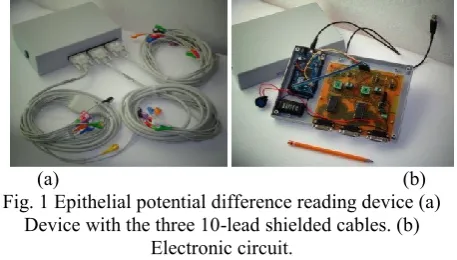
ESULTSThe developed prototype was implemented in a plate of single-sided PCB and DIP adapters for the ADS1115 converters and for I2C ADUM1250 insulator used. Figure 1 illustrates the finished device.
(a) (b)
Fig. 1 Epithelial potential difference reading device (a) Device with the three 10-lead shielded cables. (b)
Electronic circuit.
For testing we used a laptop with a microprocessor I3 to 1.20GHz with 4GB of RAM and Windows of 32-bit operating system installed with Matlab® 2012a. Computer equipment and the developed device are shown in Figure 2.
For testing the device a sinusoidal signal of 10mVpp at 1 Hz and 10 Hz in the channel is applied, while channel 2 is connected to ground, the result of the implementation of the device with the PC using the signal of 1Hz is illustrated in Figure 3 which shows that the maximum magnitude is close to that measured with the oscilloscope, and for the second channel is observed that there may be noise generated by USB port or by the voltage source, with a maximum amplitude 312.5mV and a median 152.5mV.
Fig.3 Test signal of 1Hz applied to channel 1 with channel 2 connected to ground, plotted with the GUI.
With these test signals and applying a DC input characteristics shown in Table 1 were obtained.
Table I. Characteristics of the device.
Characteristics Value
Supply voltage 9 V
Input over voltage and under voltage ±1.024 V
Maximum resolution 31.25 µV
Input impedance 2.4 MΩ
Accuracy ±0.4% + 2
Uncertainty ±0.9mV
In the BDS device [6], [10], [16], a procedure is performed after acquiring the bio electrical signal from the epithelial surface of the breast, which consists of applying a digital low pass filter with a cutoff frequency of 1Hz, thus avoiding interference of the chest movement due to breathing, then an average of the output signal obtained after filtering is applied, after taking a series of samples by a procedure called by researchers the maximum differential voltage is applied (MDV) consisting of subtracting the maximum average voltage acquired the minimum value (see equation 1), this result Is used to make the diagnosis.
𝑀𝐷𝑉 = 𝑚𝑎𝑥(𝑣) − 𝑚𝑖𝑛(𝑣) (1)
Figure 4 is a sample of the signals obtained by performing a preliminary experiment in a 53-year-old woman with size 36B to the electrode 20 of the left breast.
(a)
(b)
Fig. 4 Epithelial potential difference reading device (a) The signal acquired without applying the filter. (b) The
result of applying the IIR low pass filter of 0.25Hz.
As you can see there is a time for the signal to stabilize, the settling time is affected by the time it takes to the multiplexer to perform switching; so that the necessary information is after 300 samples. The information will be useful corresponds to a vector of 700 samples which equals to 5 second measurement. In Figure 5 the results and plotting them are shown, this was acquired after making 5 measurements on the right breast of 4 women, the age of women are: 23 years Person 1, 28 years Person 2, 20 years person 3 and 53 years person 4.
(a)
(b)
Fig. 5 Obtained values from the 4 women in the clinical trial (a) Right breast. (b) Left breast.
In the work on measuring potential differences in breast, the results are reported to be measured On the breast that has benign and malignant tumors. In them it is concluded that when a benign tumor exist, the potential difference in the region where it was located previously it has acquired a 12.4mV measurement or average values greater than this value in some other [6], [7], [16], [23]; so it is considered that there may be a breast abnormality when the reading is above this value. For Malignant tumors measurements in these studies show a value above 15mV. In the results we see that there is no value that exceeds the value that indicates the presence of a breast abnormality.
III.
D
ISCUSSIONbreast with a resolution of 31.25mV and feature a maximum measuring voltage 1.024V was developed. Test signals on the order of millivolts applied and the result was satisfactory as it has accuracy of 0.4% and an absolute uncertainty of 0.19mV. The data measured with the developed device agrees with those acquired by the oscilloscope, both amplitude and frequency.
For the device developed in this project the sampling frequency is 142Hz but according to the further processing to the acquisition concludes that the sampling frequency may be lower than that used in this project, which would have a shorter duration of the test and making a larger number of samples.
The disadvantages faced were the difficulty to place the electrodes and access for people with previously detected anomalies or people who agreed to participate in the clinical trial. To cover these problems is necessary to use other tips to fix cables and create a solid database with measurements in healthy people, to make a diagnosis with the reading device differences in breast epithelial potential.
IV.
M
ATERIALS ANDM
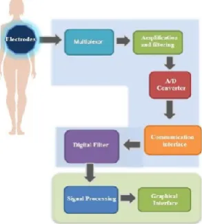
ETHODSThe key to the measurement and analysis of direct current potential (DC) of the skin is absolute maintenance of signal integrity from the skin surface to the signal processing components [9]. This is very important due to the inherent low amplitude of biologic potential. Because biological systems possess excitable cells may use bio electric signals to study and monitor the main functions of these systems. The block diagram shown in Figure 6 is proposed to measure the potential differences, which is based on the history of the development of other diagnostic equipment and detection of breast tumors [10], [11].
Fig. 6 Block diagrams of an epithelial potential difference reading device for anomaly detection in breast.
A.
Electrode Array
When bio potentials are recorded it requires an element that works as an interface between the Body and the measuring instrument, one of them is the electrode. To
measure the potential difference in the surface epithelium of the breast, the most appropriate is surface electrode and more specifically the fastener [12]. Pediatric electrodes were used in the arrangement of electrodes of the developed device, allowing you to place a greater number of them on the epithelial surface of the breast. The diameter of pediatric electrodes is 2.5 cm taking into account the adhesive surface features, but the composite Gel and which acquire the bio electrical signal through the skin surface is 1.5x1.5cm.
For the electrode array design took into account the average bra size used by Mexican women, which can give us a reference average breast size of women in the country, and the average size in Mexico is 34B. A circle with a diameter of 11.4cm can be used as a reference to prepare a cup of this size [13], [14]. So taking into account the area covered by this breast size the electrode array was proposed is shown in Figure 7, as seen two electrodes were placed near the armpit because in this region are the axillary lymph nodes; When breast cancer spreads usually does in this area [15].
Fig. 7 Twenty-four electrode arrangement to measure the potential difference of epithelial breast.
The arrangement of electrodes for a 34B cup may contain 24 electrodes in total per breast. Two electrodes plus one were placed in the palm of the hand that serve as a reference and a third L electrode serving for grounding, this according to other devices designed previously [10], [10]. In the case of the left breast, the distribution of the electrode arrangement would be the mirror of the arrangement of the right breast; this will also lead connecting the H electrode in the left palm and the L electrode in the left ankle. For connection between electrodes and multiplexing stage, three 10-lead shielded cables were used with DB15 connector commonly used in ECG.
B.
Multiplexing Stage
COM2, which in turn will be connected to the next stage of the device are used.
Fig. 8 Diagram of the multiplexing stage and its connection with the DB15 connectors.
At this stage, a ADS1115 16-bit converter with Delta Sigma architecture with I2C communication was used, has an internal PGA and has the characteristic of attenuating signals above 60Hz [18] and 16. A gain of 4 was chosen for the developed device, whereby it has a resolution of 31.25mV with the ability to measure a voltage difference at its inputs of 1.24V. The value of the input impedance depends on the selected gain for the PGA, in this case a differential input impedance of 2.4MW is obtained, this impedance is within the ANSI/AAMI standards for electrocardiographs. The ADS1115 provides 16 bits of data in the form of two’s complement binary. Table 2 summarizes the ideal codes for different input signals applied to the converter output.
Table II. Input signal against output, ideal codes [8]. Input signal, VIN
(AINP-AINN) Ideal output code
≥ 𝐹𝑆(215− 1 )/215 7FFFh
+𝐹𝑆/215 0001h
0 0
−𝐹𝑆/215 FFFFh
≤ −𝐹𝑆 8000h
If the FS to be used will be 1.024V then the maximum voltage the ADC will be able to measure is 1.0239V, which is equivalent to 7FFFh code, as shown in (2).
𝑉𝐼𝑁≥
1.024(215−1)
215 (2)
A 31.25mV is obtained for 0001h and FFFFh codes which will also be the resolution of the device (3).
𝑉𝐼𝑁=1.024215 = 31.25 𝜇𝑉 (3)
In Figure 9 you can see the connection of two analog to digital converters, it is noteworthy that only one converter per multiplexer is used to prevent problems of magnetic induction by the proximity between channels.
Fig. 9 Schematic circuit of the A/D conversion stage.
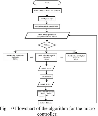
To control both the ADC and the multiplexer is necessary to use a micro controller, which also serves as a communication interface with computer equipment. The most recent card of Arduino call Arduino R DUE was chosen, this card features a micro controller Atmel 32-bit SAM3X8E elected. For the code development, a library called wire was used that is included in the
Arduino IDE 1.5.4. Figure 10 shows the flowchart of the program, a sampling frequency of 142Hz was obtained that for the device, this frequency was used because it is within the minimum requirement according to the Nyquist frequency and other developed devices [6], [10], [11].
Fig. 10 Flowchart of the algorithm for the micro controller.
Fig. 11 Graphics obtained in Matlab R of the ADS1115 converters with the test signal applied to channel 0 of each
multiplexer.
C.
Isolation Stage
It is necessary to isolate the PC voltage source and the micro controller multiplexing stage to prevent any harm to the patient, so opto couplers are used for the control lines of the multiplexer. The isolation circuit for digital terminals micro controller is shown in Figure 12.
Fig. 12 Isolation circuit of the control terminals of the multiplexer.
The I2C bus uses bidirectional lines to transmit and receive signals over a single line, in a variety of applications, drivers, and peripherals are isolated from the bus levels to avoid harmful interference or pressure [19]. To isolate the lines responsible for communication I2C, a bidirectional I2C Isolated from Analog Devices R was used, it is the ADUM1250, which provides two bidirectional channels supporting a complete isolated I2C interface [20]. The ADUM1250 isolators are based on isolating technology from couple Analog Devices, which employs integrated micro transformers semiconductor substrates, thanks to this there is a greater functionality, lower power consumption and has high immunity to external magnetic fields [20]. Figure 13 shows the circuit used to isolate the SDA and SCL lines.
Fig. 13 Circuit diagram used to isolate SDA and SCL lines
D.
Supply voltage Stage
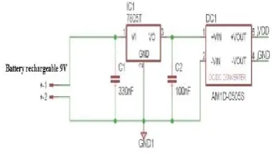
To power the device is necessary to take into account the supply requirements for each of the components that made up the device. According to the decisions taken on the magnitude of voltage necessary for each element of the device, the voltage source used was 5 V. The power stage does not include the Arduino DUE card because it is fed through the USB port and this same source voltage feed section 2 of the isolator I2C and 4 optocoupler input to control the multiplexer. The best alternative to prevent noise coming from a power line in addition to providing user safety is the use of batteries, so a 9V battery is used and for the 5V necessary a regulator voltage of 5 volts was used [21]. A DC-DC 5V AM1D-0505S converter was used to isolate the voltage source from user or patient, the converter is connected to the output of the voltage regulator and thus the 9 volt battery compound circuit will be isolated by the step of multiplexing and A/D conversion [22]. Figure 14 shows the circuit used for the power stage; it will power the steps of multiplexing, analog to digital conversion, the section 1 of the I2C isolators and the receiving part of the optocouplers.
Fig. 14 Circuit diagram of the supply voltage.
E.
Graphical user interface and signal filtering
To view the signals and to filter them or average them a GUI (Graphical User Interface, GUI) was performed using the GUI tool 2012a mathematical software MATLAB R. With this interface the 2-channel signal is acquired and with it you can control the selection analog pin multiplexer and to be swept in the array of electrodes.At the stage of the GUI it was necessary to design a digital IIR low pass filter to obtain the potential difference in the breast epithelial surface and remove frequency components that will not be useful in analyzing the signal. The designed filter has a cutoff frequency of 0.25Hz and the necessary minimum order of 3, Equation 4 corresponds to the filter function.
𝐻(𝑧) =2.5588𝑥10−6𝑧−3+ 7.6763𝑧−1+ 7.6763𝑧−2+ 2.5588𝑥10−6 1 − 2.9335𝑧−1+ 2.8687𝑧−2− 0.9352𝑧−3
The filter response is shown in Figure 15, one can observe the attenuation of -20(dB/decade) and unity gain for CD component, the phase shift is not important for the development of the device since the interest is in the magnitude of the measured signal.
Fig. 15 Frequency and phase response of the filter. After filtering the arithmetic mean of the signal is obtained by Matlab®, as it has been done in previous studies [6], [10].
V.
C
ONCLUSIONThe objective settled at the beginning of the project was fulfilled, since a device was developed and is able to measure the potential differenceson the epithelial surface
of the female breast,with a resolution of 31.25uV and with the characteristic of measuring a maximum voltage of 1.024V. The test signals in the order of millivolts were applied, and the result was satisfactory since it has an accuracy of 0.4% and an absolute uncertainty of 0.19mV. The data measured with the developed device matched with those acquired by the oscilloscope, both in amplitude and frequency.
For the device developed in this project, a sampling frequency of 142 Hz was used, but according to the post-acquisition processing, it is concluded that the sampling frequency may be lower, allowing a shorter duration of the test and take a larger number of samples.
The disadvantages encountered were, the difficulty in placing the electrodes and the cooperation of people with detected anomalies, who agreed to participate in the clinical trial. To cover these drawbacks, it is necessary to use other types of cables for the array and to create a solid database with measurements of healthy people, in order to be able to make a diagnosis with the device and detect to differences in the epithelial potential in the breast.
R
EFERENCES[1] ROBLES-CASTILLO, Javier, et al. Cáncer de mama en mujeres mexicanas menores de 40 años. GinecolObstet Mex 2011, vol. 79, no 8, 482-488.
[2] KERLIKOWSKE, Karla, et al. Effect of age, breast density, and family history on the sensitivity of first screening mammography. Jama 1996, vol. 276, no 1, 33-38. Version December 1, 2016 submitted to Sensors 12 of 12
[3] SREE, SubbhuraamVinitha, et al. The use of skin surface electropotentials for breast cancer detection—Preliminary clinical trial results obtained using the biofield diagnostic system. Journal of medical systems 2011, vol. 35, no 1, 79-86.
[4] SMITH, Susan Rae; FOSTER, Kenneth R.; WOLF, Gerald L. Dielectric properties of VX-2 carcinoma versus normal liver tissue. Biomedical Engineering, IEEE Transactions on 1986, no 5, 522-524.
[5] CUZICK, Jack, et al. Electropotential measurements as a new diagnostic modality for breast cancer. The Lancet 1998, vol. 352, no 9125, 359-363.
[6] FAUPEL, Mark L.; HSU, Yu-Sheng. Dedicated systems for surface electropotential evaluation in the detection and diagnosis of neoplasia. EnElectropotentials in the clinical assessment of breast neoplasia. Springer Berlin Heidelberg 1996, 37-44.
[7] FUKUDA,Mamoru, et al. Prospective evaluation of skin surface electropotentials in Japanese patients with suspicious breast lesions. Japanese journal of cancer research 1996, vol. 87, no 10, 1092-1096.
[8] LONG JR, David M., et al. Initial preclinical experiments with the electropotential differential diagnosis of mammary cancer. EnElectropotentials in the Clinical Assessment of Breast Neoplasia. Springer Berlin Heidelberg 1996, 23-35.
[9] ARONSON, S.; GEDDES, L. A. Electrode potential stability. Biomedical Engineering, IEEE Transactions on 1985, no 11, 987-988.
[10] M. L. Faupel, C. Gordon, J. Stephens y S. D. Nathanson.
Apparatus for sensing bodily conditions. WO 96/33651 PCT/US96/05331, (Cl. A61B 5/05), October 31, 1996. Patent application: April 25, 1996, 38 p.
[11] B. H. Hirschowitz, Kwok-Leung Li. Device and Method for
Detecting the Potential Level of the Electromagnetic Field of a Living Organism. US4328809A, May 11, 1982. Patent application June 26, 1979, 12p.
[12] WEBSTER, John. Medical instrumentation: application and
design. John Wiley & Sons, 2009.
[13] Journal for Sex Research (vol. 24, pp. 177-183), World Health Organization January 2014. Available in:
http://www.statisticbrain.com/breast-size-statistics/
[14] Mc CunnDonald, Custom–Bras 2008. Available in:
http://deofsf.com/Resources/Bra-Underwire-Charts.pdf
[15] Breast cáncer.org. “Afectación de los ganglios linfáticos 2014”.
Available in:
http://www.breastcancer.org/es/sintomas/diagnostico/ganglios_li nfaticos
[16] CROWE JR, Joseph P.; FAUPEL, Mark L. Use of non-directed
(screening) arrays in the evaluation of symptomatic and asymptomatic breast patients. EnElectropotentials in the Clinical Assessment of Breast Neoplasia. Springer Berlin Heidelberg 1996, 57-62.
[17] Data sheet of the CD4067 Multiplexer. Available in:
http://www.ti.com/lit/ds/symlink/cd4067b.pdf
[18] Data sheet of the ADS1115 converter, Texas InstrumentsR .
Available in: http://www.ti.com/lit/ds/symlink/ads1115.pdf
[19] Isolating I2C Interfaces by RonnKliger, AN-913
APPLICATION NOTE. Analog devices. Available in: http://www.analog.com/static/importedfiles/application_notes/A N_913.pdf
[20] Data sheet of the isolator I2C AUM1250 Analog devicesR .
Available in:
http://www.analog.com/static/importedfiles/data_sheets/ADUM1 250_1251.pdf
[21] Data sheet of the UA7805 voltage regulator, Texas InstrumentsR
. Available in: http://www.ti.com/lit/ds/symlink/ua7805.pdf
[22] Data sheet of the DC-DC Converter AimtecR . Available in:
http://www.aimtec.com/site/aimtec/files/datasheet/highresolution /am1d-n-z.pdf
[23] SACCHINI, V., et al. Utility of biopotentials measured with the
Biofield Diagnostic System for distinguishing malignant from benign lesions and proliferative from nonproliferative benign
lesions. En BREAST CANCER RESEARCH AND
TREATMENT. VAN GODEWIJCKSTRAAT 30, 3311 GZ DORDRECHT, NETHERLANDS: KLUWER ACADEMIC PUBL 2002, S41-S41.
A
UTHORSP
ROFILE’
She is a career professor in the Department of Electronics at the Faculty of Engineering of the National Autonomous University of Mexico (UNAM).
Her research interests include the areas of radiofrequency and biomedical, has published articles in congresses, national and international magazines and has a recognition as responsible of the thesis "Mobile Wireless Electrocardiograph Warning System", the winner of TR35 Mexico Magazine 2012 by MIT Innovation.
Email Id. [email protected]
Sergio MeléndezArmas (Mexican, 1986), Electronics and Electrical Engineering, Autonomous University of the State of Hidalgo (UAEH), Mexico. Master of Engineering, National Autonomus University of Mexico (UNAM),Support Technician to equipment of local television and radio company, Support Enginer of Bank TPV´s, Science Teacher of secondary education and Member of Mathematics Academy in Mexico. Fields of Interest: Biomedical Instrumentation, applications of microcontrollers, electronic signal processing computational Mechanics and Software Engineering Method. E-mail: [email protected]
Jorge Naude (Mexican, 1968), is a mechanical engineer, with a Master and PhD. In Thermal fluids engineering.
He works as a full time professor at the faculty of Engineering at the National University of Mexico in Mexico City.
His mayor research is in bubble mechanics, ultrasound theoretical cavitation and thermal bubble collapse.