IRANIAN JOURNAL OF VETERINARY SURGERY
(IJVS) WWW.IVSA.IR
Canine Periodontal Stem Cells: Isolation, Differentiation Potential and
Electronic Microscopic Characterization
Hamideh Salari Sedigh1, DVM Anna Saffarpour2, DDS Shahram Jamshidi∗1, DVM, DVSc
Mahdi Ashouri3, DDS Seyed Mahdi Nassiri4, DVM, PhD
Davood Sharifi1, PhD Parviz Torkzaban5, DDS Abdolmohammad Kajbafzadeh6, M.D
1
Department of clinical Sciences, Faculty of Veterinary Medicine, University of Tehran,Tehran, Iran.
2
Department of Periodontology, Tehran University of Medical Sciences, International Campus, Iran
3
Department of Oral and Maxillofacial Pathology, Faculty of Dentistry, Shahed University of Medical Sciences, Tehran, Iran.
4
Department of Clinical Pathology, Faculty of Veterinary Medicine, University of Tehran, Tehran, Iran.
5
Dental Research Center and Dental School,
Hamadan University of Medical Sciences, Hamadan, Iran.
6
Department of Urology, Children’s Hospital Medical Center, Tehran University of Medical Sciences, Tehran, Iran.
Abstract
Objective- Investigating of the isolation, culture, differentiation potential and electronic
microscopiccharacterization of canine periodontal ligament stem cells (PDLSCs).
Design- Experimental in vitro study
Animals- Four intact, male, mongrel dogs, 8-10 months-old were selected to collect
PDLSCs from their teeth.
Procedures- The dogs were anesthetized and the first maxillary and mandibular
premolars were extracted for the isolation of periodontal ligament cells (PDLCs). Canine PDLCs were plated and culture expanded through 3 successive subcultures. The resultant cells were then plated at differentiating conditions into bone and adipose cell lineages in order to confirm their mesenchymal stem cell character. Furthermore for the first time we
∗
Corresponding author:
Shahram Jamshidi, DVSc
studied PDLSCs ultrastructural morphology by scanning and transmission electron microscopy (SEM and TEM).
Results- based to our observations, isolated canine PDLSCs exhibited mesenchymal stem
cells (MSCs) characteristics such as fibroblastic appearance, clonogenicity, high proliferation and easily differentiation into osteocyte and adipocyte. This study determined morphologically by electron microscopy a secreted extracellular matrix (ECM) around the PDLSCs and also they contained abundant organelles, such as mitochondria, ribosome, rough endoplasm and collagen fiber as assessed by transmission electron microscopy.
Conclusion- Fibroblastic cells from canine PDLCs are capable to be expanded and
proliferated in vitro and have differentiation potential into two skeletal lineages; hence they are MSCs that are suitable for cell therapy experimentations.
Key Words- Stem cell, Periodontal ligament, canine.
Introduction
Periodontitis is an inflammatory disease of the periodontium that consist of the supporting structures of the teeth comprising periodontal ligament, cementum, alveolar bone and gingiva1. Several diseases are caused the destruction of the connective tissue matrix and cells, the loss of periodontal ligament attachment and resorption of alveolar bone which resulting in tooth loss.2,3 In recent years, a number of protocols for periodontal treatment including grafting, root surface conditioning, application of various growth factors as well as bone and enamel matrix proteins 4,5 and guided tissue regeneration have been applied. Among these methods, guided tissue regeneration is currently the most popular procedure, however it has some problems consisting variable results, limited clinical improvement, and susceptibility to infection.6 There is a high demand to achieve more predictable clinical procedures in periodontal regeneration which has long been a challenge. To date, cell-based therapies have progressed rapidly7. The defining characteristics of stem cells (SCs) are clonogenic
self-renewal cells which have the capability of production and differentiation of mature progeny cells. Embryonic SCs and adult SCs are the two major categories of SCs which have been described.8
The periodontal ligament (PDL), dental papilla, dental pulp and follicle in human are identified as easily accessible sources of adult SCs.9 The PDL is a specialized, vascular, and highly fibrous tissue that connects cementum to inner wall of alveolar bone to maintain tooth in situ. This highly supports tooth function, maintenance of homeostasis and tissue healing in response to any periodontal insults or mechanical trauma.10,11 This indicates that progenitor cells and possibly stem cells, exist within the PDL cell population.12 It has been demonstrated that human PDL which contains a heterogeneous cell populations 13, has in vitro capability of differentiation into chondrocytes, osteoblasts and adipocytes.14-17 In a recent study, human PDL-derived mesenchymal stem cells as periodontal ligament stem cells were initially identified and subsequently PDLSCs were characterized.18 The objectives of this study were
primarily to isolate and characterize for the first time canine PDL-derived mesenchymal stem cells.
Materials and Methods
Animals
vaccination and deworming were carried out. The animal research protocol was approved by the institutional Animal Care and use committee of Tehran, University of Medical Sciences, and it was according to the standards of Association for Assessment and Accreditation of Laboratory Animal Care. Throughout the experiments, the animals were kept individually in the cages, fed with Nutripet (Behintash Company, Karaj, Iran) and monitored for general appearance, activity, exertion and weight.
Isolation and culture of periodontal stem cells
Under general anesthesia using acepromazine (0.1 mg/kg, IM) and ketamine-diazepam (10 mg/kg, IV), the teeth were wiped with gauze soaked in chlorhexidine.19 The PDL cells were gently separated from the root surface of the middle third of the extracted maxillary premolars in each dog. After separation, PDLCs culture was performed in accordance with the technique described by Seo et al..18 The obtained PDL cells was enzymetically digested for 1 hour at 37°C in a solution of 3mg/ml Collagenase type I (Gibco, UK) and 4 mg/ml Dispase (Gibco, Uk). Single-cell suspensions were obtained by filtering it through a 70 µm cell strainer[1]. Single-cell suspension (1×10⁵ cells) were seeded into 25-cm2 culture flasks in Dulbecco Modified Eagle Medium (DMEM) (Gibco, Uk) supplemented with 15% fetal bovine serum (FBS) (Gibco, Uk) and 100 IU/ml penicillin G and 100 µg/ml streptomycin solution (Gibco,
Uk) and incubated in a humidified atmosphere (37°C, 5% Co2)19,21. Canine PDLSCs were
harvested with 0.25% trypsin/EDTA (Gibco, Uk) at 37°C for 3 minutes.20
Differentiation
Osteogenic induction
To investigate the potential of PDLSCs to differentiate into osteoblasts, confluent passage-3 cells were provided with DMEM supplemented with 50µg/ml ascorbic2-phosphate (Gibco, Uk), 10 nM dexamethasone (Sigma, USA) and 10 mM ß-glycerol phosphate (Sigma) for 3 weeks. At the end of this period, alizarin red staining was used to show the deposition of mineralization matrix. For staining, the cultures were first fixed by methanol for 10 minutes and then subjected to alizarin red solution 2 g in 100 ml water for 2 minutes.
Adipogenic induction
To induce adipose differentiation, passage-3 confluent culture was treated with the medium containing 100 nM dexamethasone (Sigma) and 50 mg/ml indomethasine (Sigma, USA). After 3 weeks, the cells were fixed with 4% paraformaldehyde at room temperature for 30 min, washed with 70% ethanol and stained by oil red solution (0.5 g in in 99% isopropanol) for 15 minutes.
Scanning Electron Microscopy
with isoamyl acetate. After gold coating, the samples were examined using a TESCAN VEGA scanning electron microscopy (TESCAN VEGA, USA, www.Tescan-usa.com).
Transmission Electron Microscopy
After washing with phosphate-buffered saline (PBS), the cells were fixed in 3% glutaraldehyde in 0.1 mol/L PBS at 4 °C for 1 h and post-fixed in 1% osmium tetroxide for 1 h. The samples were then dehydrated through a graded ethanol series and embedded in epoxy resin. The thin sections were mounted on copper grids and double-stained with uranyl acetate for 10 min and lead citrate for 4 min as described previously22, then ultrastructurally scoped using an electron microscopy (Zeiss, Germany).
Results
Cell culture
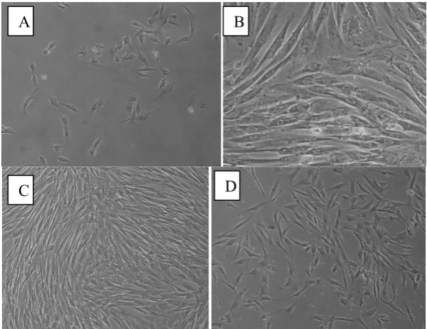
To identify the capacity to generate clonogenic clusters of adherent fibroblastic-like cells, single cell suspension of canine PDL cells was plated in 25-cm2 culture flasks under specific culture conditions. After 7 days numerous colonies were formed, in fact the ability of canine PDL-derived cells to form adherent clonogenic cell clusters of fibroblast-like cells, similar to those recorded for human PDLSCs, was shown by the formation of approximately 21 single colonies generated per 105 single cells cultured at low density and the cells were spindle-shaped (Fig. 1), morphological characteristic of the plastic-adherent colony-forming canine PDLSCs were similar to other MSCs 23 including large flattened cells (Fig. 1A), long spindle-shaped cells (Fig. 1B), and short-spindle-shaped cells (Fig. 1C) and colonies had a high proliferation rate and a more finite lifespan (data not show).
Figure 1. Morphological observation of canine primary PDLSCs. (A) Low magnification view of primary
culture on day 7. Primary PDLSCs grew in colonies with various cell morphologies comprising each colony. Small rounded cells were occasionally observed on top of adherent PDLSCs (arrow). There were at least three major types of adherent cells as observed under higher magnification views on day 14; (B) large flattened cells, (C) long spindle-shaped cells, and (D) short spindle-shaped cells. Scale bar=100 μM.
D C
Differentiation assays
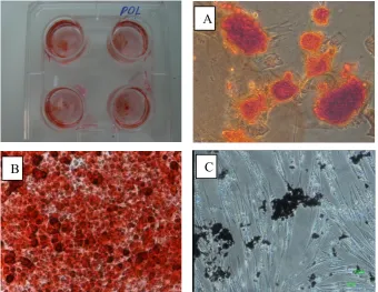
We assessed in vitro whether PDL cells, like the other mesenchymal stem cells, had similar capacity to differentiate into other cell lineages such as osteocytes and adipocytes. Mineral deposition: Alizan red staining — After 17 days of induction in mineralization medium, alizarin red staining of the PDL stem cell cultures (Fig. 2A,B) was associated with distribution of mineralized deposits throughout the tissue culture wells as well as cell clusters in both of them. Adipogenic differentiation: Oil red O staining — After 14 days of induction in adipogenic media, lipid droplets were detected in the cytoplasm of differentiated PDLSCs (Fig. 2C).
Figure 2. Canine differentiation potential: The cells were able to differentiate into osteblasts and adipocytes. A,
B- Differentiation into osteocytic, these were evident in alizarin red staining. C- Differentiation into adipocytes, this was evident in oil red staining.
SEM
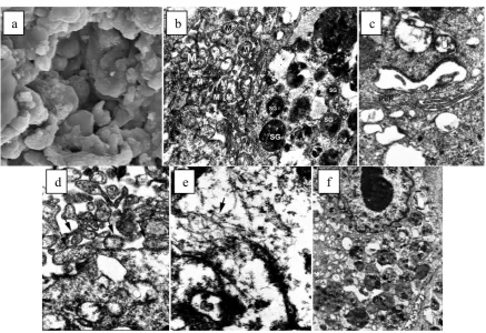
Secreted ECM, which has a critical role in regeneration of damaged tissues, was discernible by scanning electron microscopy. It seems the ECM produced by PDLSCs is comparable in thickness and prominence for the same mesenchymal stem cells from bone marrow source (Fig. 3a).
TEM
PDLSCs were readily distinguishable by electron microscopy. PDLSCs contained numerous secretary granules and relatively abundant mitochondria within the cytoplasm (Fig. 3b-f). PDLSCs were characterized by developed rough endoplasmic reticulum (RER) and free ribosomes. Granule exocytosis was also seen in some PDLSCs. Moreover, extracellular
A
collagen fibers were discernible by close ultrastructural scrutiny of PDLSCs and had round nuclei with regular outlines, a single, large nucleolus was present in the nuclei of these types cells.
Figure 3. Scanning and Transmission electron microscopy analyses of PDLSCs. (a): Scanning electron
microscopy, clusters of PDL cells, with secreted ECM. (b-f): Transmission electron microscopy; (b) A PDL cell with numerous intracytoplasmic secretory granules (SG) and many mitochondria (M). (c) Rough endoplasmic reticulum (RER), polyribosomes (PR), along with numerous free ribosomes is seen in the cytoplasm of PDL cells. (d) Granule exocytosis in PDL cells. (e) Synthesized collagen fibers organizing in the extracellular space around PDL cells. (f) PDLSCs had round nuclei with regular outlines; a single, large nucleolus was present in the nuclei of these types of cells.
Discussion
In the present study, we successfully isolated and studied morphology, proliferation capacity, differentiation ability, ultrastructural characteristics multipotent mesenchymal stem cells from canine PDL, which can serve as a unique reservoir of stem cells to apply to regenerative procedures without the necessity of more invasive procedures. PDLSCs in this report showed many characteristics similar to those already reported for human24, ovine 25, equine 26, porcine PDLSCs 27 and canine 28 and other mesenchymal stem cells 29-31 and dental pulp derived stem cells.32-35 Our findings also clearly demonstrated that canine PDL includes a population of
multipotent postnatal stem cells at different stages of differentiation and lineage commitment, Nagatomo and et al have founded the similar results for human PDL stem cells.36 As researches have earlier demonstrated, PDL cells can be cultured at low densities with duplicitous potential indicating the capacity of PDL cells to proliferate and produce colonies from a single cell.36 We observed heterogenicity in cell morphology for colony-forming unit-fibroblastic (CFU-F) derived from PDL in primary passages. Two distinct morphologies were
a b c
detected to grow concomitantly: solitary canine periodontal fibroblast (CPF) was elongated cells with oval-shaped nuclei and an extended cytoplasm that grew long cell processes; canine dental cementoblast (CDC) is round cells with a small cytoplasm and intensive nuclei. CPF in the vicinity of the CDF, became bipolar and spindle-shape.26 In addition, we induced cell differentiation along the two classical mesenchymal pathways, osteocytic and adipocytic lineages, and we confirmed differentiation PDLSCs capability for the production of mineralized deposits and lipid droplets as previously has been shown with other mesenchymal cell populations. In this study, we attempted to isolate PDLSCs purely and then maintained high purity in the expansion of them in third passage, thus we did not interfere of differentiated cell types such as fibroblasts with mineralization process.37 Morphologically, PDLSCs displayed many short and long branching cytoplasmic processes under a scanning electron microscopy, surrounded by secreted extracellular matrix the cells contained abundant organelles, such as mitochondria, ribosome, and rough endoplasm, and secreted extracellular matrix, as assessed by transmission electron microscopy. The collagen fibrils produced by PDLSCs were large and thick.
Consequently, canine model in stem cell investigations are a great importance since they are genetically more close to humans in contrast to rodents. Hence preclinical therapeutic procedures such as cell and gene therapies require an intermediate step between rodent or in vitro study and clinical trials due to safety and investigation of efficacy. For this reason, results from canine models have been much more predictive with regard to human clinical trials.
Conclusion
Recent finding reveal that periodontal ligament contains a population of multipotent postnatal stem cells that can be isolated and expanded in vitro, providing a unique source of stem cells. We hope this present study preliminary demonstrates that canine PDLSC may hold promise as the utility of using an autologous PDLSCs therapeutic approach to treat periodontitis in dogs as preclinical model.
Acknowledgment
The authors gratefully acknowledge the tremendous contribution and financially supported by Research Centre of Dentistry Faculty, Hamadan University of Medical Sciences, and we would like to thank Dr. Kajbafzadeh, Dr. Mohseni and Dr. Rouhi from Department of Urology, Children’s Hospital Medical Center, Tehran University of Medical Sciences for supporting the study.
References
1. Page RC, Offenbucher S, Schroeder HE, et al. Advanced in the pathogenesis of periodontitis: Summary of developments, Clinical implications and future direction. Periodontology 2000 1997;14:216-248.
2. Bennatti BB, Gasati MZ, Sallum GA, et al. Physiological feature of periodontal tissue engineering utilizing periodontal ligament cells. Journal of bioscience and bioengineering 2007;103(1):1-6.
4. Quinones CR, and Caffesse RG, Current status of guided periodontal tissue regeneration.Periodontology 2000 1995; 9:55-68.
5. Giannobile WV, Periodontal tissue engineering by growth factors. Bone 1996;19(1):S23-S37.
6. Kramer PR, Nares S, Kramer SF, et al. Mesenchymal Stem Cells Acquire Characteristics of Cells in the Periodontal Ligament in vitro. J Dent Res 2004;83(1):27-34.
7. Matsuda N, Shimizu T, Yamato M, et al. Tissue Engineering Based on Cell Sheet Technology. Adv Mater 2007;19:3089-3099.
8. Martin DR, Cox NR, Hathcock TL, and et al. Isolation and charaterization of multipotential mesenchymal stem cells from feline bone marrow. Experimental Hematology 2002;30:879-886.
9. Morsczeck C, Schmalz G, Reichert TE, et al. Somatic stemm cells for regenerative dentistry.Clin Oral Investig 2008;12:113-118.
10.Bartold PM, McCulloch CA, Narayanan AS, et al. Tissue Engineering: a new paradigm for periodontal regeneration based on molecular and cell biology. Periodontol 2000 24:253-269.
11.Shimono M, Ishikawa T, Ishikawa H, et al. Regulatory mechanisms of periodontal regeneration.Microsc Res Tech 2003;60:491-502.
12.Acharya, A., Shetty, SH., and Deshmukh, V., Periodontal Ligament Stem Cells: An Overview.J Oral Biosci 2010;52(3):275-282.
13.Lekic P, Rojas J, Birek C, et al. Phenotypic comparison of periodontal ligament celss in vivo and in vitro. J Periodontal Res 2001;36:71-79.
14.Gould TR, Melcher AH, Brunette DM, Migration and division of progenitor cell-population in periodontal-ligament after wounding. Journal of Periodontal Research 1980;15(1):20-42.
15.McCulloch CA, Melcher AH, Cell-density and cell generation in the periodontal ligament of mice.American Journal of Anatomy 1983;167(1):43-58.
16.McCulloch CA, Bordin S, Role of fibroblast subpopulations in periodontal physiology and pathology.Journal of Periodontal Research 1991;26(3):144-154.
17.Isaka J, Ohazama A, Kobayashi M, et al. Participation of periodontal ligament cells with regeneration of alveolar bone. Journal of Periodontology 2001;72(3):314-323. 18.Seo BM, Miura M, Gronthos S, et al. Investigation of multipotent postnatal human
dental pulp stem cels.Lancet 2004;364:149-155.
19.Akizuki T, Oda S, Komaki M, et al. Application of periodontal ligament cell sheet for periodontal regeneration: a pilot study in beagle dogs.J PeriodontalRes 2005;40:245-251.
20.Kim SH, Kim KH, Seo BM, et al. Alveolar Bone Regeneration by Transplantation of Periodintal Ligament Stem Cells and Bone Marrow Stem Cells in Canine Peri-Implant Defect Model: A Pilot Study. J Periodontol;2009:1815-1823.
21.Iwata T, Yamato M, Tsuchioka H, et al. Periodontal regeneration with multi-layered periodontal ligament-derived cell sheets in a canine model. Biomaterials 2009;30:2716-2723.
23.Kamishina H, Farese JP, Storm JA, et al. The frequency, growth kinetics, and osteogenic/adipogenic differentiation properties of canine bone marrow stromal cellc. In Vitro Cell.Dev.Biol.-Animal 2008;44:472-479.
24.Seo BM, Miura M, Gronthos S, et al. Investigation of multipotent postnatal stem cells from human periodontal ligament. Lancet 2004;364(9429):149-155.
25.Gronthos S, Akintoye SO, Wang CY, et al., Bone marrow stromal stem cells for tissue engineering. Periodontology 2000 2006;41:188-195.
26.Staszyk C, Gasse H, primary culture of fibroblasts and cementoblasts of the equine periodontium.Research in veterinary science 2007;82:150-157.
27.Liu Y, Zheng Y, Ding G, et al. Periodontal ligament stem cell-mediated treatment for periodontitis in miniature swine.Stem Cells 2008;26(4): p. 1065-1073.
28.Iwata T, Yamato M, Tsuchioka H, et al. Periodontal regeneration with multi-layered periodontal ligament-derived cell sheets in a canine model. Bimaterials 2009;30:2716-2723.
29.Owen M, Lineage of osteogenic cells and their relationship to the stromal system. In: Peck WA, editor.Bone and Mineral Research 1985; 3:1-25.
30.Burder S, Fink DJ, Caplan AI., Mesenchymal stem cells in bone marrow development, bone repair, and skeletal regeneration.J Cell Biochem 1994;56:283-94.
31.Caplan AI, Mesenchymal stem cells. J Ortho Res 1991. 9:641-50.
32.Gronthos S, Mankani M, Brahim J, et al. postnatal human dental pulp stem cells (DPSCs) in vitro and in vivo. Proc Natl Acad Sci 2000;97:13625-30.
33.Gronthos S, Brahim J, Li W, et al. Stem cells properties of human dental pulp stem cells. J Dent Res 2002;81:531-35.
34.Shi S, Robery PG, Gronthos S, comparison of human dental pulp nd bone marrow stromal stem cells by cDNA microarray analysis.Bone 2001;29:532-39.
35.Miura M, Gronthos S, Zhao M, et al. SHED: stem cells from human exfoliated decidous teeth.PNAS 2003;100:5807-12.
36.Nagatomo K, Komaki M, Sekiya I, et al. Stem cell properties of human peridodontial ligament cells. J Periodont Res 2006;41:303-310.
هﺪﻴﻜﭼ
ﻲﮕﺳ
لﺎﺘﻧدﻮﻳﺮﭘ
نﺎﻣﺎﮕﻴﻟ
ﻲﻤﻴﺸﻧاﺰﻣ
يدﺎﻴﻨﺑ
يﺎﻬﻟﻮﻠﺳ
:
و
ﺰﻳﺎﻤﺗ
ﻞﻴﺴﻧﺎﺘﭘ
،ﺖﺸﻛ
،يزﺎﺳاﺪﺟ
ﻲﻧوﺮﺘﻜﻟا
پﻮﻜﺳوﺮﻜﻴﻣ
تﺎﺼﺨﺸﻣ
ﻖﻳﺪﺻيرﻻﺎﺳهﺪﻴﻤﺣ
1
، رﻮﭘرﺎﻔﺻﺎﻧآ
2
يﺪﻴﺸﻤﺟماﺮﻬﺷ،
1
* يرﻮﺷﺎﻋيﺪﻬﻣ،
3
يﺮﻴﺼﻧيﺪﻬﻣ،
4
ﻲﻔﻳﺮﺷدواد،
1 ، كﺮﺗﺰﻳوﺮﭘ نﺎﺑز 5
فﺎﺒﺠﻛﺪﻤﺤﻤﻟاﺪﺒﻋ، هداز
6
1
ناﺮﻳا،ناﺮﻬﺗ،ناﺮﻬﺗهﺎﮕﺸﻧادﻲﻜﺷﺰﭙﻣادهﺪﻜﺸﻧاد،ﻲﻫﺎﮕﻧﺎﻣردمﻮﻠﻋهوﺮﮔ
2
ﻮﺘﻧدﻮﻳﺮﭘهوﺮﮔ ﻟ
ﻦﻴﺑﻪﺧﺎﺷ،يژﻮ ﻤﻟا
ﻠ هﺪﻜﺸﻧادﻞ مﻮﻠﻋهﺎﮕﺸﻧادﻲﻜﺷﺰﭙﻧاﺪﻧد
هﺎﮕﺸﻧادﻲﻜﺷﺰﭘ ﺗ
ناﺮﻬ
3
ناﺮﻳا،ناﺮﻬﺗ،ﺪﻫﺎﺷهﺎﮕﺸﻧادﻲﻜﺷﺰﭙﻧاﺪﻧدهﺪﻜﺸﻧادناﺪﻧدونﺎﻫديژﻮﻟﻮﺗﺎﭘهوﺮﮔ
4
ناﺮﻳا،ناﺮﻬﺗ،ناﺮﻬﺗهﺎﮕﺸﻧادﻲﻜﺷﺰﭙﻣادهﺪﻜﺸﻧاد،يژﻮﻟﻮﺗﺎﭘلﺎﻜﻴﻨﻴﻠﻛهوﺮﮔ
5
ناﺮﻳا،ناﺪﻤﻫ،ناﺪﻤﻫﻲﻜﺷﺰﭘمﻮﻠﻋهﺎﮕﺸﻧادﻲﻜﺷﺰﭙﻧاﺪﻧدهﺪﻜﺸﻧادتﺎﻘﻴﻘﺤﺗﺰﻛﺮﻣ
6
اﺮﻬﺗﻲﻜﺷﺰﭘهﺎﮕﺸﻧاد،يژﻮﻟورواﺶﺨﺑ،ناﺮﻬﺗهﺎﮕﺸﻧاد،ﻲﻨﻴﻤﺧمﺎﻣانﺎﺘﺳرﺎﻤﻴﺑلﺎﻔﻃاﻲﻧﺎﻣردﺰﻛﺮﻣ ناﺮﻳا،ناﺮﻬﺗ،ن
فﺪﻫ
-لﺎﺘﻧدﻮﻳﺮﭘنﺎﻣﺎﮕﻴﻟﻲﻤﻴﺸﻧاﺰﻣيدﺎﻴﻨﺑيﺎﻬﻟﻮﻠﺳتﺎﻴﺻﻮﺼﺧوﺰﻳﺎﻤﺗﻞﻴﺴﻧﺎﺘﭘ،ﺖﺸﻛ،يزﺎﺳاﺪﺟ ﻪﻌﻟﺎﻄﻣحﺮﻃ -ﻲﺑﺮﺠﺗﻪﻌﻟﺎﻄﻣ -ﻲﻫﺎﮕﺸﻳﺎﻣزآ تﺎﻧاﻮﻴﺣ -4 هﺪﺸﻧﻪﺘﺧا،ﺮﻧ،ﻲﻧاﺮﻳاداﮋﻧﮓﺳهدﻼﻗ 10 -8 ﻪﻫﺎﻣ رﺎﻛشور -ﻪﻌﻟﺎﻄﻣﻦﻳارد رﻻﻮﻣﺮﭘناﺪﻧدﻞﻣﺎﻛ ﻲﺷﻮﻬﻴﺑﺖﺤﺗ اﺪﺘﺑا
لﺎﺘﻧدﻮﻳﺮﭘ يﺎﻬﻟﻮﻠﺳيزﺎﺳاﺪﺟياﺮﺑﺎﻬﮕﺳ ﻦﻴﻳﺎﭘوﻻﺎﺑﻚﻓ
ﺪﻧﺪﺷ هﺪﻴﺸﻛ ﺖﻨﻣﺎﮕﻴﻟ .
ﺪﻧﺪﻳدﺮﮔ ﺮﻴﺜﻜﺗ ﻲﻟاﻮﺘﻣ ژﺎﺳﺎﭘ ﻪﺳ ﻲﻃو ﺖﺸﻛ ﻲﮕﺳ لﺎﺘﻧدﻮﻳﺮﭘنﺎﻣﺎﮕﻴﻟ يﺎﻬﻟﻮﻠﺳ .
رد ﻞﺻﺎﺣ يﺎﻬﻟﻮﻠﺳ
دﻮﺷﺪﻴﻳﺄﺗﺎﻬﻧآﻲﻤﻴﺸﻧاﺰﻣ يدﺎﻴﻨﺑﺖﻴﻫﺎﻣ ﺎﺗﺪﻨﺘﻓﺮﮔ راﺮﻗﻲﺑﺮﭼو ناﻮﺨﺘﺳا ﻪﺑ ﺰﻳﺎﻤﺗﻂﻳاﺮﺷ .
اﺮﺑﻦﻳاﺮﺑ هوﻼﻋ ﺮﻈﻧ زا رﺎﺑﻦﻴﻟوا ي
ﺪﻨﺘﻓﺮﮔراﺮﻗﻲﺳرﺮﺑدرﻮﻣﻲﻧوﺮﺘﻜﻟاپﻮﻜﺳوﺮﻜﻴﻣﻂﺳﻮﺗﻲﮕﺳلﺎﺘﻧدﻮﻳﺮﭘنﺎﻣﺎﮕﻴﻟﻲﻤﻴﺸﻧاﺰﻣيدﺎﻴﻨﺑيﺎﻬﻟﻮﻠﺳﻲﻜﻳژﻮﻟﻮﻓرﻮﻣ .
ﺞﻳﺎﺘﻧ
-ﺨﺸﻣ ﻲﮕﺳ لﺎﺘﻧدﻮﻳﺮﭘ نﺎﻣﺎﮕﻴﻟ ﻲﻤﻴﺸﻧاﺰﻣ يدﺎﻴﻨﺑ يﺎﻬﻟﻮﻠﺳ ،ﺎﻣ تاﺪﻫﺎﺸﻣ سﺎﺳا ﺮﺑ ﺼ
يدﺎﻴﻨﺑ يﺎﻬﻟﻮﻠﺳ صﻮﺼﺨﻣ تﺎ
ﺪﻨﻧﺎﻣﻲﻤﻴﺸﻧاﺰﻣ
:
ﺘﺳﻼﺑوﺮﺒﻴﻓﻞﻜﺷ ار ﺖﻴﺳﻮﭙﻳدآوﺖﻴﺳﻮﺌﺘﺳايﺎﻬﻟﻮﻠﺳﻪﺑﺖﺣارﺰﻳﺎﻤﺗوﻊﻳﺮﺳﺮﻴﺜﻜﺗ،نﺪﺷهﺰﻴﻧﻮﻠﻛﻪﺑ ﻞﻴﻣ،ﻲ
ﺪﻧداد نﺎﺸﻧ .
رد ﻲﺳرﺮﺑ يدﺎﻴﻨﺑ يﺎﻬﻟﻮﻠﺳ فاﺮﻃا رد ﻲﻟﻮﻠﺳ جرﺎﺧ ﺲﻜﻳﺮﺗﺎﻣ ﻚﻳ ﻲﻧوﺮﺘﻜﻟا پﻮﻜﺳوﺮﻜﻴﻣ ﻂﺳﻮﺗ ﻲﻜﻳژﻮﻟﻮﻓرﻮﻣ
ﺎﺑ ﻲﺳرﺮﺑ رد ﻦﻴﻨﭽﻤﻫ و ﺪﻳدﺮﮔ ﺺﺨﺸﻣ ﻲﮕﺳ لﺎﺘﻧدﻮﻳﺮﭘ نﺎﻣﺎﮕﻴﻟ ﻲﻤﻴﺸﻧاﺰﻣ پﻮﻜﺳوﺮﻜﻴﻣ
ﻦﺸﻴﻤﺴﻧاﺮﺗ ﻲﻧوﺮﺘﻜﻟا ﺎﻬﻟﻮﻠﺳ ﻦﻳا
ﻞﻧﺎﮔرايوﺎﺣ ﻪﻠﻤﺟزاﻲﻧاواﺮﻓيﺎﻫ
: ﺪﻧدﻮﺑﻲﻧژﻼﻛيﺎﻫﺮﺒﻴﻓوﻦﺸﺧﻢﺳﻼﭘوﺪﻧا،موزﻮﺒﻳر،يرﺪﻨﻛﻮﺘﻴﻣ .
ﻪﺠﻴﺘﻧ ﻲﻨﻴﻟﺎﺑدﺮﺑرﺎﻛويﺮﻴﮔ
-ﻂﻴﺤﻣ ردﺮﻴﺜﻜﺗوﺖﺸﻛترﺪﻗلﺎﺘﻧدﻮﻳﺮﭘنﺎﻣﺎﮕﻴﻟيﺎﻬﻟﻮﻠﺳزاهﺪﺷاﺪﺟﻲﺘﺳﻼﺑوﺮﺒﻴﻓيﺎﻬﻟﻮﻠﺳ
ﻪﺑﺰﻳﺎﻤﺗﻞﻴﺴﻧﺎﺘﭘوﺖﺸﻛ ﺪﻨﺘﺴﻫارادارﻲﺘﻠﻜﺳا هدرود
. ﻲﻧﺎﻣردلﻮﻠﺳياﺮﺑﻲﻤﻴﺸﻧاﺰﻣيدﺎﻴﻨﺑيﺎﻬﻟﻮﻠﺳناﻮﻨﻋﻪﺑﺎﻬﻧآورﻦﻳازا
ﻲﻣﺐﺳﺎﻨﻣﻲﺑﺮﺠﺗيﺎﻫرﺎﻛرد ﺪﻨﺷﺎﺑ
.
نﺎﮔژاوﺪﻴﻠﻛ
-ﮓﺳ،لﺎﺘﻧدﻮﻳﺮﭘنﺎﻣﺎﮕﻴﻟ،يدﺎﻴﻨﺑلﻮﻠﺳ .