Review Article
ULTRASOUND BONE SURGERY: A BOON FOR THE BONE
Ajeya Kumara EG,* NehaToshniwal,** Vinayak S Gowda ***
*Senior Lecturer, Department of Periodontology, Kannur Dental College, Anjarakandy, Kerala, India **Consultant Periodontist, Mumbai, India
***Professor, Department of Periodontology. V S Dental College and Hospital, Bangalore, Karnataka, India
________________________________________________________________________
INTRODUCTION
The ultrasonic or ultrasound frequency, as the name implies, is a frequency above the audible range for humans, usually above 20 kHz. In dental applications the ultrasound frequency used ranges from 24 kHz to 36 kHz the frequency range capable of cutting mineralized tissue. Ultrasound has been used for decades to cut tissue. At present, the use of power ultrasonics is becoming very popular in the field of dentistry. Ultrasonic scalers driven by magnetostrictive, or piezoelectric ultrasonic transducers have been used to remove tartar and plaque on teeth. In the last decade; a revolutionary surgical technique, known as piezosurgery, was invented by Professor Tomaso Vercellotti in 1998. The Piezosurgery device consists of a novel piezoelectric ultrasonic transducer powered by an ultrasonic generator, capable of driving a range of resonant cutting inserts. This innovative device was first designed and commercialised by Mectron® Medical Technology.[1] Piezosurgery was invented and developed to reach increasingly
higher levels of precision, safety and rapidity in recovery in bone surgery.
MECHANISM
With piezoelectric ultrasonic the frequency is created by driving an electric current from a generator over piezoceramic rings, which leads to their deformation. The resulting movement from the deformation of the ring sets up a vibration in a transducer and/or amplifier, which creates the ultrasound output. These waves transmitted to the handpiece tip, also called an insert, where the longitudinal movement results in cutting of osseous tissue by microscopic shattering of bone. The active element is basically a piece of polarized material with electrodes attached to two of its opposite faces. Application of electric field to this material causes electrostriction of molecules. The active element of most acoustic transducers used today is a piezoelectric ceramic, which can be cut in various ways to produce different wave modes. Early 1950's, piezoelectric crystals made from quartz crystals and magnetostrictive materials were primarily used. Piezoeletric ceramic soon became the dominant ABSTRACT
Piezoelectric ultrasonic units have been used to power periodontal scalers and endodontic instruments for many years. Piezoelectric bone surgery known simply as piezosurgery is a new technique of osteotomy and osteoplasty which requires the use of micro-vibrations of ultrasonic frequency scalpels. The principle of piezosurgery is ultrasonic transduction obtained by piezoelectric ceramic contraction and expansion. The vibrations thus obtained are amplified and transferred onto the insert of a drill which then has mechanical cutting effect exclusively on mineralized tissues. With piezoelectric surgery it has been possible to perform precise osteotomy lines, micrometric and curvilinear particularly in close proximity to the vessels and nerves. Though this new ultrasound cutting method involves a different learning curve compared to other techniques, it has been increasingly used in various fields of dentistry with improvements in power and geometry of the inserts which has a selective action upon the mineralized tissues.
KEYWORDS: Ultrasound; Piezosurgery; Bone surgery
molecular cohesion in liquids and the appearance of zones of depression that fill up with vapour until they form bubbles about to implode. In case of detartrating tools, cavitation occurs when the water spray comes in contact with the insert vibrating to intermediate frequencies.[1-3]
INSTRUMENT
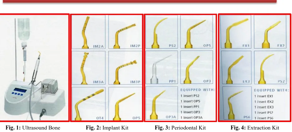
Piezoelectric device is with a functional frequency of 25-29 kHz and the possibility of 30 Hz digital modulation and a series of inserts of different forms with a linear vibration ranging from 60 to 200 μm. The power of the device is 5 W (Ultrasonic scaler 2 W). Each part of the instrument (Fig. 1) is described below.[1,4,5] 1. Control Panel
There are two basic programs BONE and ROOT. In the BONE program, it is possible to adapt the power to any of four levels depending upon bone quality. In the ROOT program, the power can be set to either PERIO or ENDO. The CLEAN function is activated by pressing a button and the footswitch together to start the cleaning cycle of the unit’s main tubes.
2. The Dynamometric Wrench
The insert tips are tightened to the handpiece with the dynamometric wrench.
3. The Liquid
Liquid is drawn from a bag or bottle which hangs from the rod provided. All parts of the unit through which the liquid passes, including the handpiece cord and the handpiece itself, are fully sterilizable.
4. The Peristaltic Pump
The peristaltic pump is for cooling with a jet of solution that discharges from the insert with an adjustable flow of 0 – 60 ml/min and removes detritus from cutting area. For cooling effect, the solution is refrigerated at 400C.
5. The Handpiece
The handpiece is connected permanently to the handpiece cord and the two are sterilized together. Each Piezosurgery unit comes with two handpieces.
osteoplasty.
The Ultrasonic bone surgery device differs from conventional tools by 4 parameters: the generator frequencies, and insert’s weight, hardness and form. It is made of a generator of intermediate frequencies. Inserts used are those whose vibrations can enter in resonance with the piezoelectric ceramic chips of shaft. This resonance enables us to increase the energetic output, making the insert more efficient.
Inserts Available Can Be Classified As
- Sharp, covered with titanium nitrate (gold colour), which offers a harder surface, and, in turn, maximum efficacy in cutting;
- Diamonds, which are used in the case of thin
bone osteotomy or to complete osteotomies, close to important anatomical structures. These offer a clinically less efficacious cut, are histologically more traumatic than the cutting inserts, but much safer.
INSERT TIPS COLOR
- Gold For all insert tips used to treat bone. - Steel For all insert tips used to treat soft tissue
or delicate surfaces such as the roots of teeth. Many different tips are available for different purposes. Four major categories of tip kits depending on the type of surgery are Implant Kit (Fig. 2), Periodontal kit (Fig. 3), Extraction Kit
(Fig. 4), Sinus lift Kit (Fig. 5). Tips for Ridge
expansion, Corticotomy technique, Bone block grafting, Bone chip grafting, Scraper, Apicectomy and retro surgical procedures, Osteotomy close to nerves are also available.
BIOLOGICAL EFFECTS ON BONE CUT BY PIEZOELECTRIC DEVICE
drills demonstrated that more inflammatory cells were present in samples from drilled sites. Also, neo-osteogenesis was consistently more active in bone samples from the implant sites that were prepared using piezoelectric bone surgery. It was also noted that bone around the implants treated with piezoelectric bone surgery technique showed an earlier increase in BMP-4 and TGF-β2 proteins as well as a reduction in proinflammatory cytokines.[7]
APPLICATIONS
Ultrasonic instruments are used for soft tissue incisions.
Applications of Piezosurgery devices[1,8- 23] 1. Cranial Osteoplasty
2. ENT surgery, neurosurgery, paediatric surgery and orthopaedics
3. Rhinoplasty 4. Otologic surgery
5. Orthodontic Applications - Corticotomy.
- Exposure of impacted canines. 6. Oral surgery
- To treat TMJ ankylosis.
- Nerve mobilization or nerve transposition procedures.
- Atraumatic extractions.[24] 7. Periodontology
- Resective surgeries.
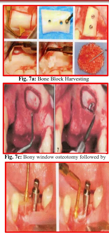
- Regenerative surgeries - To obtain autogenous grafts for treatment of periodontal intrabony defects (Fig. 7a). 8. Implantology (Fig. 7b, Fig. 7c, Fig. 7d)
- For harvesting block (bone) grafts and eventually implant placement in the recipient sites.
- Osteotomy procedures.
- Distraction osteogenesis followed by Implant placement.
- For Retrieval of blade implants.
- Ridge Expansion and implant placement. - Maxillary sinus elevation procedures. - Drilling hole in the bone for implant
placement.
- For insertion of implant. ADVANTAGES
UBS device have the advantages of being3,22,23 1. Safe
2. Selective for the mineralized tissues
3. Micrometric - the insert vibrates with a range of 60-200 μm at a modulated US frequency which while cutting maintains the bone constantly clean, thus avoiding excessive temperatures;
4. Surgical control with piezosurgery is maximum as the strength required by the surgeon to affect a cut is far less compared to that with a drill or with oscillating saws. 5. It is possible to have direct visibility over
whole osteotomies since there is minimal intra-operative bleeding.
6. The slightly increased time for operation using the Piezosurgery instrument, compared with that of the conventional drill, is negligible.
Fig. 1: Ultrasound Bone Surgery Unit
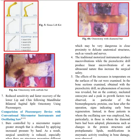
Fig. 6a: Osteotomy with carbide bur
Fig. 6b: Osteotomy with diamond bur
7. Reduced sensitivity and faster recovery of the lower Lip and Chin following Mandibular Bilateral Sagittal Split Osteotomy Using Piezosurgery.
Comparision of Piezosurgery Device with Conventional Micromotor Instruments and Oscillating Saw[5,22]
1. Burs controlled by a micromotor require greater strength that is obtained by applying increased pressure by hand. As a result, surgical sensitivity is reduced, especially when there are structures presenting different mineralization.
2. Oscillating saws though guaranteeing excellent linearity, they do not allow control of the depth of the cutting, at the sides or in the centre, and, therefore, it is often necessary to complete the incision with a scalpel and hammer.
3. When it is necessary to perform an osteotomy starting from the cortex, clearly the strength necessary to make use of the “torque”, in the more mineralized bone structure, this suddenly becomes excessive when passing to the spongiosum or when the cortical bone is finished. This causes an immediate loss of control of the surgical instrument, a condition
which may be very dangerous in close proximity to delicate anatomical structures, such as vessels and nerves.
4. The traditional motorized instruments produce macrovibrations while the piezoelectric drill produce linear microvibrations of an ultrasound nature thus increase the surgical safety.
5. The effects of the increases in temperature on the surfaces of the cut were examined. In the bone sections examined, obtained with the piezoelectric drill, no phenomenon of necrosis was revealed, but on the contrary, nucleated osteocytes and a peak in growth factors was observed, in particular of the biomorphogenetic proteins, one hour after the operation, signs indicating early bone regeneration. Instead, in those patients in whom the oscillating saw was employed, but, particularly, in those in whom the diamond burrs had been adopted, phenomena such as changes in the protein structures and protoplasmatic lipids, modifications in enzymatic activity resulting in bone damage, were observed.
A recent randomized controlled clinical trial done to compare the use of UBS and conventional rotary instruments for the bone removal for impacted third molar removal demonstrated reduced post operative pain, discomfort, trismus and swelling in the UBS treated sites.[24]
Limitations
Limitations of UBS device include5 1. Different learning curve.
2. Increasing the working pressure above a certain limit impedes the vibrations of the insert, and the energy is transformed into heat. 3. Increase in the operating time, compared with
Fig. 7d: Implant Site Preparation
4. The difficulty or impossibility to perform the deeper osteotomies (e.g.: maxillo-pterygoid disjunction), due to lack of inserts of the appropriate length.
5. The economic aspects. Piezosurgery inserts get worn away very rapidly. It is recommended never to go beyond ten uses in bone surgery.
CONCLUSION
Ultrasound Bone surgery provides many advantages over the conventional motorized instruments and oscillating saws. UBS with its various ranges of tips has a variety of applications in Dentistry like in the fields of Periodontics and Implantology and Oral and Maxillofacial surgery. With few limitations this invention of Dr. Tomaso Vercelloti has helped surgeons make the results of bone surgery more predictable, has improved healing, minimized the trauma and provided greater safety for patients. Thus, we conclude that Piezosurgery is an advantageous osteotomy technique for delicate structures in the oral and maxillofacial region. With respect to osteotomies of thin and fragile bones, the application of ultrasound is superior to other mechanical instruments because of the extremely precise and virtually arbitrary cut geometries, easy handling, efficient bone ablation and minimal accidental damage to adjacent soft tissue structures.
BIBLIOGRAPHY
1. Cardoni A. Power Ultrasonics in Oral Implantology. 2007 IEEE Ultrasonics Symposium.
2. http://www.ndted.org/EducationResources/C ommunityCollege/Ultrasonics/EquipmentTr ans/piezotransducers.htm
3. Leclercq P, Zenati C, Amr S, Dohan D. Ultrasonic Bone cut Part 1: State-of-art technologies and common applications. J Oral Maxillofac Surg. 2008;66:177-82. 4. www.mectron.com
5. Crosetti E, Battiston B, SuccoG. Piezosurgery in head and neck oncological and reconstructive surgery: personal experience on 127 cases. Acta Otorhino laryngol Ital. 2009;29:1-9.
6. Vercellotti T, Nevins ML, Kim DM, Nevins M, Wada K, Schenk RK et al. Osseous Response following Resective Therapy with
Fig. 6c: Osteotomy with UBS Fig. 7a: Bone Block Harvesting
Fig. 7b: Ridge Expansion Followed by Implant Placement
8. Kotrikova B, Wirtz R, Krempien R, Blank J, Eggers G, Samiotis A et al. Piezosurgery - a new safe technique in cranial osteoplasty? Int J Oral Maxillofac Surg. 2006;35:461-5. 9. Salami A, Dellepiane M, Proto E.
Piezosurgery in otologic surgery: four years of experience. Otol Head Neck Surg. 2009;140(140);412-8.
10. Vercellotti T, Podesta A. Orthodontic Microsurgery: A New Surgically Guided Technique for Dental Movement. Int J periodontics Restorative Dent. 2007;27:325-31.
11. Grenga V, Bovi M. Piezoelectric Surgery for Exposure of Palatally Impacted Canines. J Clin Orthod. 2004;38(8):446-8.
12. Sembronio S, Albiero AM, Polini F, Robiony M, Politi M. Intraoral endoscopically assisted treatment of temporomandibular joint ankylosis: Preliminary report. Oral Surg Oral Med Oral Pathol Oral Radiol Endod. 2007;104(1):e7-e10.
13. Bovi M. Mobilization of the Inferior Alveolar Nerve with simultaneous implant insertion: A New Technique. A Case Report. Int J Periodontics Restorative Dent. 2005;25(4):375-83.
14. Sakkas N, Otten JE, Gutwald R, Schmelzeisen R. Transposition of the mental nerve by piezosurgery followed by postoperative neurosensory control: a case report. Br J Oral Maxillofac Surg. 2008;46(4):270-1.
15. Vercellotti T, Pollack AS. A New Bone Surgery Device: Sinus Grafting and Periodontal Surgery. Compend Contin Educ Dent. 2006;27(5):319-25.
16. Sohn DS, Ahn MR, Lee WH, Yeo DS, Lim SY. Piezoelectric Osteotomy for Intraoral Harvesting of Bone Blocks. Int J Periodontics Restorative Dent 2007;27:3-7.
Distraction Osteogenesis in the Atrophic Maxillary Anterior Area: A Case Report. Implant Dent. 2007;16:227-34.
19. Sivolella S, Berengo M, Fiorot M, Mazzuchin M. Retrieval of blade implants with piezosurgery: two clinical cases. Minerva Stomatol. 2007;56(1-2):53-61. 20. Vercellotti T. Piezoelectric Surgery in
Implantology: A Case Report - A New Piezoelectric Ridge Expansion Technique. Int J Periodontics Restorative Dent. 2000;20(4):359-65.
21. Wallace SS, Mazor Z, Froum SJ, Cho SC, Tarnow DP. Schneiderian membrane perforation rate during sinus elevation using piezosurgery: clinical results of 100 consecutive cases. Int J Periodontics Restorative Dent. 2007;27(5):413-9.
22. Schlee M, Steigmann M, Bratu E, Garg AK. Piezosurgery: basics and possibilities. Implant Dent. 2006; 15(4):334-340.
23. Geha H, Gleizal A, Nimeskern N, Beziat JL. Sensitivity of the Inferior Lip and Chin following Mandibular Bilateral Sagittal Split Osteotomy Using Piezosurgery. Plast Reconstr Surg. 2006;118(7):1598-1607. 24. Barone A, Marconcini S, Giacomelli L,
Rispoli L, Calvo JL, Covani U. A Randomized Clinical Evaluation of Ultrasound Bone Surgery versus Traditional Rotary Instruments in Lower Third Molar Extraction. J Oral Maxillofac Surg. 2010;68:330-6.