case report opeN access
A case report of osteochondroma of the mandibular condyle
Icoz Derya, Akgunlu Faruk, Demir Esin, Karabağlı Pınar,
Ozturk Kayhan
AbstrAct
Introduction: Osteochondroma is one of the most common benign bone tumors of axial skeleton but it is rarely seen in maxillofacial region. Only about 1% of these occur within the head and neck region but when it presents more than half of these appear in coronoid process. Osteochondroma of the mandibular condyle is very rare. the clinical presentation of condylar osteochondroma mostly includes malocclusion, facial asymmetry, temporomandibular joint dysfunctions and disturbances during mouth opening and radiographically, the lesion presents an enlargement of the affected structure and a slight radiopacity increase. case report: We describe a case of osteochondroma affecting the right mandibular condyle of a male patient with the chief complaints of facial asymmetry and temporomandibular joint dysfunctions. conclusion: Although osteochondroma is not a frequent lesion for maxillofacial area, it should
Icoz Derya1, Akgunlu Faruk1, Demir Esin2, Karabağlı
Pınar3, Ozturk Kayhan4
Affiliations: 1Selcuk University, Faculty of Dentistry, De-partment of Oral Diagnosis and Radiology, Konya, Turkey; 2PhD Student, Selcuk University, Faculty of Dentistry, De-partment of Oral and Maxillofacial Surgery, Konya, Turkey; 3Assistant professor, Selcuk University, Faculty of Medi-cine, Department of Pathology, Konya, Turkey; 4Professor, Selcuk University, Faculty of Medicine, Department of Oto-laryngology, Konya, Turkey.
Corresponding Author: Icoz Derya, Selçuk University, Fac-ulty of Dentistry Department of Oral Diagnosis and Radiol-ogy Campus Selçuklu/konya, Turkey; Derya Icoz, Konya, Turkey, 42074; Email: [email protected]
Received: 06 October 2015 Accepted: 14 January 2016 Published: 25 February 2016
be considered in the differential diagnosis of masses in the temporomandibular joint region.
Keywords: Mandibular condyle, Osteochondroma, Panoramic radiography, temporomandibular joint
How to cite this article
Derya I, Faruk A, Esin D, Pınar K, Kayhan O. A case report of osteochondroma of the mandibular condyle. J Case Rep Images Dent 2016;2:1–5.
Article ID: 100003Z07ID2016
*********
doi:10.5348/Z07-2016-3-CR-1
INtrODUctION
Osteochondroma is a benign, slow-growing osseous protuberance with cartilagenous growth potential, projects from surface of the bone usually near its growth center [1, 2]. Although osteochondromas are mostly osseous, the entity is usually regarded as one of the cartilagenous lesions due to the endochondral ossification of cartilage cap [1].
The etiology of the lesion is unclear but discussions about its neoplastic, develomental and reperative nature is frequent [3]. It may occur at any age and females are more commonly affected than males [4, 5]. It is one of the most common benign tumors of the axial skeleton, it constitutes approximately 20–50% of all benign tumors and 10–15% of all bone tumors, but only about 1% of these occur within the head and neck region [6, 7]. The reason for the rare occurence of osteochondroma in this anatomical area is the intramembranous development of
grows slowly, and so, the symptoms may develop over a long period [1, 8]. The most common clinical symptoms include malocclusion, progressive facial asymmetry, restricted mandibular movements, clicking, popping and crepitation of the affected joint and changes in the condylar morphology [1, 5]. Condylectomy is usually curative for this lesion, the possibility of recurrence is a disadvantage although only one recurrence of a case has been reported for mandibular condyle which occured one year after the excision [1, 10].
The current report describes the diagnostic features and treatment planning of an osteochondroma affecting the right mandibular condyle which is a rare location for osteochondroma.
cAsE rEPOrt
A 34-year-old male was referred to our department with chief complaints of asymmetry and difficulty in opening and closing of the mouth and mastication approximately for two years. He did not seek any medical therapy until he admitted to our clinic, there was no history of facial trauma and he had no systemic diseases.
On clinical examination there was a painless bony, hard swelling on the right preauricular region. The symptoms were restricted inter-incisal range, malocclusion, deviation during mouth opening and prominent facial asymmetry (Figure 1). The overlying skin was in normal color and texture. No evidence of soft tissue involvement was noted. The lesion was fixed to the underlying bone and was non-mobile. Intraoral examination revealed contralateral cross-bite and restricted mandibular movements.
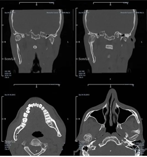
Panoramic radiograph of the patient revealed mandibular asymmetry and a well-defined bone enlargement on the right condylar head with slight radiopacity increase. Slow displacement of mandible results with adaptation of bony structures for compensation. Therefore, compared with the contralateral side increase in height of condylar neck and thickness of mandibular body were observed on panoramic radiography (Figure 2). For more detailed radiographic examination patient was advised a computed tomography (CT) scan. The axial and coronal CT sections revealed a clearly distinguishable cartilaginous/bony outgrowth arising from the right condylar head and length increase of the condylar neck. The lesion was extending from anteromedial aspect of
cap and mature bone tissue beneath, confirming the diagnosis of osteochondroma of the mandibular condyle (Figure 4). After two years follow-up of the patient mouth opening and TMJ functions were in normal limits but slight facial asymmetry and occlusal cant still remained. However, occlusion was in acceptable range (Figure 5). Postoperative panoramic radiography of the patient proved condylar remodeling and a more acceptable appearance in terms of symmetry (Figure 6).
DIscUssION
Although osteochondromas of the mandibular condyle is rare, early identification is essential to provide timely treatment and to prevent dramatic impacts on TMJ such as severe pain, hypomobility, clicking and locking of the TMJ, as well as headaches and cervical pain [8, 9]. Trauma and inflammation have been implicated as predisposing or initiating factors but this is not valid for all cases of osteochondroma including the present case, the patient had no history of trauma to related region. Review of literature reveals osteochondromas are frequently seen in second and third decades of the life and males are more commonly affected than males, however osteochondroma of the mandibular condyle is mainly seen in fourth decade of life with a mean age of 39.7 years and females are more commonly affected than males [1,
9]. The symptoms of the tumor vary depending on the location and the most common locations of the condylar osteochondroma is the medial aspect of the condyle (52%), followed by anterior location (20%) and rarely in the lateral or superior positions (<1%). The growth of an osteochondroma is usually slow, causing gradual displacement and elongation of the mandible [8, 9]. Meng et al. reviewed that hyperplasia of ramus and body of mandible on the affected side may be seen in condylar osteochondroma cases. Horizontal mandibular deviation caused by tumor is compensated by maxillary vertical overdevelopment and/or mandibular enlargement [4]. In our case hemimandibular hypertrophy with increased vertical height of entire mandible and slight compensatory vertical overdevelopment of maxilla was seen.
Figure 2: Panoramic radiography of the patient reveals
asymmetry on the right side, well defined bone enlargement on the condylar head, length increase on condylar neck and
hemimandibular hypertrophy.
Figure 3: Coronal and axial CT sections showing the bony/ cartilagenous enlargement of the condyle.
Figure 4: The histopathologic image is showing the
osteochondroma with cartilaginous cap, mature bone tissue and
marrow tissue (H&E stain, x50).
Figure 5: Postoperative images of the patient show acceptable
mouth opening and occlusion although a slight facial asymmetry
and occlusal cant remained.
mass of the condyle and CT is an invaluable imaging tool for treatment planning and differential diagnosis of the tumor especially with the unilateral condylar hyperplasia. Computed tomography scan clearly depicts the continuation of the cortex and medulla of the parent bone with the tumor and is the best imaging option for calcified cartilage cap [9]. Common clinical manifestations of the condylar osteochondroma were compatible with the present case [4]. Despite the integration of the clinical signs with osteochondroma the diagnosis of the present case report was done based on the combination of clinical, radiological and histopathological findings for definite diagnosis.
The aim of the treatment of a condylar osteochondroma should be achieving the acceptable mouth opening, recover facial asymmetry, establish facial harmony and occlusion [8]. In the present case, the treatment was curative and acceptable as functional but facial asymmetry could not be provided completely due to the enlargement of the hemi-mandible but patient refused corrective surgery for facial asymmetry.
cONcLUsION
Although osteochondroma is not a frequent lesion for maxillofacial area, it should be considered in the differential diagnosis of masses in the temporomandibular joint region, mandibular symphysis, maxillary sinuses, mandibular corpus and ramus. For more appropriate treatment method, a reliable diagnosis of the osteochondroma is necessary. Panoramic radiography and computed tomography evaluation should be performed for suspected condylar osteochondroma cases and for the final decision the diagnosis should be supported with histopathologic examination.
*********
Author contributions
Derya Icoz – Substantial contributions to conception and design; Acquisition of data, Drafting the article, Revising it critically for important intellectual content, Final approval of the version to be published
Faruk Akgunlu – Substantial contributions to conception and design, Acquisition of data, Drafting the article,
Kayhan Ozturk – Substantial contributions to conception and design, Acquisition of data, Drafting the article, Revising it critically for important intellectual content, Final approval of the version to be published
Guarantor
The corresponding author is the guarantor of submission.
conflict of Interest
Authors declare no conflict of interest.
copyright
© 2016 Derya Icoz et al. This article is distributed under the terms of Creative Commons Attribution License which permits unrestricted use, distribution and reproduction in any medium provided the original author(s) and original publisher are properly credited. Please see the copyright policy on the journal website for more information.
rEFErENcEs
1. Ongole R, Pillai RS, Ahsan AK, Pai KM. Osteochondroma of the mandibular condyle. Saudi Med J 2003 Feb;24(2):213–6.
2. Nanda Kishore D, Shiva Kumar HR, Umashankara KV, Rai KK. Osteochondroma of the mandible: a rare case report. Case Rep Pathol 2013;2013:167862. 3. Yamamoto FP, Silva BSF, Modes RW, Fonseca FP,
Pontes FSC, Sousa SC. Computed tomography and
scintigraphy for diagnosis and treatment planning
of the condylar osteochondroma: A case report. Rev Odonto Cienc 2010;25(4):422–6.
4. Meng Q, Chen S, Long X, Cheng Y, Deng M, Cai H. The clinical and radiographic characteristics of condylar osteochondroma. Oral Surg Oral Med Oral Pathol Oral Radiol 2012 Jul;114(1):e66–74.
5. Kumar KRA, Omprakash TL, Goudar GN, Kumar RR. Osteochondroma of Mandibular Condyle. Journal of Dental Sciences & Research 2010;1(2):57–66. 6. Murphey MD, Choi JJ, Kransdorf MJ, Flemming DJ,
Gannon FH. Imaging of osteochondroma: variants and complications with radiologic-pathologic correlation. Radiographics 2000 Sep-Oct;20(5):1407–34.
7. El-Mofty SK. Diagnostic Surgical Pathology of the Head and Neck. 2ed. Philadelphia, Pa, USA: Saunders; 2009.
9. Avinash KR, Rajagopal KV, Ramakrishnaiah RH, Carnelio S, Mahmood NS. Computed tomographic features of mandibular osteochondroma. Dentomaxillofac Radiol 2007 Oct;36(7):434–6. 10. González-Otero S, Navarro-Cuéllar C, Escrig-de
Teigeiro M, Fernández-Alba-Luengo J,
Navarro-Vila C. Osteochondroma of the mandibular condyle:
Resection and reconstruction using vertical sliding
osteotomy of the mandibular ramus. Med Oral Patol Oral Cir Bucal 2009 Apr 1;14(4):E194–7.
Access full text article on