IJHPD 201 3;3:1 0–1 6.
www.ijhpd.comClinicopathological significance of fatty acid synthase
expression in pancreatic ductal carcinoma
Hiroshi Maekawa, Tomoaki Ito, Hajime Orita, Koichi Sato, Ryo Wada
ABSTRACT
Introduction: Fatty Acid Synthase (FAS) expression is increased in various cancers and it contributes to tumor cellular activities. In clinical practice, pancreatic ductal carcinoma is regarded as a cancer with poor prognosis. Aims: To investigated FAS expression in pancreatic ductal carcinoma (PDC) and compare with clinicpathological features. Methods: We studied 38 patients who had resection for PDC in our hospital. We compared FAS staining with clinicopathological features, including Ki67 and p53 staining. Results: Using our criteria, out of 38 cases, 15 cases were considered FAS positive. FAS positivity correlated with Ki67 positivity (p = 0.001), but there was no significant relationship between FAS expression and p53 staining (p = 0.224). FAS positive cases showed higher rate of lymph node metastases (p = 0.0038). FAS expression was related to
differentiation of adenocarcinoma (p = 0.0347). Clinically, overall survival rate of FAS positive cases was lower than that of FAS negative cases (p = 0.0068). Conclusion: FAS expression may be related to lymph node metastases and clinical behavior in pancreatic ductal carcinoma.
Keywords: Pancreatic cancer, Fatty acid synthase, Ki67, Immunohistochemistry, Prognosis
*********
Maekawa H, Ito T, Orita H, Sato K, Wada R. Clinicopathological significance of fatty acid synthase expression in pancreatic ductal carcinoma. International Journal of Hepatobiliary and Pancreatic Diseases 2013;3:10–16.
Article ID:100011IJHPDHM2013
*********
doi:10.5348/ijhpd201311OA2
INTRODUCTION
In human tissues, fatty acid synthase (FAS) is usually expressed in hormonesensitive and high lipid metabolism cells [1]. In normal tissues, FAS expression is regulated and inhibited by several hormones [2]. FAS expression is also increased in various cancers. Breast cancer or prostate cancer is a good example of cancers expressing FAS [37]. FAS expression increases de novo biosynthesis of fatty acids which contributes to tumor cellular activities [8]; metabolism for energy, building cellular membranes and intracellular second messengers [9, 10]. It has been demonstrated that cancer cells often express FAS and the intensity of FAS expression correlates with clinical outcome. Cases of
ORIGINAL ARTICLE OPEN ACCESS
Hiroshi Maekawa
1, Tomoaki Ito
2, Hajime Orita
1, Koichi
Sato
3, Ryo Wada
4Affiliations:
1Associate professor, department of surgery,
Juntendo university school of medicine Shizuoka hospital,
Izu-no-kuni city, Shizuoka, Japan;
2Assistant professor,
department of surgery, Juntendo university school of
medicine Shizuoka hospital, Izu-no-kuni city, Shizuoka,
Japan;
3Professor, department of surgery, Juntendo
university school of medicine Shizuoka hospital,
Izu-no-kuni city, Shizuoka, Japan;
4Professor, department of
pathology, Juntendo university school of medicine
Shizuoka hospital, Izu-no-kuni city, Shizuoka, Japan.
Corresponding Author: Hiroshi Maekawa, 11 29 Nagaoka,
Izu-no-kuni, Shizuoka, Japan. 41 0-2295; Phone No. 81
55.948.3111 ; Fax No. 81
55.946.051 4; Email:
hmaekawa0201 @gmail.com
Received: 1 8 July 201 2
Accepted: 30 July 201 2
Published: 25 January 201 3
high FAS expression often show poor prognosis in various types of cancers [11–17]. In clinical practice, pancreatic ductal carcinoma (PDC) is regarded as a cancer with poor prognosis.
AIMS
To investigate the clinicopathological significance of FAS expression in pancreatic ductal carcinoma (PDC), we studied patients surgically treated for PDC in our hospital. The aims of this study were, i) to investigate the relationship between the expression of FAS and clinicopathological features using immunohistochemistry in PDC, and ii) to analyze whether FAS may be considered as a prognostic marker in PDC.
MATERIALS AND METHODS
We studied 38 patients resected for PDC in our hospital, between January 2002 and January 2011. Clinical data such as gender, age, recurrence, and prognoses were obtained from clinical records. Pathological diagnosis was performed by examining hematoxylin and eosin stained sections. We investigated expressions of FAS, Ki67, and p53 with immunohistochemical stainings on tissue sections cut from formalinfixed and paraffinembedded carcinoma specimens. We then compared FAS staining with clinical information and pathological aspects, including Ki67 and p53 staining. The study protocol conformed to the ethical guidelines of the World Medical Association Declaration of Helsinki, and was approved by the ethical committee in our hospital.
Immunohistochemical procedure for FAS expression: We used 4 µm tissue sections cut from
formalinfixed and paraffinembedded specimens. Sections were deparaffinized by treatment with xylene for three minutes (two times) and rehydrated by passage through 100% ethanol for 30 sec (three times). Antigen retrieval was performed by microwaving slides in 10 mM sodium citrate buffer (TRS) (Dako; Carpinteria, CA, USA) for eight minutes. Endogenous peroxidase activity was quenched by incubating slides for five minutes in 3% H2O2. Slides were then washed in Tris buffered saline – 0.05% Tween 20 (TBST) for five minutes (two times) and blocked with a solution containing 5% goat serum for 15 minutes at room temperature. Samples were incubated with the primary antibody (antihuman FAS rabbit IgG; ImmunoBiological Laboratories Co. Ltd., Fujioka, Gunma, Japan) for 60 minutes at room temperature. Slides were washed with TBST for five minutes (three times) and incubated with secondary antibody, (anti rabbit IgG; Envison+/HRP; DAKO; Carpenteria, CA, USA) for 60 minutes at room temperature. Slides were washed in TBST for 5 minutes (three times) and incubated with diaminobenzidine solution containing 20 mg/ml DAB4HCl in 0.01 phosphate buffer M PB and 30% HO until color production developed. Slides were
then stained with hematoxylin, dehydrated and mounted with coverslips.
Immunohistochemical procedure for Ki67 and p53 expressions: Immunoperoxidase assays for
p53 and Ki67 analysis were performed using a commercially available kit (Roche Diagnostics Co. Ltd.; Tokyo, Japan) in the same manner as described in the immunohistochemical procedure for FAS analysis. Monoclonal mouse antihuman Ki67 and p53 antibodies were used as primary antibodies (Dako, Carpinteria, CA, USA).
Scoring of immunoreactivity: We classified FAS
staining of carcinoma tissue compared with staining of adipose tissue. Scoring of cellular FAS positivity was classified as follows: i) negative carcinoma tissue not stained or stained less than adipose cell, ii) positive carcinoma tissue stained same as adipose tissue or stained more than adipose cell.
Tissue FAS expression was scored using the following scale: 0 if positive cells were less than 10% of tumor cells, 1 if positive cells were 10% to 30% of tumor cells, 2 if positive cells were 30% to 50% of tumor cells, and 3 if positive cells were more than 50% of tumor cells.
Finally, scores of 0 and 1 were combined to represent negative FAS expression and scores of 2 and 3 were combined to represent positive FAS expression.
Tissue expressions of p53 and Ki67 were classified into four groups in a similar way as done for tissue FAS expression described above. We compared FAS scores with Ki67 scores and p53 scores.
Statistical Analysis: Relationship of the scores of
tissue FAS expression with those of tissue Ki67 expression and tissue p53 expression was analyzed using the Spearman’s correlation coefficient by rank test. Cutoff endpoints were determined according to positive and negative immunohistochemical tissue FAS expressions observed. FAS positive cases were analysed for association of FAS expression with clinicopathological features using the Chisquare test, Fisher’s exact test (if available) or MannWhitney’s test. We estimated survival based on FAS expression patterns using the KaplanMeier analysis and compared survival between groups using logrank tests. A p value less than 0.05 were considered significant. All analyses were conducted using the GraphPad Prizm5®software statistic package (GraphPad Software Inc., La Jolla, CA, USA).
RESULTS
IJHPD 201 3;3:1 0–1 6.
www.ijhpd.com
Maekawa et al.
1 2
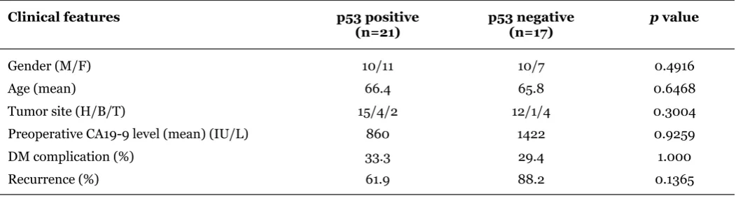
The pathological features were analyzed according to FAS expression status. FAS positive cases showed higher rates of high vessel involvement (p = 0.001), lymph node metastases (p = 0.004) and differentiation (p = 0.035) (Table 2). The clinical features of the two groups were also analyzed. There were no significant differences between the two groups in gender, age, tumor site, preoperative levels of CA199 or diabetes mellitus complication (Table 3). Clinicopathological findings are shown according to Ki67 or p53 positivity (Tables 4, 5). There were no differences in the clinicopathological features according to positivity of Ki 67 or p53. Although recurrences were often detected in both groups, there was no difference in relapse free survival rates (RFS) between the two groups (p = 0.1499) (Figure 5). However, overall survival rate (OS) of FAS positive cases was lower than that of FAS negative cases (p = 0.007) (Figure 6).
DISCUSSION
Human FAS is a 270kDa cytosolic enzyme that synthesizes longchain fatty acids, palmitate, myristate, and stearate, using acetylCoA, malonylCoA, and NADPH [9]. Recently, it has been suggested that FAS plays an important role not only in tumor growth, but also in tumor survival and drug resistance with de novo lipogenesis [18, 19]. Therefore, increase in FAS expression may be associated with cancer progression, high risk of cancer recurrence, and shorter survival. Pancreatic ductal cancer is known to be a cancer with poor prognosis because recurrences are often seen even though curative resections are performed. Recently, several studies of FAS expression in cases of pancreatic ductal cancer have been reported [17, 19, 20]. In these studies, FAS was reported to have relationship to some
Table 1: Clinical features of pancreatic ductal carcinoma cases (n=38).
Figure 1: Sample of FAS positive case. In FAS positive cases, A) Cellular cytoplasm was well stained, B) 30% to 50% of cancer cells stained positively. (Immunohistochemistry for FAS; A x200; Bx100).
Figure 2: Samples of FAS negative case. A) Cellular cytoplasms were slightly stained, B) Less than 10% of cancer cells were stained positively. (Immunohistochemistry; A x200; x100.
Figure 3: Correlation between scores of tissue FAS expression and scores of tissue Ki67 expression. FAS positivity correlated with Ki67 positivity (p = 0.001).
Figure 4: Correlation between scores of tissue FAS expression and scores of tissue p53 expression, There were no relationship between scores of tissue FAS expression and scores of tissue p53 expression (p = 0.224).
Clinical features
Gender (male/female) 20/18
Age (years) 66.1
Table 2: Pathological features of pancreatic ductal carcinoma according to FAS immunohistochemical staining.
Note: High vessel involvement means ly 2, 3 or v 2, 3 grade. High perineural invasion means ne 2, 3 grade. Abbreviations: ly lymphatic vessel involvement, v venous involvement, ne perineural tumor invasion.
Table 3: Pathological features of pancreatic ductal carcinoma according to FAS immunohistochemical staining.
Abbreviaions: DM diabetes mellitus, H – head, B – Body. T Tail
IJHPD 201 3;3:1 0–1 6.
www.ijhpd.comTable 5: Clinical features of pancreatic ductal carcinoma according to Ki67.
pathological characteristics such as histological type of cancer, lymph node metastases, p53 expression or clinical outcome. Alo et al. [17] reported that FAS was overexpressed in poorly differentiated adenocarcinomas and was associated with patients’ outcome in pancreatic adenocarcinoma. Witkiewicz et al. [20] reported that FAS expression strongly correlated with tumor size, tumor grade and the presence of lymph node metastasis in pancreatic cancer. Our results were similar to their
conclusions, but, interestingly, FAS positivity had no corelation with p53 positivity, though FAS positivity corelated with Ki67 positivity.
Positivity for p53 immunohistochemical staining is reported to be related to clinical outcome of different types of cancers. D’Erchia et al. [21] demonstrated that p53 family of proteins control FASN expression, the same as the SREBP1c signalling pathway. In contrast, Ogino et al. [16] reported that FAS expression was
Table 6: Pathological features of pancreatic ductal carcinoma according to p53.
Table 7: Pathological features of pancreatic ductal carcinoma according to p53.
Figure 5: KaplanMeier curves related to FAS expression in relapse free survival. There was no significant difference between FAS positive case and FAS negative cases in relapse free survival (p = 0.149). The red line shows FAS positive cases and the blue line shows FAS negative cases.
Figure 6: KaplanMeier curves related to FAS expression in overall survival. The overall survival rate of FAS positive cases was lower than that of FAS negative cases (p = 0.007). The red line shows FAS positive cases and the blue line shows FAS negative cases.
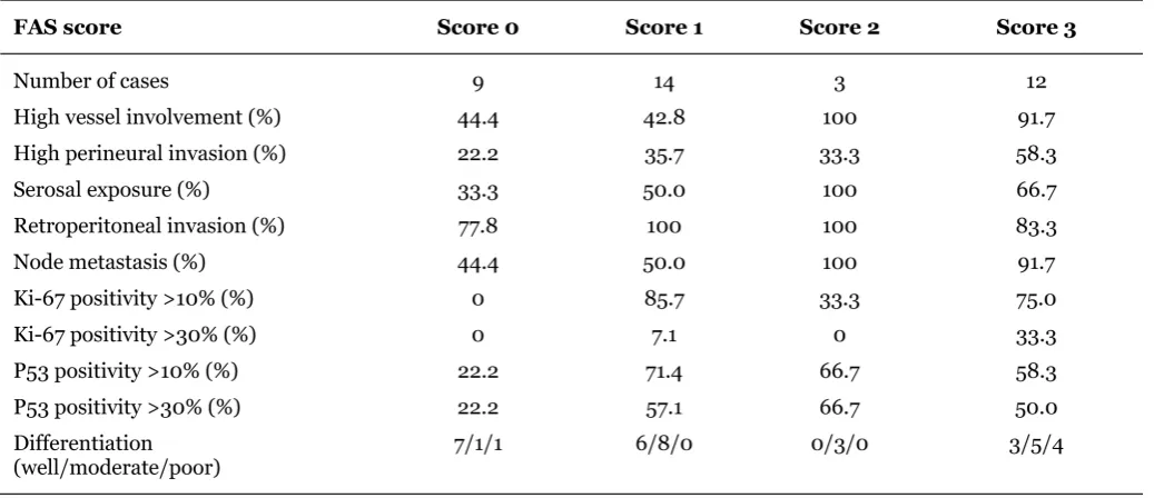
Table 8: Pathological features according to FAS intensity.
associated with clinical outcome, but it had no significant relationship with p53 positivity. Rashid et al. [22] also reported FAS staining in 139 cases of colorectal cancer, and suggested that FAS expression could correlate with poor prognosis through an association with lymph node metastases and aggressive histopathological subtypes, however, no significant association was observed between lymph node metastases, patient survival, and FAS expression. Our results of correlation between FAS expression and p53 expression was similar to Ogino’s et al.
From the previous reports and our results, it is proposed that FAS expression is related to cancer aggressiveness. FAS positivity may be considered to be one of the prognostic markers. However, the results of FAS expression are slightly different among previous reports. We suspect that these differences may be due to differences in criteria for positivity for FAS and p53 staining. More studies are needed to analyze FAS expression for establishing the exact singnificance of FAS expression.
CONCLUSION
FAS expression may be related to clinical behavior in pancreatic ductal carcinoma with high incidence of lymph node metastasis. FAS positive cases of pancreatic ductal carcinoma may have shorter survival time compared to FAS negative cases.
*********
Author Contributions
IJHPD 201 3;3:1 0–1 6.
www.ijhpd.comarticle, Critical revision of the article, Final approval of the version to be published
Tomoaki Ito – Conception and design, Acquisition of data, Analysis and interpretation of data, Drafting the article, Critical revision of the article, Final approval of the version to be published
Hajime Orita – Conception and design, Acquisition of data, Analysis and interpretation of data, Drafting the article, Critical revision of the article, Final approval of the version to be published
Koichi Sato – Conception and design, Acquisition of data, Analysis and interpretation of data, Drafting the article, Critical revision of the article, Final approval of the version to be published
Ryo Wada – Conception and design, Acquisition of data, Analysis and interpretation of data, Drafting the article, Critical revision of the article, Final approval of the version to be published
Guarantor
The corresponding author is the guarantor of submission.
Conflict of Interest
Authors declare no conflict of interest.
Copyright
© Hiroshi Maekawa et al. 2013; This article is distributed under the terms of Creative Commons Attribution 3.0 License which permits unrestricted use, distribution and reproduction in any means provided the original authors and original publisher are properly credited. (Please see www.ijhpd.com/copyright policy.php for more information.)
REFERENCES
1. Kusakabe T, Maeda M, Hoshino H, et al. Fatty acid synthase is expressed mainly in adult hormone sensitive cells or cells with high lipid metabolism and in proliferating fetal cell. J Histochem Cytochem 2000;48(5):613–622.
2. Sul HS, Wang D. Nutritonal and hormonal regulation of enzymes in fat synthesis: studies of fatty acid synthase and mitochondrial glycerol3 phosphate acyltransferase gene transcription. Annu Rev Nutr 1998;18:331–51.
3. Kuhajda FP, Piantadosi S, Pasternack GR. Haptoglobinrelated protein (Hpr) epitopes in breast cancer as a predictor of recurrence of the disease. N Engl J Med 1989;321(10):636–41. 4. Alo PL, Visca P, Marci A, Mangoni A, Botti C, Di
Tondo U. Expression of fatty acid synthase (FAS) as a predictor of recurrence in stage I breast carcinoma patients. Cancer 1996;77(3):474–82.
5. Wang YY, Kuhajda FP, Li J, et al. Fatty acid synthase as a tumor marker: its extracellular expression in human breast cancer. J Exp Ther Oncol 2004;4(2):101–10.
6. Van de Sande T, Roskams T, Lerut E, et al. High level expression of fatty acid synthase in human
prostate cancer tissues is linked to activation and nuclear localization of Akt/PKB. J Pathol 2005;206(2):214–9.
7. Shah US, Dhir R, Gollin SM, et al. Fatty acid synthase gene overexpression and copy number gain in prostate adenocarcinoma. Hum pathol 2006;37(4):401–9.
8. Kuhajda FP. Fatty acid synthase and cancer: new application of an old pathway. Cancer Res 2006;66(12):5977–80.
9. Liu H, Liu JY, Wu X, Zhang JT. Biochemistry, molecular biology, and pharmacology of fatty acid synthase, an emerging therapeutic target and diagnosis/prognosis marker. Int J Biochem Mol Biol 2010;1(1):69–89.
10. Mashima T, Seimiya H, Tsuruo T. De novo fattyacid synyhesis and related pathways as molecular targets
for cancer therapy. Br J Cancer
2009;100(9):1369–72.
11. Gansler TS, Hardman W 3rd, Hunt DA, Schaffel S, Hennigar RA. Increased expression of fatty acid synthase (OA519) in ovarian neoplasms predicts shorter survival. Hum Pathol 1997;28(6):686–92. 12. Alo PL, Visca P, Marci A, Mangoni A, Botti C, Di
Tondo U. Expression of fatty acid synthase (FAS) as a predictor of recurrence in stage I breast carcinoma patients. Cancer 1996;77(3):474–82.
13. Shurbaji MS, Kalbfleisch JH, Thurmond TS. Immunohistochemical detection of a fatty acid synthase (OA519) as a predictor of progression of prostate cancer. Hum Pathol 1996;27(9):917–21. 14. Visca P, Sebastiani V, Botti C, et al. Fatty acid
synthase (FAS) is a marker of increased risk of reccurence in lung carcinoma. Anticancer Res 2004;24(6):4169–73.
15. Sebastiani V, Visca P, Botti C, et al. Fatty acid synthase is a marker of increased recurrence in endometrial carcinoma. Gynecol Oncol 2004;92(1):101–5.
16. Ogino S, Nosho K, Meyerhardt JA, et al. Cohort study of fatty acid synthase expression and patient survival in colon cancer. J Clin Oncol 2008;26(35):5713–20. 17. Alo PL, Amini M, Piro F, et al. Immunohistochemical
expression and prognositic significance of fatty acid synthase in pancreatic carcinoma. Anticancer Res 2007;27(4B):2523–7.
18. Liu H, Liu Y, Zhang JT. A new mechanism of drug resistance in breast cancer cells: fatty acid snthase overexpressionmediated palmitate overproduction. Mol Cancer Ther 2008;7(2):263–70.
19. Yang Y, Liu H, Li Z, et al. Role of fatty acid synthase in gemcitabine and radiation resistance of pancreatic cancers. Int J Biochem Mol Biol 2011;2(1):89–8. 20. Witkiewicz AK, Nguyen KH, Dasgupta A, et al. Co
expression of fatty acid syunthase and caveolin1 in pancreatic ductal adenocarcinoma. Implication for tumor progression and clinical outcome. Cell Cycle 2008;7(19):3021–5.
21. D’Erchia AM, Tullo A, Lefkimmiatis K, Saccone C, Sbisa E. The fatty acid synthase is a conversed p53 family target from worm to human. Cell Cycle 2006;5(7):750–8.
22. Rashid A, Pizer ES, Moga M, et al. Elevted expression of fatty acid synthetic activity in colorectal neoplasia. Am J Pathol 1997;150(1):201–8.