ISSN 0015–5659 www.fm.viamedica.pl
Histomorphometric study of the effect of
methionine on small intestine parameters in rat:
an applied histologic study
S. Seyyedin
1, M.N. Nazem
2Shahid Bahonar University of Kerman, Kerman, Iran
[Received: 26 January 2017; Accepted: 11 April 2017]
Background: Assessment of morphological changes has more often been used in the diagnosis and assessment of intestinal pathology and development. Since methionine is widely used in nutritional and sports supplements and also there is not enough information about the effect of this amino acid on the gastrointestinal histomorphometry, the aim of this study was to assess the effect of methionine on the small intestine histomorphometry.
Materials and methods: Thirty male Wistar rats were randomly divided to three equal groups. Two treatment groups received 100 and 200 mg/kg L-methionine solution respectively via intraperitoneal injection while the control group received normal saline. On day 21, all rats were euthanised and segments from three parts of small intestine were taken to histomorphometrical study. Paraffin sections were stained with haematoxylin and eosin, alcian blue (AB) and periodic acid Schiff (PAS) methods separately. In order to analyse histomorphometric features of each segment, villus height, width, area, crypt depth, villus height to crypt depth ratio, goblet cell number, and muscle layer thickness were measured.
Results and Conclusions: Obtained results revealed that methionine may change the histomorphometric parameters of small intestine. (Folia Morphol 2017; 76, 4: 620–629)
Key words: histomorphometry, small intestine, methionine, rat
IntroductIon
The small intestine is the long muscular tube of the gastrointestinal tract that is the major site for absorp-tion and digesabsorp-tion of nutrients and drugs. It is made up of three structural segments: duodenum, jejunum and ileum [17–19]. All of the absorptive and also a part of the digestive capacity of the small intestine oc- cur around and near villi and crypts [21]. Actually the crypt-villus is the main functional unit of absorption in the rat’s small intestine. Various substances and also different physiological or pathological conditions
may change the proliferation rate of enterocytes in the crypt and affect their migration rate to the top of the villi [46]. The relation between small intestine and nutritional factors is very complicated. Nutrients may directly cause changes or adaptations in the intestinal mucosa [4, 47].
Methionine (Met), a sulphur-containing essential amino acid, serves as the initiating amino acid in the synthesis of many proteins. It plays an important role in the synthesis of vital molecules such as cysteine, carnitine, taurine, lecithin, phosphotidyl choline and
other phospholipids. It has a significant effect on
improving cartilage formation and is therefore used in arthritis treatment [49].
Histomorphometric analysis is greatly used in studies about gastrointestinal pathophysiology. The availability of animal models and using morphometric analysis as a quantitative assessment have facilitated evaluating the morphological alterations of intestinal mucosa under various experimental conditions such as special diets and drugs [10].
The laboratory rat, as an animal model, has been used in several histological and histomorphometrical studies [1, 53]. Some of these studies have described the qualitative and quantitative changes that occur in the small intestine under various physiological and pathological conditions [10, 11, 13, 31, 32]. Many studies have investigated the effect of methionine supplementation on the skin [30], teeth [25], liver disorders [23], feather follicles [28], female reproduc-tive system [3, 29] and neural tube in the embryos [24] but there is not enough information about its effect on the small intestine in the mammals.
Methionine is widely used in nutritional and sports supplements and on the other hand the effects of these supplements have not been investigated on the gastrointestinal histomorphometry. So the aim of this study was to assess the effect of injected methionine on histomorphometric aspects of small intestine in the rat.
MaterIals and MetHods
The experiment in this study was approved by the Animal Ethics Committee, a branch of the Research Council of the Veterinary School in Shahid Bahonar University, Kerman Province, Iran.
A total of thirty male Wistar rats weighing 190– –210 g were used for this research. The rats were housed in clean plastic cages and kept under the following conditions: 12 h light/dark cycle, controlled temperature of 22–25oC and humidity of 60–70%.
The rats were allowed an adaptation period of 5 days with free access to commercial rodent diet and water ad libitum. The rats were randomly divided into three groups containing ten rats. All three groups received intraperitoneal injections for 20 consecutive days. Rats in the group A served as the control group and were administered with normal saline. Group B and C rats were treated with L-methionine (Merck, Germany) 100 mg/kg and 200 mg/kg body weight, respectively. Dose regimen, duration and route of
administra-tion in this study were based on a previous research that investigated the histomorphometric effect of methionine on the rat’s skin [30]. At the end of the treatment period, all rats were euthanised. The ab-domen was opened through a midline incision and the small intestine of each rat was gently removed. Then small segments of intestine, about 1 cm in size, were removed from the duodenum (below the pyloro-duodenal junction), jejunum (2 cm anterior to Meckel’s diverticulum) and ileum (2 cm anterior to
ileocecal junction). All intestinal portions were fixed
in 10% neutral buffered formalin for 10 days [4, 36], dehydrated in graded anhydrous absolute ethanol
and xylol, embedded in paraffin blocks and then cut
into 5 µm sections. In each animal, one section that represented the best view of villus and crypts was selected to be analysed. The sections were stained using haematoxylin and eosin (H&E) (for standard histological evaluation), periodic acid Schiff (PAS) (in order to count the number of neutral goblet cells) and alcian blue (AB) (in order to count the number of acidic goblet cells) techniques. Slides were examined using an Olympus light microscope and photomicro-graphs were taken by an attached eyepiece camera (Dino-eye, AM-7023, 5Mp, Taiwan). The following intestinal histomorphometric parameters were evalu-ated in all sections stained with H&E method: villous height, villous width, crypt depth and the muscular layer thickness. These parameters were measured on 20 well-aligned villi and corresponding crypts from each section of all intestinal segments. The heights
of the villi were defined from their tip to the base
and the widths were measured at the half height point. Calculations using villous height and width at half height gave the villus surface area. The depth of intestinal crypts was measured as the distance from top of villus crypt to muscularis mucosa.
The number of goblet cells per villus was counted in ten well-oriented and adjacent crypt villus on each sec-tion stained by PAS and AB histochemical techniques. All
measurements were made in µm, at ×100 magnification.
Statistical analysis
Results were expressed as mean values ± standard error. Data were analysed using one-way analysis of variance (ANOVA) followed by post hoc, Tukey’s HSD test. Statistical analysis was performed using SPSS (SPSS for Windows, version 16.0, SPSS Inc., Chicago, Illinois). Differences were considered statistically
Table 1. The effect of intraperitoneal administration of methionine on the histomorphometric parameters of rat duodenum (values are mean ± standard error)
Control Met 100 Met 200
VH [µm] 389.47 ± 33.84a 533.84 ± 23.05b 525.82 ± 14.61b
VW [µm] 69.47 ± 5.58a 103.22 ± 6.41b 106.66 ± 6.56b
VA [µm2] 27056.65 ± 2350.8a 55102.96 ± 2379.22b 56084.15 ± 1557.7b
CD [µm] 264.28 ± 13.74a 264.32 ± 32.31a 166.72 ± 9.79b
VH/CD 1.47 ± 0.13a 2.01 ± 0.09b 3.15 ± 0.09c
Goblet cells (AB) 8.67 ± 1.05a 15.83 ± 1.89b 12.57 ± 2.01ab
Goblet cells (PAS) 8.9 ± 0.86a 16.11 ± 1.43b 12.13 ± 1.11ab
Muscle layer thickness [µm] 77.94 ± 5.38a 101.26 ± 8.49b 157.17 ± 6.99c
a, b, c Means in the same row with no common superscript differ (p < 0.05).
AB — alcian blue; CD — crypt depth; PAS — periodic acid Schiff; VA — villus area; VH — villus height; VW — villus width
Table 2. The effect of intraperitoneal administration of methionine on the histomorphometric parameters of rat jejunum (values are mean ± standard error)
Control Met 100 Met 200
VH [µm] 353.04 ± 10.64a 405.53 ± 8.8b 423.16 ± 16.05b
VW [µm] 87.26 ± 5.78a 120.72 ± 2.79b 107.24 ± 5.54b
VA [µm2] 30406.09 ± 1101.46a 38287.1 ± 1782.05b 45380.1 ± 1721.66c
CD [µm] 214.61 ± 7.19a 208.15 ± 6.07a 161.86 ± 10.64b
VH/CD 1.64 ± 0.05a 1.52 ± 0.07a 2.61 ± 0.1b
Goblet cells (AB) 12 ± 1.21a 20 ± 1.37b 18.6 ± 1.63b
Goblet cells (PAS) 14 ± 1.31a 23.2 ± 1.88b 19.8 ± 0.86b
Muscle layer thickness [µm] 61.33 ± 5.12a 81.35 ± 5.01b
88.06 ± 6.09
ba, b, c Means in the same column with no common superscript differ (p < 0.05).
AB — alcian blue; CD — crypt depth; PAS — periodic acid Schiff; VA — villus area; VH — villus height; VW — villus width
Table 3. The effect of intraperitoneal administration of methionine on the histomorphometric parameters of rat ileum (values are mean ± standard error)
Control Met 100 Met 200
VH [µm] 199.47 ± 5.11a 272.2 ± 7.46b 265.56 ± 2.78b
VW [µm] 94.53 ± 2.51a 130.73 ± 3.15b 118.58 ± 4.5b
VA [µm2] 18856.89 ± 482.22a 33811.26 ± 2356.84b 31490.1 ± 329.83b
CD [µm] 221.72 ± 5.01a 186.87 ± 3.66b 206.68 ± 0.73a
VH/CD 0.9 ± 0.02a 1.45 ± 0.04b 1.28 ± 0.01c
Goblet cells (AB) 19.8 ± 2.08a 27.67 ± 1.48b 25 ± 1.63ab
Goblet cells (PAS) 24.86 ± 1.35a 31 ± 1.68b 24.56 ± 1.55a
Muscle layer thickness [µm] 55.13 ± 4.54a 66.85 ± 3.94b 72.66 ± 4.15b
a, b, cMeans in the same column with no common superscript differ (p < 0.05).
AB — alcian blue; CD — crypt depth; PAS — periodic acid Schiff; VA — villus area; VH — villus height; VW — villus width
results
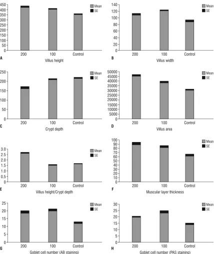
According to our findings, the villus height and
width in the duodenum (Table 1, Fig. 1) jejunum
significant difference between treatment groups in
all intestinal segments (Tables 1–3). It was observed that in all small intestine portions, the groups treated
with methionine showed significant increase in villus
area compared with the control groups (p < 0.05). It was noticeable that the difference in the villus area
Figure 1. Histomorphometric measurements of the duodenum of control (Control) and treatment groups: 100 (Met 100) and 200 (Met 200). Bars are mean ± standard error (SE). The unit of villus height, villus width, crypt depth and muscular layer thickness is µm. The unit of villus
area is µm2; A. Villus height; B. Villus width; C. Crypt depth; D. Villus area; E. Villus height/crypt depth; F. Muscular layer thickness; G. Goblet
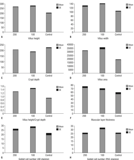
in the jejunum was significant between two treat -ment groups (Fig. 2, Table 2). The results of this study showed that the depth of the crypts in duodenum
and jejunum were significantly decreased in the Met
200 group as compared to both Met 100 and control
groups (p < 0.05), but there was no significant dif -ference between the Met 100 and control groups.
However in the ileum, the depth of the crypts of Met 100 group were decreased compared to the other groups (p < 0.05) (Fig. 4; Tables 1–3).
On histological observation, in the duodenum,
significantly increased villus height to crypt depth
ratio was observed in methionine treated groups as compared to the control group (p < 0.05). It should
Figure 2. Histomorphometric measurements of the jejunum of control (Control) and treatment groups: 100 (Met 100) and 200 (Met 200). Bars are mean ± standard error (SE). The unit of villus height, villus width, crypt depth and muscular layer thickness is µm. The unit of villus area
is µm2; A. Villus height; B. Villus width; C. Crypt depth; D. Villus area; E. Villus height/crypt depth; F. Muscular layer thickness; G. Goblet cell
also be noted that a significant increase in villus
height to crypt depth ratio was observed in the Met 200 group compared to Met 100 group (Table 1). In the jejunum, this ratio was greater in the Met 200 group compared with the other groups (p < 0.05). No
significant difference was observed the Met 100 and
the control groups (Table 2). In the ileum, the villus
height to crypt depth ratio was significantly increased
in treatment groups compared to the control group and also the villus height to crypt depth ratio was greater in Met 100 group compared with Met 200 group (Table 3).
Figure 3. Histomorphometric measurements of the ileum of control (Control) and treatment groups: 100 (Met 100) and 200 (Met 200). Bars are mean ± standard error (SE). The unit of villus height, villus width, crypt depth and muscular layer thickness is µm. The unit of villus area
is µm2; A. Villus height; B. Villus width; C. Crypt depth; D. Villus area; E. Villus height/crypt depth; F. Muscular layer thickness; G. Goblet cell
In all small intestine segments, the thickness of the muscular layer was greater in the methionine treated groups than in the controls (p < 0.05). Also, in the duodenum the Met 200 groups had thicker muscular layer compared with the Met 100 group (p < 0.05) (Tables 1–3).
In the duodenum, jejunum and ileum the count of goblet cells that were stained by AB and PAS methods, showed a growth in the treatment groups compared to controls. In all small intestine parts, goblet cells in the Met 100 group were more numerous than in the
other groups and this increase was significant in com -parison with the control groups (p < 0.05) (Figs. 5, 6). All data are shown in Tables 1–3 and Figures 1–3.
dIscussIon
The morphology of small intestine is considered as the main indicator of normal gut histology [22]. Ab-sorption of nutrients and drugs is facilitated through the enormous surface area available in this organ [18]. A part of the functional status of the small intestine
is defined by villus height and crypt depth [22]. The
villi and crypts of the absorptive epithelium play an
important role in the final stage of nutrient diges -tion and assimila-tion [54]. Altera-tions in intestinal morphology may affect nutrient metabolisability and performance [22]. For instance, an increase in villi height may result in a growth in total luminal villus absorptive area and leads to satisfactory digestive
Figure 5. Periodic acid Schiff (PAS) staining of the jejunum of control (A), Met 100 (B) and Met 200 (C) groups. The greatest number of
PAS-positive goblet cells are visible in the Met 100 group (B); PAS; 40×.
Figure 6. Alcian blue (AB) staining shows an increase in the acidic goblet cellsof the jejunum of control (A), Met 100 (B) and Met 200 (C)
groups. The greatest number of AB-positive goblet cells are visible in the Met 200 group (C); AB; 40×.
Figure 4. Histological features of the jejunum of control (A), Met 100 (B) and Met 200 (C) groups. Based on these figures, villi heigh and
enzyme action and higher transport of nutrients at the villus surface [48].
The development of small intestine can be evalu-ated through measurements of the crypt depth, villus
height and surface area to define the available area
for digestion and absorption [9, 44].
Nowadays methionine supplements are widely used for the treatment of some skin and hair diseases [45], arthritis [49], tooth growth [25] and also protect the kidney from some drugs’ nephrotoxicity [5]. Due to the important side effects of methionine [29, 35, 37, 38], the consumption of this amino acid must be
under controlled conditions. This is the first study to
investigate the effect of different methionine dosages on the histomorphometric parameters of the small intestine of rat. Also to the authors’ knowledge, there have been no studies done on the effects of amino acids on the histology and morphology of the rat’s small intestine.
Based on the results of this study, administration of methionine caused an increase in villus height,
villus width and finally villus area of all intestinal
segments. It has been suggested that increased villi height is correlated with improved normal gut his-tology and may increase performance by improving nutrient absorption [2, 50]. The measurement of the villus area correlates very well with the total number of epithelial cells in the villus [12]. Therefore, it seems that following the injection of methionine could lead to an increase in digestion and absorption of the nutrients in all small intestine parts. The duodenum has the major role in nutrient absorption and the decrease in villi size from the duodenum to ileum is due to the lower absorptive capacity in the last por-tion of small intestine [41]. This is consistent with our
findings in this study that showed the villi height in
the duodenum was greater than those in the jejunum and ileum. It is also noticeable that the villi width was increased from the duodenum, through the jejunum to the ileum. Depth of the crypts was decreased in methionine treated rats compared to the controls. However, the crypt depth of the duodenum in the Met 100 group was not decreased as compared to the control group. Kelly et al. [20] reported that de-crease in the crypt depth caused an inde-crease in the enzymatic activity of the small intestine of pigs. So we suggest that methionine supplementation may increase the intestinal digestion activity. Following the administration of methionine, the ratio of villus height to crypt depth was increased. This parameter
was not increased in the jejunum of Met 100 group compared to the control group. A higher villus height to crypt depth ratio results in a decreased turnover of the intestinal mucosa. A slower turnover rate of the intestinal epithelium leads to a lower
mainte-nance requirement and finally can result in a higher growth efficiency of the animal [52]. Overall it can
be said that villus height, crypt depth and the ratio of villus height to crypt depth are considered as
a criterion to reflect the small intestine morphology
and absorption capacity [27]. Therefore, an increase in villus height, villus height to crypt depth ratio or decrease in the crypt depth is correlated with an improvement in the digestion and absorption of nutrients [14–16, 55].
Villus height to crypt depth ratio is one of the most important histomorphometric parameters of the small intestine. Based on the obtained results in the duodenum and jejunum, the villus height to crypt
depth ratio was significantly increased in Met 200
group compared to Met 100 group. Since the duode-num and jejuduode-num are the most important absorptive parts of the small intestine, it may be concluded that the 200 mg/kg dosage of methionine has a better effect on the absorption of the nutrients than the 100 mg/kg dosage. So it can be interpreted that 200 mg/kg dosage does not have toxic effects on
the small intestine. This finding is in contrast with
a previous study conducted by Nazem et al. [29] which showed that 200 mg/kg methionine has more toxic effects than 100 mg/kg dosage in rat’s uterus. Unlike the obtained results in duodenum and jejunum, the villus height to crypt depth ratio was greater in Met 100 compare to Met 200 in the ileum. It may be due to an imbalance in amount of amino acids in this small intestine part. The similar pattern between the histomorphometry of the ileum and the two other parts of small intestine was also reported in the pregnant rats [36].
The epithelial cells of the small intestine are cov-ered by a protective mucus blanket composed of high molecular weight glycoproteins known as mucins [34]. Mucins which are synthesised and secreted from
goblet cells act as the first line of defence against intestinal pathogens and play a significant role in
mucin secretion under different conditions such as varied diet, surgery and altered microbiota [7, 33, 39, 40, 51]. Obtained results in our study showed that in all intestinal segments, the number of goblet cells that were stained by PAS and AB methods were increased in methionine treated rats compared to controls, which may be indicative of an increase in mucin secretion. The increase in mucin secretion may favour movement of the digesta [8]. Also any changes
in the thickness of the mucus layer may influence
nutrient digestibility processes [42]. So it seems that administration of methionine may affect digestion by an increase in mucin secretion in the small intestine. Muscular layer thickness was also increased after methionine administration. A previous study con-ducted by Sabet Sarvestani et al. [36] showed that the duodenum has gained more capacity to digest food intake during pregnancy by increasing villi length and muscular layer thickness. A positive effect of methio-nine supplementation on skeletal muscle growth has been also reported in the chickens [45]. In this study, the tunica muscularis was thicker in all intestinal seg-ments in methionine treated rats, which may increase the contact between the intestinal content and the mucosa. We proposed that methionine may have a positive effect on the growth of the tunica muscularis layer and therefore improves digestion.
conclusIons
In conclusion we have shown that administration
of methionine as the first limiting amino acid may
affect the digestion and absorption of nutrients in all small intestine segments by an increase in villi length,
villi width, villi area, number of goblet cells and finally
the muscular layer thickness. Although injection of methionine caused an alteration in small intestine
histomorphometric parameters, defining the optimal dose of methionine in rat needs further studies. It is noticeable that there is not a direct relation between increased methionine dosages and their effects on some tissues in rats [30].
Acknowledgements
This research was financially supported by a grant
(No: 1/5/1395) from the Technology and Research Council of Shahid Bahonar University of Kerman, Iran. The authors would like to thank Mr. Mazhab Jafari for providing histological laboratory methods and histology slides.
reFerences
1. Al-Qudah MM. Histological effects of aging on male albino rats duodenum. World J Med Sci. 2014; 10: 174–178.
2. Baurhoo B, Phillip L, Ruiz-Feria CA. Effects of purified lignin and
mannan oligosaccharides on intestinal integrity and microbial populations in the ceca and litter of broiler chickens. Poult Sci.
2007; 86(6): 1070–1078, indexed in Pubmed: 17495075. 3. Berker B, Kaya C, Aytac R, et al. Homocysteine concentrations
in follicular fluid are associated with poor oocyte and embryo
qualities in polycystic ovary syndrome patients undergoing assisted
reproduction. Hum Reprod. 2009; 24(9): 2293–2302, doi: 10.1093/ humrep/dep069, indexed in Pubmed: 19443458.
4. Caruso M, Demonte A, Neves VA. Histomorphomet-ric study of role of lactoferrin in atrophy of the intestinal
mucosa of rats. Health. 2012; 04(12): 1362–1370, doi: 10.4236/health.2012.412198.
5. Derakhshanfar A, Bidadkosh A, Hashempour Sadeghian M. L-me-thionine attenuates gentamicin nephrotoxicity in male Wistar rat:
pathological and biochemical findings. IJVR. 2009; 10(4): 323–328.
6. Dharmani P, Srivastava V, Kissoon-Singh V, et al. Role of intestinal mucins in innate host defense mechanisms against pathogens.
J Innate Immun. 2009; 1(2): 123–135, doi: 10.1159/000163037,
indexed in Pubmed: 20375571.
7. Dunsford BR, Haensly WE, Knabe DA. Effects of diet on acidic and neutral goblet cell populations in the small intestine of early weaned pigs. Am J Vet Res. 1991; 52(10): 1743–1746, indexed
in Pubmed: 1768000.
8. Forstner JF, Oliver MG, Sylvester FA. Production, structure and biologic relevance of gastrointestinal mucins. In: Guerrant RL. Infections of the Gastrointestinal Tract. Raven Press, New York 1995: 71–88.
9. Franco J, Murakami AE, Natali M, et al. Influence of delayed place -ment and dietary lysine levels on small intestine morphometrics and performance of broilers. Braz J Poult Sci. 2006; 8(4): 233–241,
doi: 10.1590/s1516-635x2006000400006.
10. Gulbinowicz M, Berdel B, Wójcik S, et al. Morphometric analysis of the small intestine in wild type mice C57BL/6L — a develop-mental study. Folia Morphol. 2004; 63(4): 423–430, indexed in
Pubmed: 15712138.
11. Habold C, Reichardt F, Foltzer-Jourdainne C, et al. Morphologi-cal changes of the rat intestinal lining in relation to body stores
depletion during fasting and after refeeding. Pflugers Arch - Eur J Physiol. 2007; 455(2): 323–332, doi: 10.1007/s00424-007-0289-0,
indexed in Pubmed: 17638014.
12. Hasan M, Ferguson A. Measurements of intestinal villi non-specific
and ulcer-associated duodenitis-correlation between area of micro-dissected villus and villus epithelial cell count. J Clin Pathol. 1981;
34(10): 1181–1186, indexed in Pubmed: 7309898.
13. Higa OH, Parra ER, Ab’Saber AM, et al. Protective effects of ascorbic acid pretreatment in a rat model of intestinal ischemia-reperfusion injury: a histomorphometric study. Clinics (Sao Paulo). 2007; 62(3):
315–320, indexed in Pubmed: 17589673.
14. Hou Y, Wang L, Ding B, et al. Dietary alpha-ketoglutarate supple-mentation ameliorates intestinal injury in
lipopolysaccharide-chal-lenged piglets. Amino Acids. 2010; 39(2): 555–564, doi: 10.1007/ s00726-010-0473-y, indexed in Pubmed: 20127262.
15. Hou Y, Wang L, Zhang W, et al. Protective effects of N-acetylcysteine on intestinal functions of piglets challenged with
lipopolysac-charide. Amino Acids. 2012; 43(3): 1233–1242, doi: 10.1007/ s00726-011-1191-9, indexed in Pubmed: 22180025.
16. Hou Y, Wang L, Yi D, et al. N-acetylcysteine reduces inflammation
in the small intestine by regulating redox, EGF and TLR4 signaling.
Amino Acids. 2013; 45(3): 513–522, doi: 10.1007/s00726-012-1295-x, indexed in Pubmed: 22532030.
17. Huang X, Zhu HM, Deng CZ, et al. Gastroesophageal reflux: the
features in elderly patients. World J Gastroenterol. 1999; 5(5):
421–423, indexed in Pubmed: 11819480.
animals. Biopharm Drug Dispos. 1995; 16(5): 351–380, indexed
in Pubmed: 8527686.
19. Kellow JE, Borody TJ, Phillips SF, et al. Human interdigestive motil-ity: variations in patterns from esophagus to colon.
Gastroenterol-ogy. 1986; 91(2): 386–395, indexed in Pubmed: 3721125. 20. Kelly D, Smyth JA, McCracken KJ. Digestive development of the
early-weaned pig. 1. Effect of continuous nutrient supply on the development of the digestive tract and on changes in digestive
enzyme activity during the first week post-weaning. Br J Nutr.
1991; 65(2): 169–180, indexed in Pubmed: 1904270.
21. Kitt SJ, Miller PS, Lewis AJ. Factors affecting small intestine development in weanling pigs. Nebraska Swine Report. 2001: 33–35.
22. Laudadio V, Passantino L, Perillo A, et al. Productive performance and histological features of intestinal mucosa of broiler chickens fed different dietary protein levels. Poult Sci. 2012; 91(1): 265–270,
doi: 10.3382/ps.2011-01675, indexed in Pubmed: 22184453. 23. Lieber CS. S-adenosyl-L-methionine: its role in the treatment of
liver disorders. Am J Clin Nutr. 2002; 76(5): 1183S–1187S, indexed in Pubmed: 12418503.
24. Makhro AV, Mashkina AP, Solenaya OA, et al. Prenatal hyperho-mocysteinemia as a model of oxidative stress of the brain. Bull Exp
Biol Med. 2008; 146(1): 33–35, indexed in Pubmed: 19145343. 25. McGrath KR, Nakamoto T. Orally administered methionine alters
the growth of tooth germs in newborn rats. Ann Nutr Metab.
1985; 29(6): 374–380, indexed in Pubmed: 4062248.
26. Montagne L, Piel C, Lallès JP. Effect of diet on mucin kinetics and composition: nutrition and health implications. Nutr Rev. 2004;
62(3): 105–114, indexed in Pubmed: 15098857.
27. Montagne L, Pluske JR, Hampson DJ. A review of interactions
between dietary fibre and the intestinal mucosa, and their con -sequences on digestive health in young non-ruminant animals.
Anim Feed Sci Technol. 2003; 108(1-4): 95–117, doi: 10.1016/ s0377-8401(03)00163-9.
28. Nazem MN, Amanollahi R, Tavakoli H, et al. Effect of in ovo injected methionine on feather follicle formation and its growth in the chicken embryo. ASJ. 2015; 12: 83–88.
29. Nazem M, Kheirandish R, Babaei H, et al. Effect of short-term administration of methionine on the ovary and uterus in a rat.
Comp Clin Pathol. 2017; 26(4): 867–873, doi: 10.1007/s00580-017-2458-7.
30. Nazem M, Teymouri M, Jahantigh M. The histomorphometric and histopathologic effect of methionine on the epidermis and dermis layers of skin in rat. Comp Clin Pathol. 2016; 25(4): 699–704,
doi: 10.1007/s00580-016-2250-0.
31. Olaibi OK, Ijomone OM, Adewole SO. Histological and Histomor-phometric studies of ethanol-injured pylorus and duodenum of Wistar rats pre-treated with Moringa oliefera extract. Al Ameen J Med Sci. 2014; 7: 104–111.
32. Olaibi OK, Ijomone OM, Ajibade AJ. Histomorphometric study of stomach and duodenum of aspirin treated Wistar rats. J Exp Clin
Anat. 2014; 13(1): 12, doi: 10.4103/1596-2393.142923. 33. Olubuyide IO, Williamson RC, Bristol JB, et al. Goblet cell
hyper-plasia is a feature of the adaptive response to jejunoileal bypass
in rats. Gut. 1984; 25(1): 62–68, indexed in Pubmed: 6690374. 34. Pearson JP, Brownlee IA. Structure and function of mucosal
sur-faces. In: Nataro JP, Cohen PS, Mobley HLT, Weiser JN, ed. Coloni-zation of Mucosal Surfaces. ASM Press, Washington 2005: 3–16. 35. Peng YS, Evenson JK. Alleviation of methionine toxicity in young
male rats fed high levels of retinol. J Nutr. 1979; 109(2): 281–290, indexed in Pubmed: 430229.
36. Sabet Sarvestani F, Rahmanifar F, Tamadon A. Histomorphometric changes of small intestine in pregnant rat. Vet Res Forum. 2015;
6(1): 69–73, indexed in Pubmed: 25992254.
37. Sakino TE, Riho KA, Michiko A, et al. Screening of toxicity bio-markers for methionine excess in rats. J Nutr. 2006; 136(6 Suppl):
1716–1721, indexed in Pubmed: 16702345.
38. Sema TK, Nevhayat TG, Nevgul DT, et al. Effects of maternal hy-perhomocysteinemia induced by methionine intake on oxidative stress and apoptosis in pup rat brain. Int J Dev Neurosci. 2010;
28(4): 325–329, doi: 10.1016/j.ijdevneu.2010.02.006, indexed in
Pubmed: 20188811.
39. Sharma R, Schumacher U. Morphometric analysis of intestinal
mucins under different dietary conditions and gut flora in rats. Dig Dis Sci. 1995; 40(12): 2532–2539, indexed in Pubmed: 8536508. 40. Sharma R, Schumacher U, Ronaasen V, et al. Rat intestinal mucosal
responses to a microbial flora and different diets. Gut. 1995; 36(2): 209–214, indexed in Pubmed: 7883219.
41. Sharp P, Villano J. The Laboratory Rat. 2nd ed. CRC Press, California 2012. 42. Smirnov A, Sklan D, Uni Z. Mucin dynamics in the chick small
intestine are altered by starvation. J Nutr. 2004; 134(4): 736–742, indexed in Pubmed:15051819.
43. Specian RD, Oliver MG. Functional biology of intestinal goblet cells. Am J Physiol. 1991; 260: C183–93.
44. Swatson H, Gous R, Iji P, et al. Effect of dietary protein level, amino acid balance and feeding level on growth, gastrointestinal tract, and mucosal structure of the small intestine in broiler chickens.
Anim Res. 2002; 51(6): 501–515, doi: 10.1051/animres:2002038. 45. Tesseraud S, Métayer Coustard S, Collin A, et al. Role of sul-fur amino acids in controlling nutrient metabolism and cell functions: implications for nutrition. Br J Nutr. 2009; 101(8):
1132–1139, doi: 10.1017/S0007114508159025, indexed in
Pubmed: 19079841.
46. Thomson A, Keelan M. The development of the small intestine. Can
J Physiol Pharmacol. 1986; 64(1): 13–29, doi: 10.1139/y86-003. 47. Thomson AB, Keelan M, Wild GE. Nutrients and intestinal
ada-ptation. Clin Invest Med. 1996; 19(5): 331–345, indexed in
Pubmed: 8889271.
48. Tufarelli V, Desantis S, Zizza S, et al. Performance, gut morphology and carcass characteristics of fattening rabbits as affected by particle
size of pelleted diets. Arch Anim Nutr. 2010; 64(5): 373–382, doi: 10.1080/1745039X.2010.496945, indexed in Pubmed: 21114233. 49. Ursini F, Pipicelli G. Nutritional Supplementation for
Osteoar-thritis. Alter Compl Ther. 2009; 15(4): 173–177, doi: 10.1089/ act.2009.15404.
50. Vaezi GH, Teshfam M, Bahodaran SH, et al. Effects of different levels of lysine on small intestinal villous morphology in starter diet of broiler chickens. Glob Vet. 2011; 7: 523–526.
51. Vahouny GV, Le T, Ifrim I, et al. Stimulation of intestinal cytokinetics and mucin turnover in rats fed wheat bran or cellulose. Am
J Clin Nutr. 1985; 41(5): 895–900, indexed in Pubmed: 2986445. 52. Van Nevel CJ, Decuypere JA, Dierick NA, et al. Incorporation
of galactomannans in the diet of newly weaned piglets: effect on bacteriological and some morphological characteristics of
the small intestine. Arch Anim Nutr. 2005; 59(2): 123–138, doi: 10.1080/17450390512331387936, indexed in Pubmed: 16080306.
53. Viguera RM, Rojas-Castañeda J, Hernández R, et al. Histological
characteristics of the intestinal mucosa of the rat during the first year of life. Lab Anim. 1999; 33(4): 393–400, doi: 10.1258/0023 67799780487814, indexed in Pubmed: 10778790.
54. Wang JX, Peng KM. Developmental morphology of the small intes-tine of african ostrich chicks. Poult Sci. 2008; 87(12): 2629–2635, doi:10.3382/ps.2008-00163.
55. Yao K, Yin Y, Li X, et al. Alpha-ketoglutarate inhibits glutamine deg-radation and enhances protein synthesis in intestinal porcine