O R I G I N A L A R T I C L E
Open Access
Diagnostic performance of hybrid cardiac
SPECT/CT imaging for patients with
takotsubo cardiomyopathy
Yasuro Sugihara
1*, Yoshimitsu Fukushima
1, Shin-ichiro Kumita
1, Hitoshi Takano
2and Wataru Shimizu
2* Correspondence: yasuro-sugihara1001@nms.ac.jp 1Department of Clinical Radiology,
Nippon Medical School Graduate School of Medicine, 1-1-5 Sendagi, Bunkyo-ku, Tokyo 113-0022, Japan Full list of author information is available at the end of the article
Abstract
Background:The diagnostic performance of SPECT-only imaging for takotsubo cardiomyopathy (TC) is limited due to the lack of coronary artery distribution information. The aim of this study was to evaluate the diagnostic performance of hybrid cardiac SPECT/CT for patients with TC or acute coronary syndrome (ACS). Methods:Twenty-two patients with apical ballooning left ventricular (LV)
dysfunction who underwent cardiac perfusion SPECT using99m Tc-methoxy-isobutyl-isonitrile (MIBI), cardiac fatty-acid metabolism SPECT using123I– beta-methyl-P-iodophenyl-pentadecanoic acid (BMIPP), cardiac CT, and hybrid cardiac SPECT/CT imaging were analyzed. On the SPECT images, MIBI and BMIPP defects were quantified using a 17-segment model with a 5-point grading system and a summed MIBI defect score (SMDS), summed BMIPP defect score (SBDS), and summed
mismatch score (SMS) were calculated. Furthermore, apical and non-apical MDS, BDS, and mismatch scores (A- and NA-MDS, A- and NA-BDS, and A- and NA-MS) were calculated. These quantitative scores were compared between the TC (n= 11) and ACS (n = 11) groups, and the diagnostic performances of SPECT-only imaging and hybrid SPECT/CT imaging were compared. For all patients, the diagnoses of both SPECT-only and SPECT/CT imaging were categorized as TC: SPECT accumulation defects around apical LV wall deviated from the actual coronary artery territories, equivocal: unclear relationship of accumulation defects and the coronary artery territories, or non-TC: accumulation defects coincided with the coronary artery territories.
Results:SMDS and SBDS did not significantly differ between TC and ACS patients [SMDS: 5 (3–7) vs. 8 (4–16),p= 0.216; SBDS: 10 (8–12) vs. 18 (9–24),p= 0.354]. While A-MDS and A-BDS did not significantly differ between patients with TC and ACS (p= 0.567 andp= 0.386, respectively), NA-MDS and NA-BDS were significantly lower for patients with TC (p= 0.022 andp= 0.022, respectively). Compared with SPECT-only imaging (sensitivity: 30% and specificity: 81%), hybrid SPECT/CT imaging showed a higher accuracy (sensitivity: 90% and specificity: 100%) for the diagnosis of TC. Conclusions:Hybrid cardiac SPECT/CT imaging may have superior diagnostic performance compared with SPECT-only imaging for patients with TC.
Keywords:Diagnostic performance, Takotsubo cardiomyopathy, Hybrid cardiac SPECT/CT
Background
Takotsubo cardiomyopathy (TC) is a common cardiac syndrome characterized by a tran-sient left ventricular (LV) wall motion abnormality with apical hypokinesis and basal hyperkinesis, ST-segment elevation in electrocardiograms (ECG), slight myocardial enzymatic release, and no involvement of obstructive coronary artery disease (CAD) (Kawai et al. 2000, 2007; Kurisu and Kihara 2012). While the distinct pathophysiology of TC is still uncertain, it is likely multifactorial, including microvascular dysfunction, endo-crine abnormality, and abnormal nervous responses to stressful events (Barletta et al. 2009). Furthermore, the diagnostic strategy for patients with TC is not adequately estab-lished (Ito et al. 2003; Kurisu et al. 2003; Matsuo et al. 2014).
Invasive coronary angiography (ICA), the most common diagnostic imaging modality for TC, is useful for ruling out obstructive CAD. However, this modality cannot reliably distinguish TC from acute coronary syndrome (ACS) caused by distal left anterior descending artery (LAD) vasospasms (Tsuchihashi et al. 2001; Prasad et al. 2008). Other modalities, cardiac perfusion single-photon emission computed tomography (SPECT) and cardiac fatty-acid metabolism SPECT with 123I– beta-methyl-P-iodophe-nyl-pentadecanoic acid (BMIPP), a branched-chain fatty-acid, can detect myocardial damage in TC (Hachamovitch et al. 2003; Tamaki et al. 1992; Kawai et al. 2001) and ACS (Matsuo et al. 1998; Yoshida et al. 2013). Cardiac BMIPP SPECT can show more accumulation defects in damaged myocardium than cardiac perfusion SPECT due to the slow recovery of myocardial fatty-acid metabolism impairment in patients with myocardial damage, including TC and ACS (Miyachi et al. 2013; Hambye et al. 2000; Ito et al. 2005). However, since SPECT-only imaging does not show coronary artery dis-tribution, the diagnostic performance of this modality is not sufficient. In addition, sim-ultaneous use of 99mTc perfusion tracers, such as 99mTc-methoxy-isobutyl-isonitrile (MIBI), and BMIPP under conventional NaI gamma cameras has still been limited due to their mutual crosstalk rates.
Hybrid cardiac SPECT/computed tomography (CT) imaging simultaneously shows myocardial damage distribution and coronary artery distribution as well as their rela-tionship. This imaging can easily be performed using stand-alone systems with dedi-cated fusion software (Gaemperli et al. 2007, 2007, 2008; Rispler et al. 2007). Several reports have shown that hybrid cardiac SPECT/CT imaging has a higher diagnostic performance than SPECT-only imaging for CAD (Gaemperli et al. 2007, 2008; Rispler et al. 2007). Similarly, hybrid cardiac SPECT/CT may have higher diagnostic perform-ance compared with SPECT-only imaging for the differential diagnosis of TC or ACS (Miyachi et al. 2013).
The aims of this study were to reveal the feasibility of dual-isotope MIBI and BMIPP imaging using conventional NaI gamma cameras, and to evaluate the diagnostic performance of hybrid cardiac perfusion and fatty-acid metabolism SPECT/CT imaging compared with SPECT-only imaging for patients with TC.
Methods
Phantom study for dual-isotope MIBI and BMIPP SPECT imaging
Kyoto, Japan) to determine the most suitable energy windows for the dual-isotope acquisition. The cardiac phantom was filled with 99mTc: 6.66 MBq/175 ml (38.06 kBq/ml) for one acquisition, and 123I: 5.99 MBq/175 ml (34.23 kBq/ml) for the other. The doses of radioactive tracers were determined based on reports about biodistribution of MIBI and BMIPP (Kubo et al. 1992; Torizuka et al. 1991).
SPECT data acquisitions of the phantoms were performed 60 min after the construc-tion of the phantom model using a dual-headed gamma camera: Infinia (GE Healthcare Japan, Tokyo, Japan). The energy windows at 140 keV were set at the ranges −5% to +5%, −6% to +4%, −7% to +3%,−8% to +2%, and −9% to +1%, and those at 159 keV were set at the ranges−5% to +5%,−4% to +6%,−3% to +7%,−2% to +8%, and−1% to +9%. Within these energy windows, gamma-ray counts of 99mTc and 123I were then measured, respectively. Within the 140 keV windows, crosstalk rates of123I were calcu-lated dividing the radioactive count of 123I by that of 99mTc; within the 159 keV windows, the crosstalk rates of99mTc were calculated dividing the radioactive count of 99m
Tc by that of123I. For all tests, 30 projection images were obtained in 6-degree in-crements over an orbit of 180 degrees at a rate of 45 s per projection. The image matrix size was 64, and an extended low energy, general-purpose collimator was used.
Clinical study
Patient population
A total of 88 consecutive patients with acute heart failure and apical-ballooning type LV dysfunction, admitted to the coronary care unit between January 2010 and June 2016, were included in this study. Patients with ACS who underwent emer-gent percutaneous coronary intervention (PCI) (n= 35) were excluded. Further-more, patients with hypertrophic cardiomyopathy (n= 7) and those with complete left bundle branch block (n= 5) were also excluded. Out of the remaining 41 pa-tients, 22 patients who underwent cardiac dual-isotope MIBI and BMIPP SPECT and cardiac CT (CCT) were analyzed [8 men and 14 women, 74 (66–84) years] (Fig. 1). Clinical characteristics of the final study cohort are shown in Table 1. The
88 consecutive patients with acute heart failure and takotsubo-like LV dysfunction included in this study.
Patients with ACS undergoing emergent PCI (n = 35) excluded.
Patients with HCM (n = 7) or CLBBB (n = 5) excluded.
22 patients who underwent cardiac MIBI and BMIPP SPECT, and cardiac CT analyzed.
Patients who could not undergo cardiac MIBI and BMIPP SPECT or cardiac CT (n = 19) excluded.
study protocol was received and approved by the institutional review board, and written informed consent was obtained from all study participants.
Cardiac dual-isotope MIBI and BMIPP SPECT imaging
All analyzed participants underwent a cardiac dual-isotope MIBI and BMIPP SPECT test. SPECT data were acquired 60 min after the simultaneous intravenous administra-tion of MIBI 600 MBq and BMIPP 148 MBq using the same method as in the phantom study, with the addition of ECG-gated acquisition. MIBI and BMIPP data were acquired using the most suitable energy window for each radioactive tracer derived from the phantom study. Collected data were reformatted into non-ECG-gated short axial, hori-zontal long axial, and vertical long axial SPECT images via reconstruction with ordered-subsets expectation maximization, without attenuation correction, using an it-eration of 4 and subset of 10.
CCT imaging
All CT scans were performed on a 64-section CT scanner: Light Speed VCT (GE Healthcare Japan, Tokyo, Japan) with helical scanning and retrospective ECG-gated re-construction. An optimal dose of metoprolol was administered orally 90 min before CT scans to achieve target heart rate (< 65 beats/min). CT data were obtained using bolus tracking method after an intravenous injection of 0.8 ml/kg of iodine contrast material Iopamiron 370 (Bayer Healthcare Pharmaceuticals, Tokyo, Japan) and reconstructed into 0.625-mm slice transaxial images, multiplanar reformatted images, and maximum-intensity projection images.
Hybrid cardiac SPECT/CT imaging
For all analyzed patients, the fused images of cardiac dual-isotope MIBI and BMIPP SPECT with CCT were created using a dedicated software, CardIQ Fusion (GE Health-care Japan, Tokyo, Japan), installed on a diagnostic image viewer. This software allows the projection of cardiac SPECT images onto the LV epicardial surface of CCT images and the creation of 3D volume-rendered hybrid cardiac SPECT/CT images.
Table 1Patient characteristics
Number of patients 22
Age (years) 74 (66–84)
Female 14 (64%)
Coronary risk factor
Smoking habit 9 (41%)
Hypertension 18 (82%)
Dyslipidemia 10 (45%)
Diabetes mellitus 5 (23%)
Family history 3 (14%)
Blood exam
CK (IU/l) 140 (98–373)
CK-MB (ng/ml) 10 (6–21)
Troponin T (ng/dl) 0.051 (0.020–0.599)
BNP (pg/ml) 185 (61–501)
Onset to SPECT (days) 11 (8–15)
Onset to CCT (days) 9 (6–14)
Data acquisition
Image interpretations of cardiac SPECT, CT, and hybrid SPECT/CT images were per-formed under the consensus of two experienced nuclear medicine specialists with no preexisting knowledge of the other modality findings.
Cardiac SPECT
Accumulation defects were visually quantified using a 17-segment model of the LV with a 5-point grading system (0, normal uptake; 1, slightly-reduced uptake; 2, moderately-reduced uptake; 3, severely-reduced uptake; and 4, absent uptake) (Fig. 2a) (Cerqueira et al. 2002). Summed MIBI and BMIPP defect scores (SMDS and SBDS) were calculated by summating the scores in all segments. SMDS and SBDS reflect the severity of myocardial perfusion and fatty-acid metabolism impairment, respectively. The summed mismatch score (SMS) was then calculated by deducting SMDS from SBDS. SMS reflects the severity of acute myocardial damage, commonly caused by myocardial ischemia. In order to prevent underestimation of the accumulation in the LV apical wall, the accumulation and thickness were compared between end-diastolic and end-systolic images to distinguish true myocardial damage from apical thinness. Furthermore, apical MIBI and BMIPP defect scores (A-MDS and A-BDS) and non-apical MIBI and BMIPP defect scores (NA-MDS and NA-BDS) were calculated by summating the defect scores in segments 13–17 and segments 1–12, respectively (Fig. 2b). The apical mismatch score (A-MS) and non-apical mismatch score (NA-MS) were also calculated using the same procedure as with SMS.
Diagnoses by SPECT-only imaging were determined based on the most standard cor-onary artery territories. The diagnoses were categorized as TC: SPECT accumulation defects around apical LV wall deviated from the most standard coronary artery territor-ies (Fig. 2a), equivocal: unclear relationship of accumulation defects and coronary artery territories, or non-TC: accumulation defects coincided with coronary artery territories. In the ECG-gated MIBI SPECT images, LV ejection fraction, LV end-diastolic volume, LV end-systolic volume, and the percentages of wall thickening in the apical and non-apical wall (A-%WT and NA-%WT) were calculated using Quantitative Gated SPECT (Cedars Sinai Medical Center, Los Angeles, USA).
CCT
The presence or absence of coronary artery stenoses was assessed according to the So-ciety of Cardiovascular Computed Tomography coronary segmentation diagram
LAD segments LCX segments RCA segments Apical segments Non-apical segments 1 2 3 4 5 6 7 8 9 10 12 14 13 11 15 17 16 Polar map
a
Polar map 1 2 3 4 5 6 7 8 9 10 12 14 13 11 15 17 16b
(Leipsic et al. 2014). A diameter stenosis≥70% was considered a significant coronary le-sion. The findings of CCT were compared with those of ICA to confirm the CCT findings.
Hybrid cardiac SPECT/CT
Diagnoses by hybrid cardiac SPECT/CT imaging were determined based on the accord-ance of the locations of myocardial damage and coronary artery territories. The diagno-ses were categorized as TC: SPECT accumulation defects around apical LV wall deviated from the actual coronary artery territories, equivocal: unclear relationship of accumulation defects and the coronary artery territories, or non-TC: accumulation defects coincided with the coronary artery territories.
Diagnostic confirmation of TC
Definitive diagnoses of TC, as opposed to ACS, were confirmed under the consensus of two experienced cardiologists according to the proposed diagnostic criteria for TC, including (1) sudden occurrence of heart failure, (2) transient systolic dysfunction of the LV apical segments extending beyond a single coronary territory, (3) absence of sig-nificant (≥ 70%) obstructive coronary artery disease excluding preexisting conditions, and (4) complete normalization of the LV dysfunction confirmed by echocardiography three or more weeks after onset (Kawai et al. 2001). Diagnostic accuracies of SPECT-only analysis and SPECT/CT fused analysis were evaluated based on the definitive diag-noses of TC.
Statistical analysis
As all continuous variables were not distributed normally, the data were expressed as medians with 25th and 75th percentiles. Categorical variables were presented as counts (%).
In a comparison of clinical profiles between TC and ACS groups, non-normally dis-tributed continuous variables were compared using a Mann-Whitney U-test, and cat-egorical variables were compared using a Mann-Whitney U-test or a Fisher’s exact probability test. The difference in image interpretation (TC, equivocal, or non-TC) between SPECT-only and SPECT/CT fused analyses was evaluated using aχ2test with William’s correction.
A p-value <0.05 was considered statistically significant. All statistical analyses were performed using StatMate IV software version 4.01 (Advanced Technology for Medi-cine and Science, Tokyo, Japan).
Results
Phantom study
and −1% to +9% were 8.9%, 6.6%, 5.3%, 4.8%, and 4.7%, respectively. Based on these re-sults, the most suitable window ranges for 99mTc at 140 keV and for 123I at 159 keV were confirmed to be −7% to +3% and−4% to +6%, respectively.
Clinical study
Patient population
Twenty-two patients with apical-ballooning type acute heart failure who underwent cardiac dual-isotope MIBI and BMIPP SPECT test and CCT were included in the ana-lyses (Table 1). As determined by the diagnostic criteria for TC, both the TC and ACS groups consisted of 11 participants. The clinical characteristics of the two groups are presented in Table 2.
a
b
Fig. 3Total gamma-ray counts of99mTc (white circles) and123I (white squares) in each energy window. Within 140 keV windows (a). Within 159 keV windows (b)
Table 2Patient characteristics in TC and ACS groups
TC (n= 11) ACS (n = 11) Pvalue
Age (years) 75 (69–86) 72 (63–81) 0.216
Female 8 (73%) 6 (55%) 0.658
Coronary risk factor
Smoking habit 1 (9%) 8 (73%) 0.009
Hypertension 10 (90%) 8 (73%) 0.580
Dyslipidemia 2 (18%) 8 (73%) 0.032
Diabetes mellitus 2 (18%) 3 (27%) 1.000
Family history 0 (0%) 3 (27%) 0.214
Blood exam
CK (IU/l) 122 (94–155) 298 (114–540) 0.177
CK-MB (ng/ml) 9 (6–17) 12 (6–23) 0.533
Troponin T (ng/dl) 0.038 (0.016–0.280) 0.052 (0.041–0.834) 0.503
BNP (pg/ml) 176 (119–695) 191 (44–327) 0.514
Onset to SPECT (days) 10 (7–14) 13 (9–18) 0.289
Onset to CCT (days) 8 (6–10) 9 (7–18) 0.503
Cardiac SPECT findings
SMDS was 5 (3–7) for patients with TC and 8 (4–16) for patients with ACS (p= 0.216). SBDS was 10 (8–12) in the TC group and 18 (9–24) in the ACS group (p = 0.354) (Table 3). AMDS and ABDS did not differ between TC and ACS patients for (p= 0.567 and p= 0.386, respectively), while NA-MDS and NA-BDS were sig-nificantly higher for patients with ACS (p= 0.022 and p= 0.022, respectively) (Table 3). Both A-%WT and NA-%WT did not significantly differ between TC and ACS patients (p= 0.546 and p= 0.302, respectively) (Table 3).
CCT findings
A total of 334 coronary artery segments in 22 patients were assessed by CCT and the results are shown in Table 3. ICA was performed in all patients. In 19 out of 22 patients, the diagnosis of significant coronary lesion obtained from CCT was consistent with that obtained from ICA, indicating the excellent diagnostic per-formance of CCT for patients with TC or ACS. Compared with ICA, the sensitiv-ity, specificsensitiv-ity, positive predictive value, and negative predictive value of CCT were 92%, 99%, 88%, and 93%, respectively.
Table 3Cardiac SPECT and CT findings
TC (n= 11) ACS (n= 11) Pvalue
Cardiac SPECT
SMDS 5 (3–7) 8 (4–16) 0.216
A-MDS 3 (2–6) 3 (1–7) 0.567
NA-MDS 0 (0–2) 5 (3–7) 0.022
SBDS 10 (8–12) 18 (9–24) 0.354
A-BDS 8 (5–11) 7 (2–10) 0.386
NA-BDS 0 (0–5) 9 (6–13) 0.022
SMS 5 (3–7) 6 (3–8) 0.531
A-MS 3 (2–6) 1 (1–3) 0.136
NA-MS 0 (0–2) 3 (2–6) 0.080
ECG-gated cardiac SPECT
LVEF (%) 69 (54–74) 62 (43–71) 0.503
LVEDV (ml) 69 (62–83) 93 (58–119) 0.497
LVESV (ml) 23 (16–39) 35 (20–65) 0.460
A-%WT 48 (32–56) 48 (27–57) 0.546
NA-%WT 39 (28–45) 28 (21–44) 0.302
Cardiac CT
CAD (none/1VD/2VD/3VD) 7/2/1/1 5/3/1/2 0.900
LAD stenosis 3 (27%) 3 (27%) 1.000
Dx stenosis 1 (9%) 3 (27%) 0.586
LCX stenosis 1 (9%) 5 (45%) 0.149
RCA stenosis 2 (18%) 3 (36%) 1.000
SMDSSummed MIBI defect score,A-MDSApical MIBI defect score,NA-MDSNon-apical MIBI defect score,SBDSSummed BMIPP defect score,A-BDSApical BMIPP defect score,NA-BDSNon-apical BMIPP defect score,SMSSummed mismatch score,A-MSApical mismatch score,NA-MSNon-apical mismatch score,LVEFLeft ventricular ejection fraction,LVEDVLeft ventricular end-diastolic volume,LVESVLeft ventricular end-systolic volume,A-%WTApical % wall thickening,NA-%WT
Diagnoses with SPECT-only and SPECT/CT fused analyses
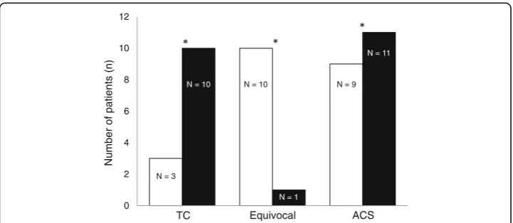
All patients underwent hybrid cardiac SPECT/CT imaging. Out of the 10 patients with equivocal results on SPECT-only analysis, 7 were diagnosed with TC and 2 were diag-nosed with ACS based on SPECT/CT fused analysis. Consequently, the number of equivocal results was significantly decreased by SPECT/CT fused analysis (p= 0.040) (Fig. 4). Concerning the diagnosis of TC, SPECT/CT fused analysis showed a superior diagnostic performance (sensitivity, 90%; specificity, 100%; and accuracy, 95%) com-pared with SPECT-only analysis (sensitivity, 30%; specificity, 81%; and accuracy, 57%) (Table 4).
Case presentations
Figure 5 shows a representative case of a patient with TC. In this 68-year-old woman with chest pain, negative T in V3–6 and ST-depression in V5 and V6 were observed on ECG upon admission. A blood test showed slightly high troponin T (0.038 ng/ml) and CCT showed no coronary artery stenoses. Cardiac MIBI and BMIPP SPECT were performed 8 days after the onset. MIBI images showed slightly reduced accumulation in the distal anterolateral to apical wall in the LV myocar-dium, while BMIPP images showed moderately reduced accumulation in the same region. Since the myocardial damage only appears to be slightly congruent with typical diagonal branch territory, SPECT-only imaging could not differentiate be-tween TC and ACS. In the SPECT/CT fused images, the accumulation defect ex-tended through distal LAD and first diagonal branch territories and this patient was therefore diagnosed with TC.
Figure 6 shows a typical case of a patient with ACS. In this 60-year-old woman with chest pain, ST-depression in V4–6 was observed on ECG. A blood test showed slightly high troponin T (0.052 ng/ml) and CCT showed no coronary artery stenoses. Cardiac MIBI and BMIPP SPECT were performed 7 days after the onset. MIBI images showed slightly reduced accumulation in the anterior to apical wall in
0 2 4 6 8 10 12
TC Equivocal ACS
Number of patients (n)
N = 9 N = 10
N = 10
N = 3
N = 1
N = 11
Fig. 4Alteration of image interpretation (TC or non-TC) between SPECT-only (white columns) and SPECT/ CT fused (black columns) analyses
Table 4Diagnostic accuracies of SPECT-only imaging and hybrid SPECT/CT imaging
Sensitivity (%) Specificity (%) Accuracy (%)
SPECT-only imaging 30 81 57
the LV myocardium, while BMIPP images showed slightly to moderately reduced accumulation in the same region. Since the myocardial damage appeared to be congruent with typical LAD territory, this patient was thought to have ACS in LAD territory. In the SPECT/CT fused images, the accumulation defect was con-gruent with first diagonal branch territory, and this patient was therefore diagnosed with ACS due to first diagonal branch vasospasm.
a
c
b
d
Fig. 568-year-old woman with TC. Cardiac MIBI and BMIPP SPECT images show accumulation defects in the distal anterolateral to apical wall (a,b). Due to the unclear alignment of accumulation defects with coronary artery territories, SPECT-only imaging could not differentiate between TC and ACS. SPECT/CT fused images revealed that the accumulation defect extended through distal LAD and first diagonal branch territories, confirming TC (c,d)
a
c
b
d
Discussion
In this study, the feasibility of dual-isotope MIBI and BMIPP imaging using con-ventional NaI gamma cameras and the diagnostic performance of hybrid cardiac SPECT/CT imaging for patients with TC was examined. The results showed that the crosstalk rates of 123I at 140 keV and of 99mTc at 159 keV were 25.4% and 6.6%, respectively, and SPECT/CT fused analysis had improved diagnostic perform-ance compared with SPECT-only analysis.
Cardiac SPECT for diagnosing TC
ICA is the most common modality for diagnosing TC, eliminating obstructive cor-onary artery lesions. In contrast, cardiac perfusion and fatty-acid metabolism SPECT are applied for diagnosing TC, detecting the myocardial damage instead. However, cardiac dual-isotope MIBI and BMIPP imaging using conventional NaI gamma cameras has been limited for use even though several investigators reported positively about the imaging procedures (Yoshida et al. 2013; Hirata et al. 2009). Recently, the dual-isotope SPECT imaging technique using semiconductor gamma cameras was introduced. The crosstalk rate of 123I at 140 keV and that of 99mTc at 159 keV using a semiconductor gamma camera are reported to be >20% and <1%, respectively (Takahashi et al. 2015). Varying slightly, in this study, using an NaI gamma camera and suitable energy windows, the crosstalk rate of 123I at 140 keV and that of 99mTc at 159 keV were 25.4% and 6.6%, respectively. While semicon-ductor detectors have superior energy resolution and sensitivity compared with NaI gamma cameras, this study shows that dual-isotope SPECT imaging using an NaI gamma camera is feasible without significant influence on the diagnosis. Using this method, diagnosing TC using cardiac dual-isotope SPECT will be more accessible, even in facilities that do not have a semiconductor detector.
For the clinical study, the patients with TC underwent both cardiac MIBI and BMIPP SPECT tests 10 (7–14) days from the onset of TC, mostly within the subacute phase. Ito et al. reported that SBDS was significantly higher than summed perfusion score (3.2 ± 3.0 vs. 12.6 ± 3.7) (Ito et al. 2005). In this study, SBDS was significantly higher than SMDS (5 (3–7) vs. 10 (8–12), respectively,p= 0.003) similarly to Ito et al. Further-more, myocardial regional accumulation of MIBI and BMIPP was observed in this study. While A-MDS and A-BDS did not differ between TC and ACS, NA-MDS and NA-BDS were higher in ACS than TC. However, Matsuo et al. reported that both A-BDS and NA-BDS were significantly higher in ACS than TC. The discrepancy of the results between Matsuo et al. and this study is due to the difference in the populations of the ACS groups, which of this study had myocardial ischemia exclu-sively in the distal LAD territory, resulting in a comparatively mild myocardial is-chemia (Matsuo et al. 2014).
CCT for diagnosing TC
(Gaemperli et al. 2007). As an alternative, CCT can reveal obstructive coronary artery lesions even in patients with suspected TC.
In this study, 4 out of 11 patients in the TC group (36%) had significant coronary ar-tery stenoses and no ischemic myocardial damage in the corresponding coronary arar-tery territories. Patients with TC and incidental significant coronary artery stenoses have the risk of being misdiagnosed with ACS. Also, 5 out of 11 patients in the ACS group (45%) did not have significant coronary artery stenoses and were diagnosed with ACS caused by coronary vasospasm via SPECT/CT imaging. In the general clinical setting, patients with ACS often have no significant coronary artery stenoses (Libby and Ther-oux 2005), and identification of the ischemic myocardial damage with cardiac SPECT is preferable for such patients (Hambye et al. 2000).
Clinical application of hybrid cardiac SPECT/CT imaging for TC
Hybrid cardiac SPECT/CT imaging can easily be conducted with a stand-alone system and dedicated fusion software, directly correlating the distribution of LV myocardial damage with the distribution of coronary arteries. Several studies have revealed that the use of hybrid cardiac SPECT/CT imaging increased the conclusive diagnostic rate com-pared with side-by-side interpretations of SPECT and CCT images in patients with CAD (Gaemperli et al. 2007, 2008; Rispler et al. 2007). In this study, hybrid cardiac SPECT/CT imaging was also used for diagnosing TC. While the indeterminate diagnos-tic rate for TC was remarkably high in the SPECT-only analysis (45%), the indetermin-ate diagnostic rindetermin-ate significantly decreased (5%) in the SPECT/CT fused analysis. Hybrid cardiac SPECT/CT imaging shows the relationship between the distributions of myo-cardial damage and coronary arteries and thus the cause of myomyo-cardial damage, TC or ACS, can easily be resolved. The detailed relationship of the distributions indicated by this hybrid imaging also permitted the differentiation of TC with ACS from solely ACS. Therefore, hybrid cardiac SPECT/CT imaging may help prevent the misdiagnosis of these patients.
Study limitations
to diagnose other causes of congestive heart failure by eliminating ischemic heart failure.
Conclusions
In this study, the feasibility of dual-isotope MIBI and BMIPP imaging using conven-tional NaI gamma cameras was revealed, resulting from the crosstalk rates of 123I at 140 keV and99mTc at 159 keV of 25.4% and 6.6%, respectively. Furthermore, hybrid car-diac SPECT/CT imaging using MIBI and BMIPP allows comprehensive assessment of coronary artery distribution and myocardial damage distribution and may have superior diagnostic performance compared with SPECT-only imaging for patients with TC.
Abbreviations
A-BDS:Apical BMIPP defect score; ACS: Acute coronary syndrome; A-MDS: Apical MIBI defect score; A-MS: Apical mismatch score; Apical: A-%WT and non-apical % wall thickening = NA-%WT; BMIPP:123I–beta-methyl-P-iodophenyl-pentadecanoic
acid; CAD: Coronary artery disease; CCT: Cardiac CT; CT: Computed tomography; ECG: Electrocardiograms; ICA: Invasive coronary angiography; LAD: Left anterior descending artery; LV: Left ventricular; MIBI:99mTc-methoxy-isobutyl-isonitrile;
NA-BDS: Non-apical BMIPP defect score; NA-MDS: Non-apical MIBI defect score; NA-MS: Non-apical mismatch score; PCI: Percutaneous coronary intervention; SBDS: Summed BMIPP defect score; SMDS: Summed MIBI defect score; SMS: Summed mismatch score; SPECT: Single-photon emission computed tomography; TC: Takotsubo cardiomyopathy
Acknowledgments
We are grateful to the radiology technologists Kyoji Asano, Shinjiro Yoshida, Masaya Suda, Junya Tashiro, and Toshio Maki for their technical assistance in the administration of cardiac SPECT and hybrid cardiac SPECT/CT imaging. The publication of this article was supported by funds of the European Association of Nuclear Medicine (EANM).
Funding None.
Authors’contributions
YS contributed to the design, analysis and manuscript preparation and submission. YF contributed to the design, statistical methodology, analysis and manuscript preparation. SK contributed to the design, statistical methodology and manuscript preparation. HT and WS contributed to the design, conduct and manuscript preparation. All authors read and approved the final manuscript.
Ethics approval and consent to participate
All procedures performed in studies involving human participants were in accordance with the ethical standards of the institutional and/or national research committee, and the 1964 Helsinki declaration and its later amendments or comparable ethical standards.
Consent for publication
Formal consent was not required, as the study was observational and data were generated during daily clinical practice.
Competing interests
The authors declare that they have no competing interests.
Publisher’s Note
Springer Nature remains neutral with regard to jurisdictional claims in published maps and institutional affiliations.
Author details 1
Department of Clinical Radiology, Nippon Medical School Graduate School of Medicine, 1-1-5 Sendagi, Bunkyo-ku, Tokyo 113-0022, Japan.2Department of Cardiovascular Medicine, Nippon Medical School Graduate School of Medicine, 1-1-5 Sendagi, Bunkyo-ward, Tokyo 113-0022, Japan.
Received: 2 November 2017 Accepted: 20 December 2017
References
Barletta G, Del Pace S, Boddi M, Del Bene R, Salvadori C, Bellandi B et al (2009) Abnormal coronary reserve and left ventricular wall motion during cold pressor test in patients with previous left ventricular ballooning syndrome. Eur Heart J 30:3007–3014
Gaemperli O, Schepis T, Kalff V, Namdar M, Valenta I, Stefani L et al (2007) Validation of a new cardiac image fusion software for three-dimensional integration of myocardial perfusion SPECT and stand-alone 64-slice CT angiography. Eur J Nucl Med Mol Imaging 34:1097–1106
Gaemperli O, Schepis T, Valenta I, Husmann L, Scheffel H, Duerst V et al (2007) Cardiac image fusion from stand-alone SPECT and CT: clinical experience. J Nucl Med 48:696–703
Gaemperli O, Schepis T, Valenta I, Koepfli P, Husmann L, Scheffel H et al (2008) Functionally relevant coronary artery disease: comparison of 64-section CT angiography with myocardial perfusion SPECT. Radiology 248:414–423 Hachamovitch R, Hayes SW, Friedman JD, Cohen I, Berman DS (2003) Comparison of the short-term survival benefit
associated with revascularization compared with medical therapy in patients with no prior coronary artery disease undergoing stress myocardial perfusion single photon emission computed tomography. Circulation 107:2900–2907 Hambye AS, Vervaet A, Dobbeleir A, Dendale P, Franken P (2000) Prediction of functional outcome by quantification of
sestamibi and BMIPP after acute myocardial infarction. Eur J Nucl Med 27:1494–1500
Hirata M, Monzen H, Suzuki T, Ogasawara M, Nakanishi A, Sumi N et al (2009) Evaluation of a new protocol for two-isotope123I-BMIPP/99mTc-TF single photon emission computed tomography (SPECT) to detect myocardial damage within one hour. Igaku Butsuri 29:3–11
Ito K, Sugihara H, Katoh S, Azuma A, Nakagawa M (2003) Assessment of Takotsubo (ampulla) cardiomyopathy using
99mTc-tetrofosmin myocardial SPECT–comparison with acute coronary syndrome. Ann Nucl Med 17:115–122
Ito K, Sugihara H, Kinoshita N, Azuma A, Matsubara H (2005) Assessment of Takotsubo cardiomyopathy (transient left ventricular apical ballooning) using 99mTc-tetrofosmin,123I-BMIPP,123I-MIBG and99mTc-PYP myocardial SPECT. Ann
Nucl Med 19:435–445
Kawai S, Kitabatake A, Tomoike H (2007) Takotsubo cardiomyopathy study group. Guidelines for diagnosis of takotsubo (ampulla) cardiomyopathy. Circ J 71:990–992
Kawai S, Suzuki H, Yamaguchi H, Tanaka K, Sawada H, Aizawa T et al (2000) Ampulla cardiomyopathy (‘Takotusbo’ cardiomyopathy)–reversible left ventricular dysfunction with ST segment elevation. Jpn Circ J 64:156–159 Kawai Y, Tsukamoto E, Nozaki Y, Morita K, Sakurai M, Tamaki N (2001) Significance of reduced uptake of iodinated fatty
acid analogue for the evaluation of patients with acute chest pain. J Am Coll Cardiol 38:1888–1894 Kubo A, Nakamura K, Hashimoto J, Sammiya T, Iwanaga S, Hashimoto S et al (1992) Phase I clinical trial of a new
myocardial imaging agent,99mTc-PPN1011. Kaku Igaku 29:1165–1176
Kurisu S, Inoue I, Kawagoe T, Ishihara M, Shimatani Y, Nishioka K et al (2003) Myocardial perfusion and fatty acid metabolism in patients with tako-tsubo-like left ventricular dysfunction. J Am Coll Cardiol 41:743–748
Kurisu S, Kihara Y (2012) Tako-tsubo cardiomyopathy: clinical presentation and underlying mechanism. J Cardiol 60:429–437
Leipsic J, Abbara S, Achenbach S, Cury R, Earls JP, Mancini GJ et al (2014) SCCT guidelines for the interpretation and reporting of coronary CT angiography: a report of the Society of Cardiovascular Computed Tomography Guidelines Committee. J Cardiovasc Comput Tomogr 8:342–358
Libby P, Theroux P (2005) Pathophysiology of coronary artery disease. Circulation 111:3481–3488
Matsuo S, Nakajima K, Kinuya S, Yamagishi M (2014) Diagnostic utility of123I-BMIPP imaging in patients with Takotsubo
cardiomyopathy. J Cardiol 64:49–56
Matsuo S, Nakamura Y, Takahashi M, Mitsunami K, Kinoshita M (1998) Myocardial metabolic abnormalities in hypertrophic cardiomyopathy assessed by iodine-123-labeled beta-methyl-branched fatty acid myocardial scintigraphy and its relation to exercise-induced ischemia. Jpn Circ J 62:167–172
Miyachi H, Kumita S, Tanaka K (2013) PET/CT and SPECT/CT cardiac fusion imaging in a patient with takotsubo cardiomyopathy. Eur Heart J 34:397
Prasad A, Lerman A, Rihal CS (2008) Apical ballooning syndrome (Tako-Tsubo or stress cardiomyopathy): a mimic of acute myocardial infarction. Am Heart J 155:408–417
Rispler S, Keidar Z, Ghersin E, Roguin A, Soil A, Dragu R et al (2007) Integrated single-photon emission computed tomography and computed tomography coronary angiography for the assessment of hemodynamically significant coronary artery lesions. J Am Coll Cardiol 49:1059–1067
Takahashi Y, Miyagawa M, Nishiyama Y, Kawaguchi N, Ishimura H, Mochizuki T (2015) Dual radioisotopes simultaneous SPECT of99mTc-tetrofosmin and123I-BMIPP using a semiconductor detector. Asia Ocean J Nucl Med Biol 3:43–49
Tamaki N, Kawamoto M, Yonekura Y, Fujibayashi Y, Takahashi N, Konishi J et al (1992) Regional metabolic abnormality in relation to perfusion and wall motion in patients with myocardial infarction: assessment with emission tomography using an iodinated branched fatty acid analog. J Nucl Med 33:659–667
Torizuka K, Yonekura Y, Nishimura T, Tamaki N, Uehara T, Ikekubo K et al (1991) A phase 1 study of betamethyl-p-(123
I)-iodophenyl-pentadecanoic acid (123I-BMIPP). Kaku Igaku 28:681–690
Tsuchihashi K, Ueshima K, Uchida T, Oh-mura N, Kimura K, Owa M et al (2001) Transient left ventricular apical ballooning without coronary artery stenosis: a novel heart syndrome mimicking acute myocardial infarction. Angina pectoris-myocardial infarction investigations in Japan. J Am Coll Cardiol 38:11–18
Yoshida A, Takano H, Asai K, Yasutake M, Amano Y, Kumita S et al (2013) Comparison of perfusion-metabolism mismatch in99mTc-MIBI and123I-BMIPP scintigraphy with cardiac magnetic resonance in patients with