R E S E A R C H
Open Access
Abnormally elevated USP37 expression
in breast cancer stem cells regulates
stemness, epithelial-mesenchymal
transition and cisplatin sensitivity
Tao Qin
1,2, Bai Li
1,2, Xiaoyue Feng
1,2, Shujun Fan
1,2, Lei Liu
3, Dandan Liu
1,2, Jun Mao
1,2, Ying Lu
4, Jinfeng Yang
5,
Xiaotang Yu
1, Qingqing Zhang
1, Jun Zhang
6, Bo Song
1,2, Man Li
7*and Lianhong Li
1,2*Abstract
Background:Recent studies have indicated that deubiquitinating enzymes (DUBs) are related to the stem-cell pathway network and chemo-resistance in cancer. Ubiquitin-specific peptidase 37 (USP37), a novel DUB, was identified to be a potential factor associated with tumor progression. However, the biological functions of USP37 in breast cancer remain unclear.
Methods:The distribution of USP37 expression in breast cancer and the correlation between USP37 expression and the overall survival rate were detected by The Cancer Genome Atlas (TCGA) database. Gene set enrichment analysis (GSEA) was utilized to evaluate potential mechanism of USP37 in breast cancer. The USP37 expression in breast cancer tissues and breast cancer cell lines were detected by immunohistochemistry and western blotting. Sorting of breast cancer stem cells (BCSCs) were by using MACS assay. In vitro and in vivo assays were performed to examine the biological functions of USP37 in breast cancer cells. MG132, CHX chase, immunofluorescence staining and co-immunoprecipitation assays were used to test the interaction between USP37 and Gli-1.
Results: Bioinformatics analysis demonstrated that USP37 gene was elevated in breast cancer tissues and its overexpression was strongly correlated with the increased mortality rate. GSEA analysis showed that USP37 expression was positively associated with cell growth and metastasis while negatively related to cell apoptosis in the TCGA breast cancer samples. USP37 expression was elevated in breast cancer tissues and breast cancer cell lines. Moreover, we also detected that USP37 was overexpressed in BCSCs. USP37 regulated the ability of cell invasion, epithelial-mesenchymal transition (EMT), stemness and cisplatin sensitivity in breast cancer cell lines. Additionally, USP37 knockdown inhibited tumorigenicity and increased anticancer effect of cisplatin in vivo. Knockdown of USP37 significantly decreased hedgehog (Hh) pathway components Smo and Gli-1. Gli-1 was stabilized by USP37 and they interacted with each other. Further studies indicated that USP37 knockdown could inhibit the stemness, cell invasion and EMT in breast cancer via downregulation of Hh pathway.
Conclusions:These findings reveal that USP37 is highly expressed in BCSCs and is correlated with poor prognosis in breast cancer patients. USP37 can regulate the stemness, cell invasion and EMT via Hh pathway, and decreased USP37 confers sensitivity to cisplatin in breast cancer cells. USP37 is required for the regulation of breast cancer progression, as well as a critical target for clinical treatment of breast cancer.
Keywords:USP37, Stemness, EMT, Cisplatin, Hedgehog, Breast cancer
* Correspondence:liman19890930@sina.com;lilianhong917@sina.com 7
Department of Oncology, The Second Affiliated Hospital of Dalian Medical University, Dalian 116023, Liaoning Province, People’s Republic of China 1Department of Pathology, Dalian Medical University, Dalian 116044, People’s Republic of China
Full list of author information is available at the end of the article
Introduction
Breast cancer is the deadliest form of carcinoma affecting women, with nearly a quarter-million cases diagnosed in 2016 [1]. Although there are effective treatments against some types of breast carcinoma, such as those for sub-types with abnormal overexpression of the HER2/Neu oncogene, the majority breast cancers remain incurable. Breast cancer stem cells (BCSCs) possess self-renewal and differentiation capabilities, leading to tumor recurrence, metastasis and therapeutic resistance [2, 3]. CD44+/ CD24−or aldehyde dehydrogenase1 (ALDH1) phenotypes are efficient in the identification of BCSCs from breast cancer populations. However, there is a small overlap be-tween CD44+/CD24− and ALDH1 stem phenotypes, as well as less stem markers in differentiation of different breast cancer subtypes [4]. Therefore, it is necessary to identify more discriminatory biomarkers of distinct mo-lecular subtypes for the isolation and identification of the BCSCs subpopulation.
EMT describes the process by which epithelial cells detach from neighboring cells and are transferred to other tissue sites via dissolution of basement membrane and passage through the extra-cellular matrix [5, 6]. EMT could also facilitate the generation of cancer stem cells from more differentiated cancer cells [7]. The EMT process during breast carcinogenesis is considered to be controlled by a series of signaling pathways, including Notch, Wnt/β-catenin and Hedgehog [8,9]. The Hedge-hog (Hh) pathway is responsible for the maintenance of stem cells and EMT, which can contribute to the evolu-tion of breast cancer [10].
Ubiquitination describes a highly conserved and re-versible modification process of protein degradation, which is involved in nearly all aspects of cell biology [11]. Deubiquitinating enzymes (DUBs) can prevent ubiquitin-mediated degradation of target proteins [12]. Importantly, dysregulated DUBs expression is frequently associated with the tumorigenesis process, specifically cell self-renewal, apoptosis and EMT [13, 14]. It has been confirmed that DUBs are essential for the regula-tion of stem cell-related markers and controlling various steps of metastatic progression, including invasion, dis-semination and eventual metastasis to distant organs [14,15]. Ubiquitin specific peptidase 37 (USP37), a novel DUB, is a member of ubiquitin-specific processing pro-teases family. Human USP37, localized mainly in the cytoplasm, is composed of 979 amino acids harboring three ubiquitin-interacting motifs between the Cys box and His box of the primary sequence [16,17]. The func-tion of USP37 was initially identified as a potent regula-tor of the cell cycle where it could accelerate the G1/S transition with exceptional high expression [18,19]. Pre-vious studies have found that USP37 could regulate the stem cell-related marker SOX2 by binding with its
promoter region at the transcriptional level [20]. Pan et al. reported that high levels of USP37 gene expression in lung cancer promoted cell viability as well as the Warburg effect via deubiquitination and stabilization of pluripotent factor c-Myc protein [21]. These advances implicated that USP37 gene may be associated with the stemness of tumor cells facilitating cancer progression. Recent studies indicate that USP37 is a potential factor involved in breast cancer progression [16]. However, the biological function of USP37 in the direct regulation of BCSCs and EMT remains unexplored.
In this study, we found that USP37 expression was up-regulated in breast cancer tissues compared with sur-rounding tissues and its overexpression was significantly correlated with increased rates of mortality. We demon-strated that USP37 was highly expressed in BCSCs. The knockdown of USP37 could inhibit the stemness, cell invasion and EMT via downregulation of Hedgehog pathway. USP37 also interacted with and stabilized glioma-associated oncogene 1 (Gli-1) protein. Addition-ally, USP37 knockdown enhanced the sensitivity of breast cancer cells to cisplatin in vitro and in vivo. We postulate that USP37 may represent a novel molecular target for breast cancer treatment.
Materials and methods
Bioinformatic analysis
Gene data extracted from invasive breast tumor samples was obtained from the TCGA Data Portal. Results of analysis were used to create figures via GraphPad Prism. According to PAM50 gene expression signature, infor-mation about four breast cancer subtypes (basal-like, Lu-minal A, LuLu-minal B, and enriched Her-2) were classified. A scatter plot diagram, where each dot indicated an in-dividual sample, was synthesized using the All Complete Tumors of Breast Invasive Cancer dataset [22, 23]. For survival analysis, clinical data related to invasive breast carcinoma were downloaded from the TCGA database [22]. Kaplan–Meier curves were analyzed by GraphPad
Prism.
Gene set enrichment analysis (GSEA) was utilized to evaluate potentially biological mechanism associated with USP37 mRNA expression levels in the TCGA breast cancer samples. GSEA software which was ob-tained from the Broad Institute draws the result pictures automatically.
Cell culture and animals
(100 ng/mL) (Sigma-Aldrich), EGF (20 ng/mL) (R&D systems, Wiesbaden, Germany), hydrocortisone (500 ng/ mL), and L-glutamine (Invitrogen, Gaithersburg, MD, USA). MCF-7 cells were cultured in DMEM/HIGH GLUCOSE (Hyclone, Logan, UT, USA) with 10% FBS. MDA-MB-231 cells were cultured in MEM Alpha Modi-fication medium (Hyclone) supplemented with 10% FBS. In addition, BT549 and T-47D cells were cultured in RPMI-1640 medium (Hyclone) supplemented with 10% FBS. Growth condition for all cells was 37 °C at 5% concentration of CO2.
BALB/c Nude mice (6 to 8 weeks old) were purchased from Vital River Laboratory Animal Technology Company (Beijing, China) and reared according to the Animal Care and Use Committee of Dalian Medical University.
Plasmids, antibodies and reagents
The pEZ-M35-USP37 plasmid was created by FulenGen Co. (Guangzhou, China). Anti-Gli-1 and anti-Smoothened antibodies were obtained from Abcam (Cambridge, MA, USA); all other antibodies were obtained from Proteintech Group, Inc. (Wuhan, China). Secondary antibodies were purchased from Santa Cruz Biotechnology (Dallas, TX, USA). Cycloheximide (CHX), MG132, cisplatin and pur-morphamine reagents were obtained from MedChemEx-press (Shanghai, China).
siRNA, shRNA, lentivirus
The design and synthesis of siRNAs were completed by RiboBio company (Guangzhou, China), and transfection of siRNAs against USP37 was performed with Lipofecta-mine 2000. MCF-7 and MDA-MB-231 cells were trans-fected with USP37 siRNAs or a negative control (NC) siRNA. Lentivirus vectors including short hairpin RNA against USP37 (shUSP37#2) and the negative control (shScramble) were purchased from GenePharma Com-pany (Shanghai, China). The transfected cells were treated with puromycin (Clontech, USA) for selection. To overex-press USP37 in MCF-7 cells, the pEZ-M35-USP37 plasmid (2μg) were infected into MCF-7 cells via Lipofec-tamine 2000. Expression was confirmed by RT-qPCR and western blotting. The siRNA or shRNA sequences were listed in Additional file1: Table S1.
BCSCs isolation
Here, 1 × 10⁷MCF-7 cells were grown with primary anti-body against CD24 following the manufacturer’s protocol for the CD24 Microbead Kit (Miltenyi Biotec, Bergisch Gladbach, Germany). Labeled cells were then incubated with goat anti-mouse IgG microBeads (Miltenyi Biotec) and magnetically separated with MiniMACS columns (Miltenyi Biotec). Acquired CD24− cells were incu-bated with CD44 microbeads (Miltenyi Biotec) at 4 °C
for 15 mins. Cells were again washed and magnetic-ally separated.
Colony formation assay and cell viability assay
Colony formation assay of breast cancer cells was car-ried out by plating infected cells at a density of 1000 cells/well in a 6-well plate and then different concentra-tions of cisplatin were added. After 2 weeks of incuba-tion, cells were washed three times with phosphate buffered saline, fixed with methanol and stained with 1% crystal violet. Visible colonies were stained violet and counted for data analysis. The detection of cell viability was performed in accordance with the Cell Counting Kit 8 assay (Dojindo Laboratories, Kumamoto, Japan).
Cell invasion assay
Cells were similarly cultured in MEM Alpha Modifica-tion medium for 24 hours. Briefly, 1 × 10⁵ cells were seeded without serum into 24-well insert Transwell chambers (8 μm pore size, Corning, USA) and pre-treated with Matrigel (BD, Bioscience, San Jose, CA, USA). Medium supplemented with 20% serum was added into the lower chamber. After 12–16 hours of seeding, cotton swabs were used to clean the upper cells. The cells on the other side of the membrane were stained with 1% crystal violet. A randomly selected area was counted with an optical microscope.
Wound healing assay
Cells were incubated in 12-well plates. When cellular density reached nearly 100%, the cell monolayer was wounded with a 200 μl micro-pipette tip. The wound areas were washed three times with phosphate buffered saline (PBS). Then the medium was changed to MEM Alpha Modification without FBS. The wound areas were micrographed at 0, 12, 24 and 48 h. All assays were per-formed in triplicate.
Western blotting
Snail1, 1:500; N-cadherin, 1:1000; Vimentin, 1:500 and GAPDH (loading control), 1:1000. Finally, the transfer membranes were incubated with anti-IgG secondary antibodies (1:16000 in TBST) for 1 h at 37 °C. The pro-tein band images were captured with a ODYSSEY infra-red imaging system.
RNA extraction and real-time quantitative PCR analysis
Total RNA was extracted from cultured cells with Trizol reagent (Transgen, China). In total, 1 μg of total RNA was reverse-transcribed with the All-in-one First-Strand cDNA Synthesis SuperMiX kit (Transgen, China). The mRNA expression levels of USP37, ALDH1, CD24 and CD44 were quantified by real-time PCR. RT-qPCR was analyzed with the iCycler™ Real Time System and a SYBR Premix EX Tag Master mixture kit (Transgen, China) according to the manufacturer’s instructions. The relative expression levels of mRNA were evaluated by using the 2−ΔΔCtmethod. Primer sequences are listed in Additional file1: Table S1.
Immunofluorescence analysis
Cells were grown to 3 × 10³ in 24-well plates at 37 °C and fixed with 100% methanol. Next, cells were cleaned two to three times using PBST (PBS and 0.5% Triton X-100). After blocked with 3% BSA for 2 h at 4 °C, cells were incubated with the following primary antibodies (San Ying Biotechnology, China) overnight at 4 °C: USP37, 1:100; Gli-1, 1:100; Flag, 1:100; E-cadherin, 1:100 and N-cadherin, 1:100. Subsequently, cells were incu-bated with fluorescence conjugated secondary antibody (Sigma, 1:200) for 1 h and stained the cell nuclei with DAPI (Beyotime, China). Images were viewed with CKX41 Inverted Microscope (Olympus, Japan).
CHX chase assay and co-immunoprecipitation (co-IP) assay
For the CHX chase assay, cells were treated with CHX (50 μg/ml) and harvested at the indicated timepoints. Treated cells were lysed, and the lysates were analyzed by western blotting with USP37 or GAPDH anti-bodies. For the Co-IP assay, 8 × 10⁶MCF-7 cells were har-vested and lysed in IP lysis buffer containing protease inhibitor. The experiment based on manufacturer’s proto-cols (IP Kit, Proteintech Group) was performed.
Sphere formation assay
Cells (MCF-7, MDA-MB-231, BT549 or T47D) were inoculated into ultra-low attachment 6-well plates (Corning, New York, USA) at a density of 1000 cells/ well, and then grown in DMEM/F12 supplemented with B27 (1:50, Invitrogen), 20 ng/ml human recombinant EGF (Sigma-Aldrich, St. Louis, Missouri, USA), 20 ng/ ml bFGF (Sigma-Aldrich, St. Louis, Missouri, USA),
4 μg/ml heparin (Sigma-Aldrich, St. Louis, Missouri, USA), and 5 μg/ml insulin (Sigma-Aldrich, St. Louis, Missouri, USA) for 14 days. Cell colonies larger than
60 μm in diameter were counted under an inverted
microscope (Olympus Corporation, Tokyo, Japan). After cultivation for 28 days, spheroid cells were collected for western blotting.
Mouse xenograft mode
Animal experiments and procedures were conducted in accordance with the Guide for the Care and Use of La-boratory Animals (NIH). Twenty BALB/C nude mice were randomly divided into four groups. The negative control group (shScramble) and shUSP37#2-transfected MCF-7 cells (5 × 10⁵) were resuspended in 100 μl PBS and injected into mammary fat pads. When the size of tumor reached approximately 100 mm3, animals were randomly treated with cisplatin (2 mg/kg) or 0.9% saline. Mice were then retreated with their assigned treatment once every 2 days. The tumor sizes were measured with a Vernier caliper and recorded every other day. The tumor volume was calculated using the formula: tumor volume [mm]3= (length [mm]) (width [mm])2× 0.5. After the inoculation of tumors for 3 weeks, xenografted tumors were excised from sacrificed mice then analyzed by immunohistochemistry and western blotting.
Immunohistochemical analysis
Tumor tissues were obtained from mouse xenograft mode. Paraffin-embedded tissue was cut into 5μm thick slices that were fixed onto glass slides. All human breast cancer tissue arrays were purchased from Shanghai Outdo Biotech (Shanghai, China), including 60 cases of cancer tissues and surrounding tissues (HBre-Duc060CS-03; HBre-Duc060CS-04). These tissue sections were immuno-stained with corresponding antibodies, and then deparaffi-nized in xylene and rehydrated with ethanol. Tissue sections were preincubated with 10% normal goat serum, followed by incubation with primary antibody solution overnight at 4 °C. After washing with PBS, slides were in-cubated with the secondary antibody at 37 °C for 10 min, then cleaned with cold PBS and treated with peroxidase conjugated-biotin streptavidin complex for 10 min. Finally stains were examined with 3,3′-diaminobenzidine and hematoxylin. As previously described [23], the immuno-stained tissues were scored by multiplying the intensity (0–3) and extent (0–100) of staining.
Ethical approval
Statistical analysis
Statistical analysis was performed with SPSS software version 11.0. Data are expressed as mean ± SD. Differ-ences between two groups were evaluated by Student’s
t-test. One-way ANOVA was used when comparing multiple groups.P< 0.05 was considered statistically sig-nificant. Clinical data analysis of survival and relevant correlations were performed with GraphPad Prism.
Results
USP37 is commonly overexpressed in breast cancer
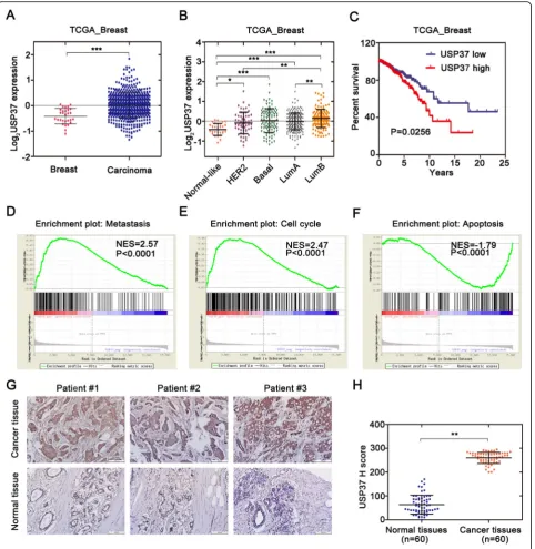
USP37 has been previously confirmed to be overex-pressed in lung cancers cells and tissues [21]. In order to investigate the role of USP37 in tumorigenesis, we examined the breast cancer database of The Cancer Genome Atlas (TCGA) to evaluate the differential ex-pression of USP37 [22]. Analysis of the TCGA database indicated that cancer with USP37 transcripts (n= 517) had a significantly higher expression level than normal samples (n= 29) (P< 0.0001) (Fig. 1a). Similar results were also found via immunohistochemistry analysis (Fig.
1g). To further confirm the overexpression of USP37 gene in breast cancer, we utilized a series of human breast cancer cells (MCF-7, MDA-MB-231, BT549 and T47D) and human normal breast epithelial cells (MCF-10A) to examine USP37 protein expression by western blotting (Fig. 2a). These results confirmed that USP37 was also overexpressed in breast cancer cells compared to normal breast epithelial cells.
For investigation of the correlation between USP37 gene and the breast cancer heterogeneity, we also tested USP37 expression in the four cell subtypes using PAM50 gene expression profiling. First, we observed an starkly different tendency within USP37 gene expression among different pathological subtypes of breast cancer cells, including the normal-like subtype having the low-est, and Luminal B type endowed with the highest ex-pression levels of USP37 (p< 0.0001) (Fig. 1b). We next estimated the effect of USP37 as an oncogenic bio-marker for overall survival of patients diagnosed with breast cancer. Clinical data from the TCGA database were divided into two groups according to the differen-tial expression of USP37 gene. The results indicated that cancer with higher expression levels of USP37 was sig-nificantly correlated with the elevated rates of mortality (P< 0.05) (Fig. 1c). Samples with high USP37 expression also had shorter survival duration than those with low USP37 expression. Moreover, GSEA analysis showed that high USP37 expression was positively associated with metastasis, cell growth and anti-apoptosis in the TCGA breast cancer samples (Fig. 1d–f).In brief, these data confirmed that USP37 gene could act as a cancer biomarker in predicting a worse outcome for breast can-cer patients. Collectively, these data suggest that USP37
is abnormally overexpressed in human breast cancer pa-tients and cell lines.
USP37 is highly expressed in breast cancer stem cells
CD24−/CD44+cells and ALDH1+cells are widely consid-ered to be breast cancer stem cells [24]. To validate the expression levels of the USP37 gene in breast cancer stem cells, we isolated CD24−/CD44+ cell populations from MCF-7 cell lines by magnetic activated cell sorting (MACS). We detected that USP37 was significantly over-expressed in CD24−/CD44+ cells compared to their counterpart CD24+ or CD24−CD44− cells in regards to mRNA levels (Fig.2c). Immunofluorescence staining fur-ther confirmed the upregulation of USP37 expression in BCSCs (Fig.2d). Additionally, we used breast cancer cell lines to perform sphere-formation experiments and mea-sured the differential expression of the USP37 gene. We confirmed that protein levels of USP37 were evidently elevated in spherical cells compared to their counterpart adherent cells (Fig. 2b). Taken together, these results in-dicated that elevated expression of USP37 was enriched in breast CSCs and was a novel feature of breast cancer stem cell-like subpopulation.
USP37 knockdown suppresses breast cancer cell migration and invasion by promoting mesenchymal-epithelial transition
Currently, the underlying biological mechanism account-ing for the elevated expression of USP37 in breast tu-mors remains unclear. To investigate the effects of USP37 on breast cancer progression, we used siRNA oli-gonucleotides to knockdown endogenous USP37 in MCF-7 and MDA-MB-231 cells. USP37 mRNA expres-sion levels significantly decreased with two different siR-NAs treated breast cancer cells compared to levels in the control group cells (Fig. 3b). Moreover, USP37 pro-tein was also reduced significantly with siRNA#2 and siRNA#3 treatment of MCF-7 and MDA-MB-231 cells as evidenced by western blotting (Fig. 3a). These results confirmed that USP37 gene expression could be effect-ively downregulated by siRNA#2 and siRNA#3.
Fig. 1Overexpression of USP37 in human BC was correlated with poor-disease outcome.aCancer with USP37 transcripts apparently had higher
expression (n= 517) than normal breast tissues (n= 29) from the TCGA database [22] (***P< 0.0001). The median and interquartile range are
represented by black lines.bUSP37 transcripts among different subtypes are divided into five distinct molecular subtypes. The parameters were
analyzed with the PAM50 gene expression profiling [22]. The median with interquartile range is represented by black lines.cBreast cancer (n= 738
patients) were evaluated with USP37 mRNA levels and results were correlated with overall survival over 25 years. The red line indicates patients with
high USP37 transcript (n= 369) and blue line indicates patients with low USP37 transcript (n= 369).Pvalue was analyzed by Kaplan-Meier analysis
using GraphPad Prism.d-fGSEA analysis showed that USP37 expression was positively associated with metastasis (d) and cell growth (e) while
negatively related to cell apoptosis (f) in the TCGA breast cancer samples.gThe USP37 protein level in breast cancer tissues and surrounding tissues
are shown by immunohistochemistry (IHC) (Brown: USP37). Scale bars: 100μm.hUSP37 IHC staining scores in breast cancer tissues (n= 60) and
Fig. 2USP37 is highly expressed in breast cancer stem cells.aUSP37 expression levels were detected in human normal breast epithelial cells
(MCF-10A) and human breast cancer cells (MCF-7, MDA-MB-231, BT549 and T47D) via western blotting. *P< 0.05, **P< 0.01.bProtein expression
levels of USP37 were tested in spheroid cells and adherent cells by western blotting. *P< 0.05, ***P< 0.001.cmRNA expression levels of USP37
confirmed in MCF-7 cell groups sorted by MACS by CD24 or CD44 marker by quantitative RT-PCR. ***P< 0.001.dImmunofluorescence staining of
greater reduction of invasion capacity compared to that of the control cells (Fig.3h).
Epithelial-mesenchymal transition (EMT) is a dynamic process in which epithelial cells acquire enhanced mobil-ity and invasive properties by losing cell-cell adhesion structures and polarity. To further detect the biological mechanism of USP37 on cell migration and invasion, we focused on the identification whether USP37 affected EMT. We examined typical EMT markers (E-cadherin, N-cadherin, Snail1 and Vimentin) by western blotting. As shown in Fig.3c and d, knockdown of USP37 signifi-cantly decreased Snail1, N-cadherin and Vimentin ex-pression, but increased E-cadherin expression levels. These results indicated that downregulation of USP37 induced the mesenchymal–epithelial transition (MET) process in breast cancer cell lines. Similar results were also found via immunofluorescence analysis (Fig.3e).
Conversely, USP37 overexpression in MCF-7 cells pro-moted a mesenchymal phenotype with the downregula-tion of E-cadherin and upreguladownregula-tion of N-cadherin, Vimentin and Snail1 as evidenced by western blotting (Fig.3i). Immunofluorescence assay showed that overex-pression of USP37 further inhibited E-cadherin expres-sion and upregulated N-cadherin expresexpres-sion (Fig. 3j). Furthermore, upregulation of USP37 markedly promoted invasion and migration of breast cancer cells (Fig. 3k and l). Based on this experimental data, we were able to further demonstrate that USP37 is involved in regulating EMT in breast cancer progression and promotes the mi-gration and invasion capacity of breast cancer cells.
USP37 is essential for the maintenance of cell self-renewal and chemo-resistance
Since USP37 was highly expressed in breast cancer stem cells, we further examined whether USP37 played a role in breast cancer stem cells behavior. We knocked down USP37 in MCF-7 and MDA-MB-231 cells via infection with lentivirus expressing USP37#2 shRNA or control lentivirus (shScramble); we confirmed USP37 gene ex-pression by examining protein levels (Fig.4a, b). The Hh pathway is considered to control the self-renewal of
CSCs in breast cancer [25]; therefore, we tested the ex-pression of representative Hh signaling factors (smooth-ened and Gli-1) and main stem cell markers (ALDH1 and OCT4) in USP37 knockdown cells and control cells by western blotting analysis. We observed that expres-sion of smoothened, Gli-1, ALDH1 and OCT4 were de-creased in USP37 knockdown cells compared with their counterpart cells, indicating that inhibited stemness was accompanied by a decrease in USP37 expression (Fig.4a
and b). In addition, USP37 knockdown significantly inhibited the formation of spheroids, as well as their size and volume, compared to the control cells in MCF-7 and MDA-MB-231 cells (Fig. 4cand d). With regard to chemical sensitivity, the cytotoxic effects of different concentrations of cisplatin on USP37 knockdown cells and their respective control cells were detected after 48 h treatment of cisplatin by CCK-8 assays. The results indicated that USP37 knockdown cells had increased sensitivity to cisplatin-induced growth inhibition and as evidenced by decreased colony formation compared with the control MCF-7 and MDA-MB-231 cells (Fig. 4f–i, k
and l). Furthermore, the expression of Smo, Gli-1, ALDH1 and OCT4 were elevated in USP37 overexpres-sion plasmid transfected MCF-7 cells compared with the control cells (Fig. 5a). USP37 overexpression also in-creased the formation of spheroid and chemo-resistance to cisplatin-induced growth inhibition (Fig.5b, d).
To further explore the mechanism involved in cisplatin-induced apoptosis caused by USP37 knock-down, we detected apoptosis-related markers Bcl-2, Bax and cleaved caspase-9 in treated cells. We observed the upregulation of Bax and cleaved caspase-9 and downreg-ulation of Bcl-2 in MCF-7 cells and MDA-MB-231 cells after transfection with USP37 shRNA and/ or treatment with cisplatin for 48 h (Fig. 4e, j). In the Bcl-2 protein family, the Bcl-2/Bax ratio has been shown to affect apoptosis induction [26, 27]. Our data indicated that USP37#2 shRNA combined with cisplatin induced the cell apoptosis with an underlying decrease in the Bcl-2/ Bax ratio. Consistent with our hypothesis, an increase in the Bcl-2/Bax ratio might be involved in anti-apoptosis (See figure on previous page.)
Fig. 3Effect of USP37 expression on breast cancer cell EMT, migration and invasion.aMCF-7 and MDA-MB-231 cells were transfected with USP37 siRNAs or NC siRNA for 48 h. Western blotting analysis of USP37 knockdown efficiency in MCF-7 and MDA-MB-231 cells. Relative expression levels were
analyzed by Image-Pro Plus 6.0 software.bReal-time quantitative PCR analysis of USP37 knockdown efficiency in MCF-7 and MDA-MB-231 cells.c,d
USP37 knockdown significantly decreased N-cadherin, Snail1 and Vimentin as well as increased E-cadherin expression in MCF-7 and MDA-MB-231 cells.
eImmunofluorescence staining of E-cadherin and N-cadherin after transfection with NC siRNA or USP37 siRNA#2 (Scale bar: 50μm).f,g
Representative images of migration assays for MCF-7 and MDA-MB-231 cells after downregulation of USP37 are shown at 12, 24 and 48 h
(Scale bar: 200μm). **P< 0.01, ***P< 0.001.hRepresentative images of Matrigel-based Transwell assay for MCF-7 and MDA-MB-231 cells
after USP37 knockdown are shown at the 48 h time point (Scale bar: 200μm).*P< 0.05.iMCF-7 cells were transfected with control plasmid (CTL) and a
USP37 overexpression plasmid (USP37) for 48 h. USP37 upregulation significantly decreased E-cadherin and increased N-cadherin, Snail1 and Vimentin
expressions in both MCF-7 and MDA-MB-231 cells.jImmunofluorescence staining of E-cadherin and N-cadherin after USP37 upregulation (Scale bar:
50μm).k,lRepresentative images of migration and Matrigel-based Transwell assay for MCF-7 and MDA-MB-231 cells after USP37 upregulation (Scale
activity induced by USP37 upregulation treatment with cisplatin in MCF-7 cells (Fig. 5c). These data demon-strated that USP37 knockdown induced drug sensitivity by dislocating intracellular apoptosis-related proteins.
Downregulation of USP37 inhibits stemness, cell invasion and EMT via hedgehog signaling pathway in breast cancer
It has been verified that tightly controlled Hh pathway ensures proper development and averts tumor formation in the mammary gland [10]. In ER-positive breast cancer, Hh pathway contributes to the maintenance and regula-tion of CSC invasion and EMT [28]. We were able to further determine the effect of USP37 on the stemness, cell invasion and EMT via Hh signaling pathway.
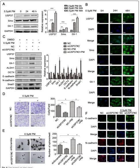
Purmorphamine (PM) is considered to be an Hh agonist. Smo, Gli-1 and USP37 expression post-PM treatment was examined by western blotting analysis and immuno-fluorescence analysis. Interestingly, our data indicated that there was a time-dependent increase in the expres-sion of Smo, Gli-1 and USP37 expresexpres-sion following 24 h, 48 h PM treatment (0.5 μM) (Fig. 6a). Immunofluores-cence assays showed similar results (Fig.6b).
These findings are further supported by the ability of USP37 to participate in Hh signaling pathway in breast cancer. Figure6c shows that silencing of USP37 expres-sion could reverse the positive effects of PM on the Hh pathway. Moreover, the effect of downregulating USP37 on CSC traits, including the formation of spheroid, BCSCs markers and cell invasion, was impaired after PM (See figure on previous page.)
Fig. 4Downregulation of USP37 weakens cell self-renewal and chemo-resistance to cisplatin.a,bMCF-7 and MDA-MB-231 cells were transfected
with shUSP37#2 and shScramble, and expressions of USP37, Smo, Gli-1, ALDH1 and OCT4 were confirmed by western blotting. *P< 0.05, **P<
0.01.c,dComparison of mammosphere formation among inoculated cells in MCF-7 and MDA-MB-231 cells (original magnification, 4× or 20×).*P
< 0.05, ***P< 0.001.eMCF-7-ShScramble and MCF-7-shUSP37#2 cells were treated with cisplatin (1μg/ml) for 48 h, then treated cells were
harvested to detect the levels of Bcl-2, Bax and cleaved caspase 9. GAPDH was examined as a loading control.jMDA-MB-231 cells transfected
with shScramble or shUSP37#2 were exposed to 3μg/ml cisplatin for 48 h, and the levels of Bcl-2, Bax and cleaved caspase 9 were detected via
western blotting.f,g,k,lCCK-8 assay and colony formation assay showed that cell viability was decreased in MCF-7 and MDA-MB-231 cells after
USP37 knockdown. Quantitation of colony formation in (h) MCF-7 and (i) MDA-MB-231. GAPDH was examined as a loading control. *P< 0.05,
**P< 0.01, ***P< 0.001
Fig. 5Overexpression USP37 elevates cell self-renewal and chemo-resistance to cisplatin.aExpressions of USP37, Smo, Gli-1, ALDH1 and OCT4
were detected in MCF-7-CTL or MCF-7-USP37 cells by western blotting. *P< 0.05, **P< 0.01.bComparison of mammosphere formation among
MCF-7-CTL or MCF-7-USP37 cells. *P< 0.05.cMCF-7-CTL and MCF-7-USP37 cells were treated with cisplatin (1μg/ml) for 48 h, then treated cells
were harvested in order to detect the levels of USP37, Bcl-2 and Bax. GAPDH was examined as a loading control. *P< 0.05.dCCK-8 assay showed
treatment (Fig.6c, dandeand Additional file2: Figure S1). Silencing of USP37 expression induced MET including upregulated E-cadherin expression and downregulated N-cadherin expression, while PM reversed MET in silenced USP37 cells. Similar results were observed with immuno-fluorescence assay (Fig.6f). Collectively, these data indicate that downregulation of USP37 expression was involved in the attenuation of the BCSCs stemness, cell invasion and EMT by suppression of the Hh pathway.
USP37 interacts with and stabilizes Gli-1 protein
Given that USP37 expression was significantly associated with the Hh pathway, we detected whether endogenous USP37 could also regulate Gli-1 stability. Endogenous USP37 was depleted by lentivirus expressing USP37#2 shRNA in MCF-7 and MDA-MB-231 cell lines. The effect of USP37 knockdown on Gli-1 was evidently reversed by MG132, which is a protease inhibitor (Fig.7a
and b). Furthermore, knockdown of USP37 expression significantly decreased the stability of Gli-1 protein both in MCF-7 and MDA-MB-231 cells (Fig. 7d and e). Additionally, the cycloheximide (CHX) chase assay was used to assess MCF-7 breast cancer cell line for the ef-fect of USP37 upregulation on endogenous Gli-1 pro-tein. As shown in Fig. 7f, upregulation of USP37 expression consistently stabilized Gli-1 protein. Im-munofluorescence staining assays showed that USP37 could regulate Gli-1 expression (Fig.7c). Co-IP assay in-dicated that USP37 interacted with Gli-1(Fig. 7h). We also found overlapping expression of exogenous USP37 and Gli-1 by immunofluorescence staining (Fig.7g). To-gether, these data suggest endogenous USP37 may regu-late Gli-1 protein stability in breast cancer.
USP37 knockdown inhibits tumorigenicity and increases sensitivity to cisplatin in vivo
To further assess the anti-tumor effect of USP37 down-regulation on tumor growth and cisplatin sensitivity in vivo, we established a xenograft tumor models by sub-cutaneously injecting breast cancer MCF-7 cells, which had been transfected with USP37#2 shRNA or scramble shRNA, into the mammary gland of nude mice (Fig.8a). Tumor volume and weight measurements were ana-lyzed. Compared with the scramble shRNA control
group, the volume and weight of USP37 shRNA group increased at slower rate (Fig. 8b, c). The average tumor volume and weight of cisplatin-treated USP37#2 shRNA tumors was evidently smaller than that of cisplatin-treated tumors or USP37#2 shRNA tumors. We found that lower expression level of USP37 obviously impaired the expression of Hh targets (Smo and Gli-1) and cell proliferation marker Ki-67 in the tumor tissues as seen by immunohistochemical staining (Fig. 8e). Consistent with these results, tissues formed from cisplatin-treated USP37 knockdown cells showed the lowest expression levels of Smo, Gli-1, ALDH1, OCT-4 and Bcl-2 than other tissues by western blotting analysis (Fig.8d). These results indicated that USP37 downregulation attenuates breast cancer progression and enhances sensitivity to cisplatin in vivo.
Discussion
DUBs have been shown to participate in ubiquitin cleav-ing from ubiquitin conjugated protein substrates [29,
30]. USP37, a novel DUB, contains an insert between its catalytic lobe and its ubiquitin-binding lobe while its function could prevent 14-3-3γ degradation, which might contribute to malignant transformation by MAPK signaling [31, 32]. Subsequently, it was verified that USP37 expression modulated the oncogenic fusion pro-tein PLZF/RARA stability and cell transformation poten-tial in PLZF/RARA-associated acute promyelocytic leukemia [33]. Recently, clinicopathological analysis con-firmed that USP37 was a poor prognostic factor in breast cancer [16]. However, there was no direct evi-dence to identify the carcinogenic mechanism of the USP37 gene in breast cancer. In this study, we have demonstrated overexpression of USP37 in Luminal B subtype breast cancer was a predictor of poor outcomes in breast cancer. Supporting our results, previous clini-copathological analysis has demonstrated that overex-pressed USP37 is considered to be a poor prognosis in breast cancer [16]. Additionally, GSEA analysis on the TCGA dataset implicated that USP37 expression was positively associated with metastasis, cell growth and anti-apoptosis. Therefore, USP37 levels could potentially serve as a specific oncogene involved in breast carcin-oma progression.
(See figure on previous page.)
Fig. 6USP37 knockdown inhibits stemness, cell invasion and EMT via Hedgehog signaling pathway in breast cancer.a,bMCF-7 cells were
incubated with 0.5μM purmorphamine for 24 and 48 h.aHedgehog pathway constituents were examined via western blotting. GAPDH was
examined as a loading control. **P< 0.01,***P< 0.001.bImmunofluorescence staining images of MCF-7 cells showed the expression of USP37
and Hedgehog pathway constituents.cProtein levels of USP37, Smo, Gli-1, ALDH1, OCT4, E-cadherin, N-cadherin, Snail1 as detected by western
blotting after the NC siRNA group or the USP37 siRNA#2 group was treated with 0.5μM purmorphamine for 48 h. GAPDH was examined as a
loading control. **P< 0.01,***P< 0.001.dCell invasion capacity of the NC siRNA group or the USP37 siRNA#2 group treated with 0.5μM
purmorphamine (Scale bar: 200μm).eSpheroid formation capacity of MCF-7-ShScramble or MCF-7-shUSP37#2 cells treated with 0.5μM
purmorphamine (original magnification, 4×).fImmunofluorescence staining of E-cadherin and N-cadherin after the NC siRNA group or the USP37
Fig. 7USP37 interacts with and stabilizes Gli-1 protein.a,bMCF-7 and MDA-MB-231 cells were transfected with shScramble and shUSP37#2,
respectively, then treated with 10μM of MG132 4 h before harvesting.cImmunofluorescence staining of Gli-1 after USP37 knockdown or USP37
overexpression (Scale bar: 50μm).d,eCells transfected with shScramble and shUSP37#2 were treated with 50μg/ml of CHX and harvested at
the indicated time point.fMCF-7-CTL and MCF-7-USP37 cells were incubated with 50μg/ml of CHX and harvested at the indicated time point.
Gli-1 expression was analyzed by western blotting and quantified with Image-Pro Plus 6.0 software.gExogenous USP37 and Gli-1 magnified
strong, positive overlapping expression in MCF-7 cells by immunofluorescent staining (Scale bar: 50μm).hCO-IP assay showed the interaction
Cancer stem cells (CSCs) are endowed with stem cell properties, which include the ability to self-renew and differentiate by symmetrical or asymmetrical cell div-ision. CSCs self-renewal of the cellular population and generation of progenitor cells have been shown to resist radiation and chemotherapy. Therefore, these cells are commonly deemed to be crucial target for cancer ther-apy [34, 35]. It was verified that CSCs could be main-tained in an undifferentiated status and induce tumor-sphere formation in defined serum-free medium [36]. However, limited markers were utilized for the identification of breast cancer stem cells. While re-searchers have demonstrated that CD44 and ALDH1 are critical biomarkers to identify BCSCs from breast cancer populations [37,38], our data suggested that the USP37 gene was significantly associated with CSC properties, such as self-renewal, treatment resistance and EMT phenotype as well. Interestingly, elevated mRNA expres-sion of USP37 was detected in CD24−/CD44+ cells and ALDH1+ cells; the protein expression of USP37 was ob-viously elevated in spheres compared to adherent cells. Knockdown of USP37 suppressed mammospheres for-mation and inhibited cancer stem markers, such as ALDH1and OCT4 in breast cancer cells. It has been re-ported that USP37 participates in regulation of CSCs-related proteins, such as SOX2 and c-myc [20,
21]. Our data further indicated that knockdown of USP37 by constructed lentiviral system weakened the stemness in MCF-7 and MDA-MB-231 cells via inhibit-ing the expression levels of ALDH1 and OCT4. CSCs are considered to be “bad seeds” due to their drug re-sistance caused by imbalanced pathway and epigenetics in cancer [39]. As shown above, we detected that knockdown of USP37 in breast cancer cells could pro-mote the sensitivity to cisplatin-induced cell death by the CCK-8 assay and colony formation assays. The Bax/ Bcl-2 ratio is a commonly used method to determine whether intracellular apoptosis system is activated [40]. We detected that USP37#2 shRNA combined with cis-platin treatment induced cell apoptosis with an under-lying decrease in Bcl-2/Bax ratio. On the contrary, the upregulation of USP37 reversed this phenomenon. USP37 knockdown suppressed stemness and chemo-resistance, which may assist clinical oncologists in designing and testing novel therapeutic strategies. In summary, we suggest that USP37 gene expression
confers the stemness and potentially acts as a critical marker of CSCs in breast cancer.
The process of EMT was involved in the acquisition of aggressive cellular traits, including motility, invasiveness and anti-apoptosis, resulting in the dissemination of cells and colonization in distance tissues [6,41,42]. Here, we showed that USP37 siRNA treatment stimulated the ex-pression of epithelial marker (E-cadherin) but decreased the expression of three known inducers of EMT (Snail1, N-cadherin and Vimentin) (Fig.6a). Moreover, we found that knockdown of USP37 suppressed cell migration and invasion in breast cancer cells. On the contrary, upregu-lation of USP37 promoted EMT, migration and invasion. Accumulation evidence has indicated that tumor cells undergoing EMT process are endowed with the trait of cancer stem-like cells [43], which further speculated USP37 as a CSC marker of breast cancer. In summary, our data showed that USP37 could regulate the migra-tion, invasion, EMT of breast cancer cells.
The Hh pathway was found to be required for the maintenance of breast CSCs traits, tumor formation and the EMT [10]. Normally, the ligand binding of Patched (Ptch1), a 12-pass transmembrane receptor of Sonic Hedgehog (SHH), can activate zinc finger transcription factor (Gli-1). It has been suggested that ectopic expres-sion of Gli-1 upregulates expresexpres-sion of the transcription
factor Snail1 accompanied with a decrease in
E-cadherin, a characteristic of EMT [24]. In fact, Snail expression promotes EMT via repressing E-cadherin [44]. Hh signaling pathway modulates EMT indirectly via Snail [45]. Therefore, it is a promising research to in-vestigate whether USP37 promoted the activation of the Hh pathway.
Our findings suggested that the post transcriptional levels of Smo and Gli-1 were decreased in vitro and in vivo. Upregulation of USP37 could enhance protein ex-pression levels of Smo and Gli-1. In order to understand the mechanism of USP37-induced EMT and stemness via the Hh pathway, we treated MCF-7 cells with PM. We found that activation of Hh signaling pathway was accom-panied by elevated expression of USP37 gene as visualized by western blotting and immunofluorescence assay. Fur-thermore, the effect USP37 downregulation on CSCs traits including the formation of spheroids, BCSCs markers and cell invasion were impaired after PM treatment. Mean-while, knockdown of USP37 reversed the effect of PM on (See figure on previous page.)
Fig. 8USP37 knockdown inhibits tumorigenicity and increases sensitivity to cisplatin in vivo.a,b,cMCF-7 cells transfected with shScramble or shUSP37#2 were subcutaneously injected into BALB/c nude mice treated with 0.9% saline or cisplatin. Tumors were excised from nude mice at
day 28.aImages of tumor xenografts andbtumor weight for tumor xenografts treated with 0.9% saline or cisplatin are shown above. *P< 0.05,
**P< 0.01.cTumor growth curves were drawn every other day up to day 25.dWestern blotting analysis of USP37, Smoothened, Gli-1, ALDH1,
OCT4 and Bcl-2 in tumor xenografts. GAPDH was used as a loading control. *P< 0.05, **P< 0.01.eHE staining and immunohistochemical detection of
the EMT markers. These data confirm that USP37 medi-ates breast cancer stem-like properties, cell invasion and EMT via the Hh pathway. Gli-1 acts exclusively as a tran-scriptional activator in the Hh pathway and signal out-comes are determined by the balance of activated and inhibitive Gli proteins [46]. Remarkably, we found that USP37 could regulate and stabilize the protein level of Gli-1 (Additional file 2: Figure S1). As a deubiquitinase, USP37 is involved in the regulation of multiple proteins by deubiquitination, including P27, Cdt1, PLZF/RARA and 14-3-3γ [31, 33, 47, 48]. Future work should aim to determine whether USP37 could stabilize Gli-1 through deubiquitination.
Conclusions
In conclusion, our research demonstrated that USP37 was highly expressed in breast CSCs and was correlated with poor prognosis in breast cancer patients. Knockdown of USP37 expression hampered cell invasion, stemness, EMT and also resulted in the drug sensitivity to cisplatin. Mech-anically, USP37 could orchestrate the stemness, cell inva-sion and EMT via activation of Hedgehog pathway. Further studies detected that USP37 also interacted with and stabilized Gli-1 protein, which is the main activator of Hedgehog target gene. These results indicate that USP37 is essential in the regulation of breast cancer progression suggesting an experimental basis for its use as a cancer biomarker and expands its potential clinical value.
Additional files
Additional file 1:Table S1.Sequences of primer, siRNA and shRNA. (DOCX 14 kb)
Additional file 2:Figure S1.Protein level of OCT4 as detected by western blotting after the NC siRNA group or the USP37 siRNA#2 group was treated with 0.5 µM purmorphamine for 48 h. GAPDH was examined as a loading control. (DOCX 80 kb)
Abbreviations
ALDH1:Aldehyde dehydrogenase1; BC: Breast cancer; BCSCs: Breast cancer stem cells; DUBs: Deubiquitinating enzymes; EMT: Epithelial-mesenchymal transition; Gli-1: Glioma-associated oncogene 1; GSEA: Gene set enrichment analysis; Hh: Hedgehog; MACS: Magnetic activated cell sorting;
MET: Mesenchymal–epithelial transition; PM: Purmorphamine;
Smo: Smoothened; USP37: Ubiquitin specific peptidase 37
Acknowledgements
None.
Funding
This work was supported by Liaoning Provincial program for Top Discipline of Basic Medical Sciences and National Natural Science Foundation of China (81272430, 81650018).
Availability of data and materials
The analyzed data sets generated during the study are available from the corresponding author on reasonable request.
Authors’contributions
QT, LM and L-LH conceived and designed the study; QT, LB and F-XY performed the experiments; Y-JF, Z-QQ and L-DD analyzed the data; LL, MJ, LY and Y-XT contributed reagents/materials/analysis tools; QT, F-SJ and ZJ discussed results and advised during the completion of the study; QT, SB and L-LH wrote and revised the paper. All authors read and approved the final manuscript.
Ethics approval and consent to participate
This research was reviewed and approved by the Ethical Committee and Institutional Review Board of Dalian Medical University. Animal experiments and procedures were approved by the Animal Care and Use Committee of Dalian Medical University and performed in accordance with the Guide for the Care and Use of Laboratory Animals (NIH).(No. AEE17046). The tissue samples of patients diagnosed with breast cancer were used in this research provided written informed consent, and the study protocol was approved by the Committee for the Ethical Review of Research, The Second Affiliated Hospital of Dalian Medical University.
Consent for publication
Not applicable.
Competing interests
The authors declare that they have no competing interests.
Publisher’s Note
Springer Nature remains neutral with regard to jurisdictional claims in published maps and institutional affiliations.
Author details
1Department of Pathology, Dalian Medical University, Dalian 116044, People’s Republic of China.2The Key Laboratory of Tumor Stem Cell Research of Liaoning Province, Dalian Medical University, Dalian 116044, People’s Republic of China.3Department of Urology, The First Affiliated Hospital of Dalian Medical University, Dalian, People’s Republic of China.4Teaching Laboratory of Morphology, Dalian Medical University, Dalian 116044, People’s Republic of China.5Department of Pathology, Xiangyang Central Hospital, Xiangyang 441000, People’s Republic of China.6Department of Dean, Dalian Medical University, Dalian 116044, People’s Republic of China.7Department of Oncology, The Second Affiliated Hospital of Dalian Medical University, Dalian 116023, Liaoning Province, People’s Republic of China.
Received: 29 August 2018 Accepted: 18 October 2018
References
1. Kraschnewski JL, Schmitz KH. Exercise in the prevention and treatment of
breast cancer: what clinicians need to tell their patients. Curr Sports Med
Rep. 2017;16(4):263–7.
2. Morel AP, Lièvre M, Thomas C, Hinkal G, Ansieau S, Puisieux A. Generation
of breast cancer stem cells through epithelial-mesenchymal transition. PLoS One. 2008;3(8):e2888.
3. Creighton CJ, Li X, Landis M, Dixon JM, Neumeister VM, Sjolund A, Rimm
DL, Wong H, Rodriguez A, Herschkowitz JI, Fan C, Zhang X, He X, Pavlick A, Gutierrez MC, Renshaw L, Larionov AA, Faratian D, Hilsenbeck SG, Perou CM, Lewis MT, Rosen JM, Chang JC. Residual breast cancers after conventional therapy display mesenchymal as well as tumor-initiating features. Proc Natl
Acad Sci U S A. 2009;106(33):13820–5.
4. Ricardo S, Vieira AF, Gerhard R, Leitão D, Pinto R, Cameselle-Teijeiro JF,
Milanezi F, Schmitt F, Paredes J. Breast cancer stem cell markers CD44, CD24 and ALDH1: expression distribution within intrinsic molecular subtype. J Clin
Pathol. 2011;64(11):937–46.
5. Hardin H, Zhang R, Helein H, Buehler D, Guo Z, Lloyd RV. The evolving
concept of cancer stem-like cells in thyroid cancer and other solid tumors.
Lab Investig. 2017;97(10):1142–51.
6. Thiery JP, Sleeman JP. Complex networks orchestrate
epithelial-mesenchymal transitions. Nat Rev Mol Cell Biol. 2006;7(2):131–42.
7. Mani SA, Guo W, Liao MJ, Eaton EN, Ayyanan A, Zhou AY, Brooks M,
Reinhard F, Zhang CC, Shipitsin M, Campbell LL, Polyak K, Brisken C, Yang J, Weinberg RA. The epithelial-mesenchymal transition generates cells with
8. Radisky ES, Radisky DC. Matrix metalloproteinase-induced epithelial-mesenchymal
transition in breast cancer. J Mammary Gland Biol Neoplasia. 2010;15(2):201–12.
9. Kuang J, Li L, Guo L, Su Y, Wang Y, Xu Y, Wang X, Meng S, Lei L, Xu L, Shao
G. RNF8 promotes epithelial-mesenchymal transition of breast cancer cells. J Exp Clin Cancer Res. 2016;35(1):88.
10. Kasper M, Jaks V, Fiaschi M, Toftgård RM. Hedgehog signaling in breast
cancer. Carcinogenesis. 2009;30(6):903–11.
11. Hershko A, Ciechanover A. The ubiquitin system. Annu Rev Biochem. 1998;
67:425–79.
12. Amerik AY, Hochstrasser M. Mechanism and function of deubiquitinating
enzymes. Biochim Biophys Acta. 2004;1695(1–3):189–207.
13. Alhosin M, Omran Z, Zamzami MA, Al-Malki AL, Choudhry H, Mousli M,
Bronner C. Signalling pathways in UHRF1-dependent regulation of tumor suppressor genes in cancer. J Exp Clin Cancer Res. 2016;35(1):174.
14. He M, Zhou Z, Wu G, Chen Q, Wan Y. Emerging role of DUBs in tumor metastasis
and apoptosis: therapeutic implication. Pharmacol Ther. 2017;177:96–107.
15. Wu Y, Wang Y, Yang XH, Kang T, Zhao Y, Wang C, Evers BM, Zhou BP. The
deubiquitinase USP28 stabilizes LSD1 and confers stem-cell-like traits to
breastcancer cells. Cell Rep. 2013;5(1):224–36.
16. Zhang QX, Wang XC, Chen SP, Qin XT. Predictive value of deubiquitination
enzymes USP37 in the prognosis of breast cancer. Zhonghua Yi Xue Za Zhi.
2016;96(12):944–8.
17. Tanno H, Shigematsu T, Nishikawa S, Hayakawa A, Denda K, Tanaka T,
Komada M. Ubiquitin-interacting motifs confer full catalytic activity, but not ubiquitin chain substratespecificity, to deubiquitinating enzyme USP37. J
Biol Chem. 2014;289(4):2415–23.
18. Huang X, Summers MK, Pham V, Lill JR, Liu J, Lee G, Kirkpatrick DS, Jackson PK,
Fang G, Dixit VM. Deubiquitinase USP37 is activated by CDK2 to antagonize
APC(CDH1) and promote Sphase entry. Mol Cell. 2011;42(4):511–23.
19. Burrows AC, Prokop J, Summers MK. Skp1-Cul1-F-box ubiquitin ligase
(SCF(βTrCP))-mediated destruction of the ubiquitin-specific protease USP37
during G2-phase promotes mitotic entry. J Biol Chem. 2012;287(46):39021–9.
20. Boyer LA, Lee TI, Cole MF, Johnstone SE, Levine SS, Zucker JP, Guenther MG,
Kumar RM, Murray HL, Jenner RG, Gifford DK, Melton DA, Jaenisch R, Young RA. Core transcriptional regulatory circuitry in human embryonic stem cells.
Cell. 2005;122(6):947–56.
21. Pan J, Deng Q, Jiang C, Wang X, Niu T, Li H, Chen T, Jin J, Pan W, Cai X,
Yang X, Lu M, Xiao J, Wang P. USP37 directly deubiquitinates and stabilizes
c-Myc in lung cancer. Oncogene. 2015;34(30):3957–67.
22. Cancer Genome Atlas Network. Comprehensive molecular portraits of
human breast tumours. Nature. 2012;490(7418):61–70.
23. Qin T, Huang G, Chi L, Sui S, Song C, Li N, Sun S, Li N, Zhang M, Zhao Z, Li
L, Li M. Exceptionally high UBE2C expression is a unique phenomenon in basal-like type breast cancer and is regulated by BRCA1. Biomed
Pharmacother. 2017;95:649–55.
24. Zhong Y, Shen S, Zhou Y, Mao F, Guan J, Lin Y, Xu Y, Sun Q. ALDH1 is a
better clinical indicator for relapse of invasive ductal breast cancer than the CD44+/CD24- phenotype. Med Oncol. 2014;31(3):864.
25. Koike Y, Ohta Y, Saitoh W, Yamashita T, Kanomata N, Moriya T, Kurebayashi
J. Anti-cell growth and anti-cancer stem cell activities of the non-canonical hedgehog inhibitor GANT61 in triple-negative breast cancer cells. Breast
Cancer. 2017;24(5):683–93.
26. Kayaselcuk F, Nursal TZ, Polat A, Noyan T, Yildirim S, Tarim A, Seydaoglu G.
Expression of survivin, bcl-2, P53 and bax in breast carcinoma and ductal
intraepithelial neoplasia (DIN 1a). J Exp Clin Cancer Res. 2004;23(1):105–12.
27. Bi D, Yang M, Zhao X, Huang S. Effect of cnidium lactone on serum mutant
P53 and BCL-2/BAX expression in human prostate cancer cells PC-3
tumor-bearing BALB/C nude mouse model. Med Sci Monit. 2015;21:2421–7.
28. Sun Y, Wang Y, Fan C, Gao P, Wang X, Wei G, Wei J. Estrogen promotes
stemness and invasiveness of ER-positive breast cancer cells through Gli1 activation. Mol Cancer. 2014;13:137.
29. Love KR, Catic A, Schlieker C, Ploegh HL. Mechanisms, biology and inhibitors
of deubiquitinating enzymes. Nat Chem Biol. 2007;3(11):697–705.
30. Nijman SM, Luna-Vargas MP, Velds A, Brummelkamp TR, Dirac AM, Sixma TK,
Bernards R. A genomic and functional inventory of deubiquitinating
enzymes. Cell. 2005;123(5):773–86.
31. Kim JO, Kim SR, Lim KH, Kim JH, Ajjappala B, Lee HJ, Choi JI, Baek KH.
Deubiquitinating enzyme USP37 regulating oncogenic function of 14-3-3γ.
Oncotarget. 2015;6(34):36551–76.
32. Dobson THW, Hatcher RJ, Swaminathan J, Das CM, Shaik S, Tao RH, Milite C,
Castellano S, Taylor PH, Sbardella G, Gopalakrishnan V. Regulation of USP37
expression by REST-associated G9a-dependent histone methylation. Mol
Cancer Res. 2017;15(8):1073–84.
33. Yang WC, Shih HM. The deubiquitinating enzyme USP37 regulates the
oncogenic fusion protein PLZF/RARA stability. Oncogene. 2013;32(43):5167–75.
34. Wang N, Wang Q, Tang H, Zhang F, Zheng Y, Wang S, Zhang J, Wang Z, Xie
X. Direct inhibition of ACTN4 by ellagic acid limits breast cancer metastasis
via regulation ofβ-catenin stabilization in cancer stem cells. J Exp Clin
Cancer Res. 2017;36(1):172.
35. Tomao F, Papa A, Rossi L, Strudel M, Vici P, Lo Russo G, Tomao S. Emerging
role of cancer stem cells in the biology and treatment of ovarian cancer: basicknowledge and therapeutic possibilities for an innovative approach. J Exp Clin Cancer Res. 2013;32:48.
36. Fletcher SJ, Sacca PA, Pistone-Creydt M, Coló FA, Serra MF, Santino FE, Sasso
CV, Lopez-Fontana CM, Carón RW, Calvo JC, Pistone-Creydt V. Human breast adipose tissue: characterization of factors that change during tumor progression in human breast cancer. J Exp Clin Cancer Res. 2017;36(1):26.
37. Charafe-Jauffret E, Ginestier C, Bertucci F, Cabaud O, Wicinski J, Finetti P,
Josselin E, Adelaide J, Nguyen TT, Monville F, Jacquemier J, Thomassin-Piana J, Pinna G, Jalaguier A, Lambaudie E, Houvenaeghel G, Xerri L, Harel-Bellan A, Chaffanet M, Viens P, Birnbaum D. ALDH1-positive cancer stem cells predict engraftment of primary breast tumors and are governed by a
common stem cell program. Cancer Res. 2013;73(24):7290–300.
38. Shima H, Yamada A, Ishikawa T, Endo I. Are breast cancer stem cells
the key to resolving clinical issues in breast cancer therapy? Gland
Surg. 2017;6(1):82–8.
39. Qiu GZ, Sun W, Jin MZ, Lin J, Lu PG, Jin WL. The bad seed gardener:
Deubiquitinases in the cancer stem-cell signaling network and therapeutic
resistance. Pharmacol Ther. 2017;172:127–38.
40. Karmakar S, Banik NL, Ray SK. Curcumin suppressed anti-apoptotic signals
and activated cysteine proteases for apoptosis in human malignant
glioblastoma U87MG cells. Neurochem Res. 2007;32(12):2103–13.
41. Wang Y, Zhou BP. Epithelial-mesenchymal transition—a hallmark of breast
cancer metastasis. Cancer Hallm. 2013;1(1):38–49.
42. Bouris P, Skandalis SS, Piperigkou Z, Afratis N, Karamanou K, Aletras AJ,
Moustakas A, Theocharis AD, Karamanos NK. Estrogen receptor alpha mediates epithelial to mesenchymal transition, expression of specific matrix effectors and functional properties of breast cancer cells. Matrix Biol. 2015;
43:42–60.
43. Huan HB, Yang DP, Wen XD, Chen XJ, Zhang L, Wu LL, Bie P, Xia F. HOXB7
accelerates the malignant progression of hepatocellular carcinoma by promoting stemness and epithelial-mesenchymal transition. J Exp Clin Cancer Res. 2017;36(1):86.
44. Li X, Deng W, Lobo-Ruppert SM, Ruppert JM. Gli1 acts through Snail and
E-cadherin to promote nuclear signaling by beta-catenin. Oncogene. 2007;
26(31):4489–98.
45. Mani SA, Yang J, Brooks M, Schwaninger G, Zhou A, Miura N, Kutok JL,
Hartwell K, Richardson AL, Weinberg RA. Mesenchyme Forkhead 1 (FOXC2) plays a key role in metastasis and is associated with aggressive basal-like
breast cancers. Proc Natl Acad Sci U S A. 2007;104(24):10069–74.
46. Wu X, Xu B, Yang C, Wang W, Zhong D, Zhao Z, He L, Hu Y, Jiang L, Li J,
Song L, Zhang W. Nucleolar and spindle associated protein 1 promotes the aggressiveness of astrocytoma by activating the Hedgehog signaling pathway. J Exp Clin Cancer Res. 2017;36(1):127.
47. Das CM, Taylor P, Gireud M, Singh A, Lee D, Fuller G, Ji L, Fangusaro J,
Rajaram V, Goldman S, Eberhart C, Gopalakrishnan V. The deubiquitylase USP37 links REST to the control of p27 stability and cell proliferation.
Oncogene. 2016;35(47):6153–4.
48. Hernández-Pérez S, Cabrera E, Amoedo H, Rodríguez-Acebes S,
Koundrioukoff S, Debatisse M, Méndez J, Freire R. USP37 deubiquitinates Cdt1 and contributes to regulate DNA replication. Mol Oncol. 2016;10(8):