Open Access
Research
Antigen-driven bystander effect accelerates epicutaneous
sensitization with a new protein allergen
Li-Fang Wang
1, Jau-Shiuh Chen
1, Chih-Jung Hsu
1, Ching-Yi Liu
1,
Jhang-Sian Yu
2and Shi-Chuen Miaw*
2Address: 1Department of Dermatology, National Taiwan University Hospital and National Taiwan University College of Medicine, Taipei, Taiwan and 2Graduate Institute of Immunology, National Taiwan University Hospital and National Taiwan University College of Medicine, Taipei, Taiwan
Email: Li-Fang Wang - [email protected]; Jau-Shiuh Chen - [email protected]; Chih-Jung Hsu - [email protected]; Ching-Yi Liu - [email protected]; Jhang-Sian Yu - [email protected]; Shi-Chuen Miaw* - [email protected]
* Corresponding author
Abstract
Exposure to protein allergen epicutaneously, inducing a Th2-dominant immune response, sensitizes the host to the development of atopic disease. Antigen-driven bystander effect demonstrates that polarized T cells could instruct naïve T cells to differentiate into T cells with similar phenotype. In this study, we aimed to determine the contribution of antigen-driven bystander effect on epicutaneous sensitization with a newly introduced protein allergen. BALB/c mice were immunized intraperitoneally with BSA emulsified in alum, known to induce a Th2 response, three weeks before given BSA and OVA epicutaneously. Lymph node cells from these mice restimulated with OVA secreted higher levels IL-4, IL-5 and IL-13 as compared with cells from mice without BSA immunization. In addition, BALB/c mice immunized subcutaneously with BSA emulsified in complete Freund's adjuvant, known to induce a Th1-predominant response, also induced higher Th1 as well as Th2 cytokine response when restimulated with OVA as compared with mice without immunization. We demonstrated that subcutaneous immunization with BSA in CFA induced Th2 as well as Th1 response. The threshold of epicutaneous sensitization to OVA was also reduced, possibly due to increased expressions of IL-4 and IL-10 in the draining lymph nodes during the early phase of sensitization. In conclusion, antigen-driven bystander effect, whether it is of Th1- or Th2-predominant nature, can accelerate epicutaneous sensitization by a newly introduced protein allergen. These results provide a possible explanation for mono- to poly-sensitization spread commonly observed in atopic children.
Background
In the past several decades, there has been a progressive increase in the prevalence of atopic disease and an associ-ated increase in the cost of medical management [1]. Atopic diseases are manifested as atopic dermatitis (AD), asthma, and allergic rhinitis. Atopy is associated with the
expression of allergen-specific immune response charac-terized by the production of Th2 cytokines such as IL-4, IL-5, and IL-13; elevated IgE production; and eosi-nophilia. In contrast, non-atopic individuals display pre-dominant Th1 immune response characterized by the production of IFN-. Genetics predisposition and
expo-Published: 6 March 2009
Journal of Biomedical Science 2009, 16:28 doi:10.1186/1423-0127-16-28
Received: 22 December 2008 Accepted: 6 March 2009
This article is available from: http://www.jbiomedsci.com/content/16/1/28
© 2009 Wang et al; licensee BioMed Central Ltd.
sure to various environmental allergens, which result in a Th2 immune response, contribute to the pathogenesis of atopic diseases [2].
AD is often the first clinical manifestation of an atopic triad and is the beginning of the "atopic march" [3]. How-ever, the route of sensitization by an allergen that results in AD is still unclear. There is compelling evidence to show that epicutaneous exposure to a protein allergen is one of the important routes to sensitize the host for AD and other atopic diseases [4]. By the presence of cutane-ous lymphocyte-associated antigen (CLA)-positive T cells, it is shown in humans, that T cells are primed or reacti-vated in the skin or its draining lymph nodes (LNs) [5,6]. In AD patients, the increased frequency of prior activation and secretion of type 2 cytokines, as well as higher prolif-eration response to allergen, are largely confined to a CLA-positive subset [7-9]. Moreover, T cell receptor skewing is only detectable within the CLA-positive subset of T cells from those subjects from whom superantigen-secreting, skin-dwelling bacteria could be identified [10]. The recent demonstration of the expression of CLA by CD8 T cells specific for a skin-tropic, but not non-skin-tropic, virus further supports this concept [11]. We and others have demonstrated in an atopic dermatitis animal model, that epicutaneous exposure of protein antigen induces a pre-dominant Th2 response with high IgE production [12,13]. Furthermore, epicutaneous sensitization with protein antigen induces AD-like skin lesions and development of asthma [14]. The epicutaneously-induced Th2 response requires IL-10 and IL-13 [15,16], while down-regulation of the response is mediated by C3a, cyclooxygenase-2, and skin scratching [17-19].
It has been demonstrated that through an antigen-driven bystander effect, polarized T cells instruct naïve T cells to differentiate into T cells with a similar phenotype. This bystander effect is observed only when the challenge inoc-ulum contains both the original antigen and a newly introduced antigen. This effect was initially shown to be a mechanism of antigen-driven peripheral tolerance after oral administration of antigens, and it can protect rats from developing experimental autoimmune encephalo-myelitis [20]. Adoptive co-transfer of two populations of T-cell receptor transgenic T cells of different specificities demonstrated that polarized Th1 or Th2 effector cells can instruct naïve T cells to differentiate into Th1 or Th2 cells, respectively [21]. Subsequently, study of a murine asthma model revealed that an ongoing Th2 response can induce antigen-specific Th2 response to a new antigen, a process termed "collateral priming" [22]. Further exploration of the underlying mechanisms of the antigen-driven bystander effect has demonstrated that conversion of a naïve T cell occurs only when it interacts with the same APC as the memory T cell, and that the orally immunized
which in turn induce naïve T cells to produce the same cytokines as those produced by the orally immunized memory T cells [23]. In this present study, we demon-strated that antigen-driven bystander effect, despite its Th1- or Th2-predominant nature, accelerates epicutane-ous sensitization with a new protein antigen.
Materials and methods
Mice and reagentsSix to 10-week-old female BALB/c and TCR-OVA-DO11.10 mice were purchased from the animal center of National Taiwan University Collage of Medicine and kept in a specific pathogen-free environment. All animal exper-iments were approved by the animal care committee of the Medical College of National Taiwan University. OVA (Grade V), BSA, CFA, and 4-nitrophenyl phosphate (pNPP) were purchased from Sigma-Aldrich (St. Louis, MO). Alum adjuvant was purchased from Pierce (Rock-ford, IL) and carboxyfluorescein succinimidyl ester (CFSE) was obtained from Invitrogen (Carlsbad, CA). Capture and biotin-conjugated detecting antibodies for IFN-, IL-4, IL-5 used in the ELISA were from PharMingen (San Diego, CA). Streptavidine-alkaline phosphatase was purchased from Southern Biotechnology (Birmingham, AL). The murine IL-13 ELISA kit purchased from R&D sys-tems (Minneapolis, MN) was used for determination of the IL-13 content of supernatants.
Sensitization
Mice were sensitized as previously described [24]. Briefly, 20 l of OVA (100 mg/ml or serial dilutions) and 20 l of BSA (100 mg/ml) were placed on the disc of a Finn cham-ber (Epitest, Tuusula, Finland). This was then applied to an area of shaved skin on the back of a mouse. For each course of sensitization, freshly prepared patches were applied daily from days 1 to 5. For pretreatment, mice received a subcutaneous (s.c.) injection of 100 g BSA emulsified in CFA over the bilateral side of tail-base or an intraperitoneal (i.p.) injection of 100 g BSA in alum adjuvant three weeks before sensitization.
Adoptive transfer
For adoptive transfer of OVA-TCR CD4 T cells, spleen cells from DO.11.10 mice were positively selected for CD4 T cells using CD4 microbeads. Then, 107 CD4 T cells/ml
were incubated with 1 M CFSE for 10 min at 37°C. Pre-warmed FCS-containing PBS was added and washed by cold PBS. Labeled OVA-TCR CD4 T cells (5 × 10⁶) were
intravenously injected into BALB/c recipients 24 h before sensitization.
LN and spleen cell cytokine production and proliferation assay
ence or absence of 100 g/ml OVA. Supernatants were harvested 48 h later and stored at -80°C. IFN-, IL-4, IL-5, and IL-13 content of supernatants was each measured by a standard sandwich ELISA. The limit of detection for IL-4, IL-5, and IL-13 were all 10 pg/ml, whereas that for
IFN- was 50 pg/ml. For spleen cells, spleens were harvested 3 weeks after sensitization, and pooled spleen cells were stimulated with 100 g/ml OVA. For the proliferation assay, graded doses of OVA were added. 48 h after the ini-tiation of culture, [3H] thymidine was added and the cells
were harvested 18 h later.
Total RNA extraction, cDNA preparation and quantitative real-time PCR
The patched skin and draining lymph node samples were obtained 24, 48, and 72 h after patch co-administration of BSA and OVA. They were frozen with liquid nitrogen and soaked in 1 ml TRIzol Reagent (Invitrogen, Carlsbad, CA, USA). After homogenization, the total RNA was extracted, cDNA was synthesized, and quantitative real-time PCR was performed according to the manufacturer's instruc-tions. Each sample was analyzed in triplicate. The relative cytokine mRNA expression level of each sample was nor-malized according to its -actin expression.
Results
Antigen-driven bystander effect enhances epicutaneously-induced Th2 immune response to a co-administered new protein allergen
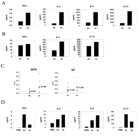
Murine protein-patch model has been established by us and others to study immune responses induced by epicu-taneous sensitization with protein antigens [12,14]. It has been shown in previous studies that patch application of OVA solution onto mouse skin induces a predominant Th2 and a weak Th1 immune response. To investigate whether polarized Th2 cells could influence the immune response induced by epicutaneous sensitization with a newly introduced protein antigen, mice were immunized with BSA emulsified in alum adjuvant through the intra-peritoneal route and were exposed to both OVA and BSA epicutaneously by patch application three weeks later. Data in Fig. 1A show that LN cells from mice previously immunized with BSA and restimulated with OVA pro-duced significantly higher IL-4, IL-5, and IL-13 in the cul-ture supernatants as compared with cells from mice that did not receive BSA immunization. There was no differ-ence in IFN- production between mice receiving immu-nization and those that did not. Interestingly, when BSA was omitted from the patch, the enhanced Th2 response
Antigen-driven bystander effect by Th2 pretreatment enhanced epicutaneously induced Th2 immune response to a new pro-tein antigen
Figure 1
Antigen-driven bystander effect by Th2 pretreatment enhanced epicutaneously induced Th2 immune
was no longer observed (data not shown). These results indicate that enhanced Th2 cytokine production was induced by antigen-driven bystander effect. We further tested whether spleen cells also mounted an enhanced Th2-dominant response at a later time point. Splenocytes from mice immunized with BSA and sensitized epicutane-ously with OVA and BSA were restimulated with OVA. Fig. 1B shows that splenocyte IL-5 and IL-13 productions were much higher than those from the mice without immuni-zation. However, OVA-specific IL-4 production was not detected in the spleen cell cultures. Next, we tested the antigen-driven bystander effect of polarized Th1 cells. Subcutaneous injection of protein antigen emulsified in CFA adjuvant is a standard way to induce Th1-predomi-nant immune response in experimental murine system. BALB/c mice were immunized subcutaneously with BSA emulsified in CFA or not three weeks before epicutaneous sensitization with OVA plus BSA. LN cells from mice receiving subcutanous immunization of BSA and patch-sensitized with BSA and OVA produced not only higher concentrations of IFN-, but also IL-4, IL-5 and IL-13 in the culture supernatants after restimulation with OVA as compared to cells from unimmunized mice (Fig. 2A). In the meantime, the splenocyte IL-5 and IL-13 but not
IFN- productions were much higher than those from the mice without immunization (Fig. 2B). Mice immunized subcu-taneously also had higher levels of serum OVA-specific IgG2a and IgE in the serum (Fig. 2C) than the mice with-out immunization, although they did not reach statistical significance. As shown in Fig. 2D, subcutaneous immuni-zation with BSA in CFA adjuvant induced both Th1 and Th2 immune responses, indicated by high concentrations of IFN- as well as significant amounts of IL-4, IL-5, and IL-13. Taken together, despite its Th1/Th2 predominant nature, the antigen-driven bystander effect enhances the Th2 immune response induced by an epicutaneously introduced new protein antigen.
Antigen-driven bystander effect decreases the threshold of epicutaneous sensitization with a new protein allergen
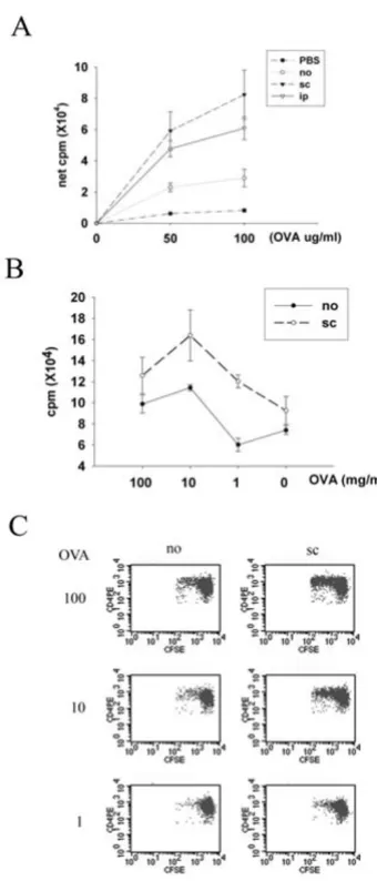
The next question was whether the antigen-driven bystander effect could modulate the threshold of epicuta-neous sensitization with a new protein allergen. LN cells from mice receiving subcutaneous or intraperitoneal immunization with BSA had higher proliferative response to OVA after patch sensitization with both BSA and OVA as compared with those without immunization (Fig. 3A). Next, serial dilutions of OVA were co-administered with a fixed concentration of BSA to explore whether immuniza-tion modulates the threshold of epicutaneous sensitiza-tion to OVA in this model system. LN cells from mice subcutaneously immunized with BSA had higher prolifer-ative response to OVA than mice without immunization when OVA was co-administered at concentrations of 100 mg/ml, 10 mg/ml or 1 mg/ml with BSA. Without
immu-nization, OVA co-administered at 100 mg/ml and 10 mg/ ml but not at 1 mg/ml with BSA induced cells to respond specifically to OVA stimulation (Fig. 3B). To examine CD4 T cell proliferation in vivo, CD4 T cells from naïve OVA-TCR transgenic mice (DO11.10) were labeled with CFSE and intravenously transferred into BALB/c mice one day before they received patch co-sensitization with OVA and BSA. Fig. 3C shows that transgenic CD4 T cells prolif-erated in mice subcutaneously immunized with BSA and sensitized epicutaneously with 10 mg/ml and 100 mg/ml OVA plus BSA, and adoptively transferred transgenic CD4 T cells had proliferative response only in unimmunized mice patched with 100 mg/ml OVA (Fig. 3C). Collec-tively, these data support the notion that the antigen-driven bystander effect decreases the threshold of epicuta-neous sensitization with a new protein allergen by at least one order of magnitude.
The role of cytokines in epicutaneously induced antigen-driven bystander effect
Among the cytokines that influence the Th1/Th2 balance, IFN-, IL-12 and IL-18 are the key cytokines to induce Th1 responses, while IL-4, IL-10 and IL-13 preferentially prime Th2 responses. Figure 4 shows that mice receiving subcu-taneous immunization had higher levels of IL-4 and IL-10 expression in their draining LNs as early as 24 h after patch sensitization as compared with mice without immunization. The expressions of other cytokines, including IL-5, IL-12, IL-13, IL-18, IFN- and TNF-, in the draining LNs were similar in mice with or without immu-nization. It is noteworthy, the expressions of all of these cytokines in the patch-applied skin were not different between mice with or without immunization groups (data not shown). Taken together, these results suggest that immunization-induced IL-4 and IL-10 expression contributes to the epicutaneously induced antigen-driven bystander effect.
Discussion
The data presented here clearly demonstrate that the anti-gen-driven bystander effect, despite its Th1- or Th2-pre-dominant nature, accelerates epicutaneous sensitization with a new protein allergen by enhancing the Th2 immune response and by lowering the sensitization threshold. To our knowledge, this is the first report to address the contribution of antigen-driven bystander effect to the epicutaneous sensitization of protein aller-gens in atopic disease.
Antigen-driven bystander effect by Th1 pretreatment also enhanced epicutaneously induced Th2 immune response to a new protein antigen
Figure 2
factors might modulate the susceptibility to allergen sen-sitization. Firstly, the dose-response relationship between allergen exposure and sensitization differs depending on allergens and locales [27]. Secondly, once sensitized to one allergen, atopic patients are more likely to become sensitized to other allergens [28]. Moreover, there are strong associations of multiple sensitizations both within and between different allergen classes [28]. Skin is contin-uously exposed to many kinds of allergens and microor-ganisms in the environment. Microormicroor-ganisms can gain access to the human body through different routes and sensitize the immune system before contact with the skin. Therefore, it is very likely that the antigen-driven bystander effect affects our skin constantly, although its effect may vary. Thus, the present study provides a possi-ble mechanistic explanation to the clinical observation of the spread of Th2 responses from mono-allergen to mul-tiple allergens in atopic individuals. Another clinical implication of our data is that our results do not conflict with "hygiene hypothesis". The "hygiene hypothesis" pre-dicts that nonspecific suppression of Th2 immune response by a predominant systemic Th1 milieu at the time of sensitization prevents the development of allergy. However, our present study demonstrated specific, but not non-specific, promotion of Th2 immune responses by antigen-driven bystander effect. Collectively, our results support the wisdom of recognizing allergen sensitization early in life and taking precautions to prevent exposure of skins to these allergens.
The mechanism underlying the antigen-driven bystander effect is still obscure. Alpan et al. suggests that orally induced memory T cells utilize IL-4 and IL-10 they pro-duced, but not CD40 ligand, to educate DCs which in turn induce conversion of naïve T cells [23]. However, since co-culturing DCs with naïve T cells plus various concentra-tions of IL-4 and IL-10 does not simulate the situation in which DC was educated by orally induced memory T cells in their studies, the authors suggested that there must be other factors involved [23]. Our current study provides more information about the underlying mechanism of antigen-driven bystander effect. As we demonstrated in this report, systemic immunization plus skin sensitization to protein antigens induce increased cytokine expression in the draining LNs, but not in the patched skin. This observation supports the notion that the draining LNs are the site where antigen-driven bystander effect takes place [29].
In summary, in this study we demonstrated that antigen-driven bystander effect contributes to accelerated epicuta-neous sensitization with a new protein allergen. These results provide a possible explanation for a mono- to poly-sensitization spread commonly observed in atopic children.
Influence of antigen-driven bystander effect on the threshold of epicutaneous sensitization with a new protein allergen
Figure 3
The role of cytokine in the epicutaneously induced antigen-driven bystander effect
Figure 4
Publish with BioMed Central and every scientist can read your work free of charge
"BioMed Central will be the most significant development for disseminating the results of biomedical researc h in our lifetime."
Sir Paul Nurse, Cancer Research UK
Your research papers will be:
available free of charge to the entire biomedical community
peer reviewed and published immediately upon acceptance
cited in PubMed and archived on PubMed Central
yours — you keep the copyright
Submit your manuscript here: BioMedcentral
Abbreviations
AD: atopic dermatitis; LN: lymph node; CLA: cutaneous lymphocyte-associated antigen.
Competing interests
The authors declare that they have no competing interests.
Authors' contributions
LFW, JSC, CJH and SCM participated in the design of the study and preparation of the manuscript. JSY carried out the measurement of cytokine mRNA expression. CYL per-formed all other experiments.
Acknowledgements
We thank Dr. Betty A. Wu-Hsieh for helpful advice and careful review of the manuscript. This work was supported by grants from National Taiwan University Hospital (94N049, 94A18-4 and 95A21-4) and from National Health Research Institutes (NHRI-EX95-9516SC).
References
1. Sublett JL: The environment and risk factors for atopy. Current allergy and asthma reports 2005, 5(6):445-450.
2. Allam J-P, Bieber T, Novak N: Recent Highlights in the patho-physiology of atopic eczema. Int Arch Allergy Appl Immunol 2005,
136:191-197.
3. Spergel JM, Paller AS: Atopic dermatitis and the atopic march. J Allergy Clin Immunol 2003, 112(6, Supplement 1):S118-S127. 4. Berard F, Marty J-P, Nicolas J-F: Allergen penetration through
the skin. Eur J Dermatol 2003, 13(4):324-330.
5. Picker LJ, Treer JR, Ferguson-Darnell B, Collins PA, Bergstresser PR, Terstappen LWMM: Control of lymphocyte recirculation in man. II. Differential regulation of the cutaneous lymphocyte-associated antigen, a tissue-selective homing receptor for skin-homing T cells. J Immunol 1993, 150(3):1122-1136. 6. Hunger RE, Yawalkar N, Braathen LR, Brand CU: The HECA-452
epitope is highly expressed on lymph cells derived from human skin. Br J Dermatol 1999, 141(3):565-569.
7. Santamaria Babi LF, Picker LJ, Perez Soler MT, Drzimalla K, Flohr P, Blaser K, Hauser C: Circulating allergen-reactive T cells from patients with atopic dermatitis and allergic contact dermati-tis express the skin-selective homing receptor, the cutane-ous lymphocyte-associated antigen. J Exp Med 1995,
181(5):1935-1940.
8. Teraki Y, Hotta T, Shiohara T: Increased circulating skin-homing cutaneous lymphocyte-associated antigen (CLA)+ type 2 cytokine-producing cells, and decreased CLA+ type 1 cytokine-producing cells in atopic dermatitis. Br J Dermatol
2000, 143(2):373-378.
9. Seneviratne SL, Jones L, King AS, Black A, Powell S, McMichael AJ, Ogg GS: Allergen-specific CD8+ T cells and atopic disease. J Clin Invest 2002, 110(9):1283-1291.
10. Strickland I, Hauk PJ, Trumble AE, Picker LJ, Leung DYM: Evidence for Superantigen Involvement in Skin Homing of T cells in Atopic Dermatitis. J Invest Dermatol 1999, 112(2):249-253. 11. Koelle DM, Liu Z, McClurkan CM, Topp MS, Riddell SR, Pamer EG,
Johnson AS, Wald A, Corey L: Expression of cutaneous lym-phocyte-associated antigen by CD8+ T cells specific for a skin-tropic virus. J Clin Invest 2002, 110(4):537-548.
12. Wang LF, Lin JY, Hsieh KH, Lin RH: Epicutaneous exposure of protein antigen induces a predominant Th2-like response with high IgE production in mice. J Immunol 1996,
156(11):4077-4082.
13. Kondo H, Ichikawa Y, Imokawa G: Percutaneous sensitization with allergens through barrier-disrupted skin elicits a Th2-dominant cytokine response. Eur J Immunol 1998, 28(3):769-779. 14. Spergel JM, Mizoguchi E, Brewer JP, Martin TR, Bhan AK, Geha RS:
Epicutaneous sensitization with protein antigen induces localized allergic dermatitis and hyperresponsiveness to methacholine after single exposure to aerosolized antigen in
15. Laouini D, Alenius H, Bryce P, Oettgen H, Tsitsikov E, Geha RS: IL-10 is critical for Th2 responses in a murine model of allergic dermatitis. J Clin Invest 2003, 112(7):1058-1066.
16. Herrick CA, Xu L, McKenzie ANJ, Tigelaar RE, Bottomly K: IL-13 is necessary, not simply sufficient, for epicutaneously induced Th2 responses to soluble protein antigen. J Immunol 2003,
170(5):2488-2495.
17. Kawamoto S, Yalcindag A, Laouini D, Brodeur S, Bryce P, Lu B, Hum-bles AA, Oettgen H, Gerard C, Geha RS: The anaphylatoxin C3a downregulates the Th2 response to epicutaneously intro-duced antigen. J Clin Invest 2004, 114(3):399-407.
18. Laouini D, ElKhal A, Yalcindag A, Kawamoto S, Oettgen H, Geha RS:
COX-2 inhibition enhances the TH2 immune response to epicutaneous sensitization. J Allergy Clin Immunol 2005,
116(2):390-396.
19. Matsushima H, Hayashi S, Shimada S: Skin scratching switches immune responses from Th2 to Th1 type in epicutaneously immunized mice. J Dermatol Sci 2003, 32(3):223-230.
20. Miller A, Lider O, Weiner HL: Antigen-driven bystander sup-pression after oral administration of antigens. J Exp Med 1991,
174(4):791-798.
21. Creusot RJ, Biswas JS, Thomsen LL, Tite JP, Mitchison NA, Chain BM:
Instruction of naive CD4+ T cells by polarized CD4+ T cells within dendritic cell clusters. Eur J Immunol 2003,
33(6):1686-1696.
22. Eisenbarth SC, Zhadkevich A, Ranney P, Herrick CA, Bottomly K: IL-4-dependent Th2 collateral priming to inhaled antigens inde-pendent of Toll-like receptor 4 and myeloid differentiation factor 88. J Immunol 2004, 172(7):4527-4534.
23. Alpan O, Bachelder E, Isil E, Arnheiter H, Matzinger P: 'Educated' dendritic cells act as messengers from memory to naive T helper cells. Nat Immunol 2004, 5(6):615-622.
24. Wang L-F, Hsu C-J, Miaw S-C, Chiu H-C, Liu C-Y, Yu H-S: Cross-priming with an epicutaneously introduced soluble protein antigen generates Tc1 cells. Eur J Immunol 2006,
36(11):2904-2911.
25. Prescott SL, Macaubas C, Smallacombe T, Holt BJ, Sly PD, Holt PG:
Development of allergen-specific T-cell memory in atopic and normal children. Lancet 1999, 353:196-200.
26. Kulig M, Bergmann R, Klettke U, Wahn V, Tacke U, Wahn U: Natu-ral course of sensitization to food and inhalant allergens dur-ing the first 6 years of life. J Allergy Clin Immunol 1999,
103(6):1173-1179.
27. Torrent M, Sunyer J, Munoz L, Cullinan P, Iturriaga MV, Figueroa C, Vall O, Taylor AN, Anto JM: Early-life domestic aeroallergen exposure and IgE sensitization at age 4 years. J Allergy Clin Immunol 2006, 118(3):742-748.
28. Roberts G, Peckitt C, Northstone K, Strachan D, Lack G, Henderson J, Golding J: Relationship between aeroallergen and food aller-gen sensitization in childhood. Clin Exp Allergy 2005,
35(7):933-940.
29. Schipf A, Heilmann A, Boue L, Mossmann H, Brocker T, Rocken M: