R E S E A R C H
Open Access
Transfer of malignant trait to
BRCA1
deficient human fibroblasts following
exposure to serum of cancer patients
Dana Hamam
1,2, Mohamed Abdouh
1, Zu-Hua Gao
3, Vincenzo Arena
4, Manuel Arena
5and Goffredo Orazio Arena
1,6*Abstract
Background:It was reported that metastases might occur via transfer of biologically active blood circulating molecules from the primary tumor to distant organs rather than only migration of cancer cells. We showed in an earlier study that exposure of immortalized human embryonic kidney cells (HEK 293) to cancer patient sera, induce their transformation into undifferentiated cancers due to a horizontal transfer of malignant traits. In the present work, we tested the hypothesis that even other human cells as long as they are deficient for a single oncosuppressor gene might undergo malignant transformation when exposed to human cancer serum.
Methods:We used the CRISPR/Cas9 system to establish a stableBRCA1knockout (KO) in human fibroblasts. The
BRCA1-KO fibroblasts were exposed to cancer patients’sera or healthy patients’sera for 2 weeks. Treated cells were analyzed for cell proliferation and transformation to study their susceptibility to the oncogenic potential of cancer patients’sera and to determine the possible mechanisms underlying their hypothesized transformation. Results:BRCA1-KO fibroblasts treated with cancer patients’ sera displayed higher proliferation and underwent malignant transformation as opposed to wild type control fibroblasts, which were not affected by exposure to cancer patients’sera. The malignant transformation was not seen whenBRCA1-KO fibroblasts were treated with healthy human sera. Histological analysis of tumors generated byBRCA1-KO fibroblasts showed that they were carcinomas with phenotypical characteristics related to the cancers of the blood donor patients. Interestingly, BRCA1-KO fibroblasts were significantly more prone to internalize serum-derived exosomes, when compared to wild type fibroblasts. This suggests that oncosuppressor genes might protect the integrity of the cell genome also by blocking integration of cancer-derived exosomes.
Conclusion:These data support the hypothesis that any human cells carrying a single oncosuppressor mutation
is capable of integrating cancer factors carried in the blood and undergo complete malignant transformation. Oncosuppressor genes might protect the cell genome by impeding the integration inside the cells of these mutating factors.
Keywords:Metastasis, Genometastasis, Fibroblasts, Tumor suppressor genes, BRCA1, Transformation, Exosomes
* Correspondence:goffredo.arena@mcgill.ca 1
Cancer Research Program, McGill University Health Centre-Research Institute, 1001 Decarie Boulevard, Montreal H4A 3J1, QC, Canada
6Department of Surgery, McGill University, St. Mary Hospital, 3830 Lacombe
Avenue, Montreal H3T 1M5, QC, Canada
Full list of author information is available at the end of the article
Background
Metastasis is considered the leading cause of morbidity and mortality related to cancer [1, 2]. It has been well accepted that metastases develop by dissemination of cancer cells, which detach from the primary tumors, travel through the circulatory system and reach the metastatic site where they start to grow [3]. A review of recent literature brought evidence that the metastatic process might not be only due to primary tumor cells spreading to distant metastatic sites. Several studies re-ported that cancer cells-derived factors could either pre-pare a niche to permit the engraftment of malignant cancer cell in distant organs or predispose target cells, located in distant organs, to their malignant transform-ation [4–9]. These factors (i.e., proteins, nucleic acids, cell-surface receptors and lipids) could be either blood floating naked entities or molecules carried as cargo in exosomes [5, 7, 10–16].
Exosomes are small (30–100 nm) extracellular membrane-enclosed vesicles, which originate from cellu-lar endosomal compartment under both physiological and pathological conditions [17–19]. Exosomes, which express distinctive surface markers, harbour substances that mirror the content of their cell of origin [20–22] and have the capability to induce different types effects, even at distance, by insuring the trafficking of different factors (i.e., survival and mitogenic signalling molecules) into target recipient cells [4, 8].
Pioneer researchers describing malignant trait transfer via blood circulating factors in immortalized mouse fibro-blasts (NIH3T3 cells) called this phenomenon “genome-tastasis” [23–25]. Recently, after exposing immortalized human embryonic kidney cells (HEK293) to cancer pa-tients’sera, we observed their transformation into malig-nant cells, confirming for the first time the validity of the genometastatic theory in human cells [26]. In our study we remarked that only HEK293 were prone to undergo malignant transformation as opposed to different types of normal cells (fibroblasts, mesenchymal stem cells and em-bryonic stem cells), which failed to acquire the malignant traits.
Our findings supported the hypothesis that the differ-ent stages of carcinogenesis such as initiation, promo-tion and progression might not represent events limited to the cells forming the primary tumor, but may actually be a process reproducible in primed cells, located in tar-get organs, through the incorporation of key factors released by the primary tumor. To strengthen our hy-pothesis that any cell with a single oncosuppressor mu-tation might be susceptible to integrate mutating factors at metastatic sites, we generated a human fibroblast cell line deficient for the oncosuppressor BRCA1 (Breast cancer susceptibility gene 1) using the CRISPR technol-ogy and we exposed it to different types of patients’
cancer sera and healthy patients’sera.BRCA1is a tumor suppressor gene that plays a significant role in DNA re-pair pathways [27]. Specific inherited mutations in BRCA1 increase the risk of breast and ovarian cancers, and it has been associated with increased risks of several additional types of cancer [28–30].
The aims of our investigations were to determine the oncogenic potential of cancer patients’sera on BRCA1 -KO human fibroblasts, to characterize their differenti-ation following serum treatments and evaluate their phenotypes, and to determine their receptiveness to integrate serum-carried factors, such as exosomes. BRCA1-KO fibroblasts treated with cancer patients’sera displayed higher proliferation and underwent malignant transformation as opposed to wild type fibroblasts, which were not affected by exposure to cancer patients’ sera. The malignant transformation was not seen when BRCA1-KO fibroblasts were treated with healthy human sera. Histological analysis of tumors generated by BRCA1-KO fibroblasts showed that they were carcin-omas with phenotypical characteristics related to the cancers of the blood donor patients. Uptake of exosomes was significantly higher in the oncosuppressor mutated cells.
Methods
Patients’recruitment and characteristics of cancers Patients for the current study were recruited form the department of General Surgery at the Royal Victoria Hospital and St-Mary’s Hospital (Montreal, Canada) and underwent a written consent for blood collection in ac-cordance to a protocol approved by the Ethics Commit-tee of our institution (SDR-10-057). Blood samples were collected from both healthy individuals and patients who underwent resection of primary cancer and who were re-admitted for metastatic disease treatment (Table 1). Healthy subjects were recruited based on three criteria: (i) age (35–45-year-old), (ii) absence of any signs and symptoms or personal history of cancer and (iii) negative family history for malignancy.
Blood collection and serum preparation from cancer patients and healthy subjects
Cell line and culture conditions
Human fibroblasts and human embryonic kidney cells (HEK-293) (ATCC, VA, USA) were maintained as per supplier’s recommendations. When cells reached 30 % confluence, they were treated with DMEM-F12 medium (Wisent, Saint-Bruno, Canada) supplemented with anti-biotics and 10 % cancer patient sera or control sera, which had been filtered through 0.2 μm filters. Cells were maintained in these conditions at 37 °C in humidi-fied atmosphere containing 95 % air and 5 % CO2with medium change every second day for 2 weeks. When cells reached 80-90 % confluence, they were passaged 1 in 6 using 0.05 % Trypsin-EDTA (Wisent, Saint-Bruno, Canada). To confirm that there was no contamination or carry-over of cells from human serum, aliquots of the culture medium were placed in a culture plate and incu-bated at 37 °C, 5 % CO2 for 4 weeks.
CRISPR/Cas9-mediatedBRCA1knockout in fibroblasts and cell sorting
We used the CRISPR/Cas9 system to establish a stable BRCA1knockout in human fibroblasts as previously de-scribed [31]. The pSpCas9(BB)-2A-GFP plasmid (PX458; Addgene, MA, USA) was used as the cloning backbone for sgBRCA1 (single-guided RNA to BRCA1). For this study, we designed two sequences targeting BRCA1 locus (Table 2). Human fibroblasts were transfected with the empty plasmid (PX458) or the plasmid containing
the guide (PX458-sgBRCA1) using Lipofectamine 3000 as per the manufacturer protocol (Invitrogen, Burlington, Canada). Transfected fibroblasts were then sorted based on the expression of the reporter GFP (green fluorescent protein) gene using a FACSAria cytometer (BD Biosci-ences, Mississauga, Canada) (Additional file 1: Figure S1). Sorted GFP positive cells were cultured and aliquots were subjected to Surveyor assay and Western blot analyses (Additional file 2: Figure S2). To minimize the off-target effects, cells were transfected with minimal amount of plasmid (500 ng). Also, the guide sequences were designed by using a web-based prediction algorithm tool [31]. We chose the highly ranked guide sequence with the least ex-onic off-target sites (Additional file 3: Table S2 and S3).
SURVEYOR nuclease assay
DNA was isolated from sorted fibroblasts using GenElute Mammalian Genomic DNA Miniprep kit according to manufacturer specifications (Sigma, Oakville, Canada). Ex-tracted DNA was amplified by PCR using Phusion High-Fidelity PCR kit (NEB, MA, USA) and set of primers for BRCA1gene (Table 2). The PCR reaction was performed in thermal cycler (Bio-Rad Laboratories, Inc., Hercules, CA, U.S.A.). Amplicons were loaded on 1 % agarose gel and corresponding bands with the expected sizes were ex-cised and purified using QIAquick Gel Extraction Kit (QIAGEN, Redwood City, CA, USA). Purified DNA sam-ples were subjected to the Indel (insertion/deletion) assay. Briefly, DNA was denatured at 95 °C for 10 min, and let anneal at decreasing temperatures (95 °C to 20 °C) for 30 min. Reannealed DNA was subjected to endonuclease digestion using IDT Surveyor Mutation Detection kit (IDT, Iowa, USA). The digestion products were run on 1 % agarose gel to quantify the Indel efficiency (Additional file 2: Figure S2) [31]. Briefly, the gel was imaged and the intensity of the bands in each lane was measured by using ImageJ Software. For each lane, we calculated the fraction of the PCR product cleaved by using the following for-mula: fcut =(B + C)/(A + B + C), where A is the intensity
Table 1Clinical features of cancer patients recruited in the present study
Cases Age (y) Gender Tumor
A 70 Female Adrenal corticocarcinoma - Lung Metastasis
B 69 Female Breast cancer - Lung & Liver Metastasis
C 60 Female Metastatic poorly differentiated neuroendocrine carcinoma
D 45 Female Breast cancer - Liver Metastasis
E 66 Male Colorectal cancer - Liver Metastasis
F 66 Male Anal squamous cell carcinoma–Liver Metastasis
G 65 Male Colorectal cancer - Liver Metastasis
H 72 Female Colorectal cancer - Liver Metastasis
I 64 Female Colorectal cancer - Liver Metastasis
J 73 Male Pancreatic cancer
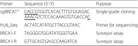
Table 2Primers sequences used in sgBRCA1 cloning and knockout validation
Primer Sequence (5’-3’) Purpose
sgBRCA1a CACCGTGGTCACACTTTGTGGAGAC
AAACGTCTCCACAAAGTGTGACCAC
Single-guide cloning
hU6_Seq ACTATCATATGCTTACCGTAAC Primer for sequencing
BRCA1-F TAGGGGTGGATATGGGTGAA Surveyor assay
BRCA1-R GTTGCAGTGAGCCAAGATCA Surveyor assay
a
of the undigested PCR product, whileBandCare the in-tensities of each cleaved band. The Indel percentage was estimated by applying the following formula:
Indelð Þ ¼% 1001−pffiffiffiffiffiffiffiffiffiffiffiffiffiffiffiffiffiffiffiffið1−f cutÞ
Population doubling level (PDL) calculation
Cells were considered at population doubling zero at the first time they are exposed to patient serum-containing culture medium. At every passage, cell number was de-termined and population doubling was calculated using the following formula; PDL = log(Nh/Ni)/log2, where Nh is the number of cells harvested at the end of the incu-bation time and Ni is the number of cells inoculated at the beginning of the incubation time. Cumulative PDL was calculated by adding the previous calculated PDL.
Immunoblotting
Cells were lysed in RIPA buffer containing protease in-hibitors (Sigma, Oakville, Canada). Equal amounts of proteins were resolved on 10 % SDS-PAGE and trans-ferred to a nitrocellulose membrane (BioRad, CA, USA). Membranes were blocked in TBS containing 5 % non-fat dry milk and exposed overnight at 4 °C to rabbit-anti-BRCA1 (ab191042 and ab131360, Abcam, MA, USA) or mouse-anti-β-Actin (A5316, Sigma, Oakville, Canada). Membranes were washed in TBST (TBS-0.05 % Tween-20) and incubated with either anti-rabbit or anti-mouse peroxidase-conjugated secondary antibody for 1 h at room temperature. After several washes in TBST, the blots were developed using Immobilon Western HRP Substrate (Millipore, Etobicoke, Canada).
Exosomes isolation and labeling
Exosomes were isolated from serum using Total Exo-some Isolation kit according to the manufacturer’s protocol (Invitrogen, Burlington, Canada). Exosomes were labeled using the PKH26 dye following the manufac-turer recommendations (Sigma, Oakville, Canada). La-beled exosomes were diluted in labeling stop solution (PBS/FBS) and pelleted by ultra-centrifugation for 80 min at 100,000 x g at 4 °C. The pellet was washed in Hank's Balanced Salt Solution (HBSS) with an ultra-centrifugation using the same parameters. The pelleted exosomes were re-suspended in HBSS and stored at −80 °C. 10 μg of labeled exosomes was added to ~5x10³BRCA1-KO Fibroblast, Control PX-458-transfected fibroblasts and HEK-293 cells cultured in 8-well chamber slides (VWR, Mont-Royal, Canada). Cells were washed, fixed for 10 min with Paraformaldehyde 4 %. Slides were mounted with coverslip in VECTASHIELD Mounting Medium with DAPI (Vector Laboratories, Burlington, Canada). Stained cells were visualized using an LSM780 confocal microscope
(Zeiss, Toronto, Canada). Exosomes internalization was quantified using ImageJ software.
Exosomes characterization
Morphological examination of isolated exosomes was done using transmission electron microscope (JEM-2010, Jeol Ltd., Tokyo, Japan). Briefly, 20μl of exosomes were loaded on a copper grid and stained with 2 % phos-photungstic acid. Samples were dried by incubating them for 10 min under an electric incandescent lamp. Samples were examined under electron microscope and imaged using a Hitachi H-600 TEM operating at 60 kV. In Parallel, an aliquot of exosome samples was run on a Nanosight NS500 system (Nanosight Ltd., Amesbury, UK), and size distribution was analyzed using the NTA 1.3 software.
In vivo tumor growth
Five-week-old female NOD-SCID mice (Jackson Labora-tory) were used in compliance with McGill University Health Centre Animal Compliance Office (Protocol 2012– 7280). Cells growing in log phase were harvested by trypsi-nization and washed twice with HBSS. Mice were injected subcutaneously with 2 million cells in 200μl HBSS/Matri-gel. Mice were euthanized one month post-injection. The resulting xenotransplants were photographed and proc-essed as indicated below.
Immunohistochemistry labelling procedures and histological analyses
Mice xenotransplants were collected, fixed in 10 % buff-ered formalin, embedded in paraffin, and stained with H&E (hematoxylin and eosin) according to standard pro-tocols or processed for immunohistochemistry. Briefly, 5 μm tissue sections were dewaxed in xylene and rehy-drated with distilled water. After antigen unmasking, and blocking of endogenous peroxidase (3 % hydrogen perox-ide), the slides were incubated with primary antibodies (Additional file 3: Table S1). Labeling was performed using iView DAB Detection Kit (Ventana) on the Ventana automated immunostainer. Sections were counterstained lightly with Hematoxylin before mounting. Histological analyses were performed by a certified pathologist who was blinded to the type of cells from which the cancerous masses, which formed in mice, had been derived.
Statistical analysis
Results
BRCA1knocking-out in human fibroblasts
A human fibroblast cell line deficient for the oncosup-pressor BRCA1was developed to study its susceptibility to the oncogenic potential of cancer patient serum (Additional file 1: Figure S1A). For this purpose, we used the CRISPR-Cas9 technology to knock-out BRCA1[31]. BRCA1-KO fibroblasts (i.e., fibroblasts transfected with a sgBRCA1-expressing vector) and control fibroblasts (empty vector transfected cells) were sorted based on the expression of the GFP reporter gene (Additional file 1: Figure S1B).BRCA1knockout was validated using the SURVEYOR nuclease assay (Additional file 2: Figure S2A). One of two guides used was efficient in knocking-outBRCA1 in fibroblasts. As described in Materials and Methods, the Indel percentage was estimated at 33 %, which is in the range of the values obtained with this assay. Moreover, protein extracts were analyzed for BRCA1 expression. BRCA1 was not detected inBRCA1 -KO fibroblasts, compared to empty vector-transfected cells (Additional file 2: Figure S2B).
Cancer patient sera increased the proliferation ofBRCA1 -KO fibroblasts
To analyze the effect of cancer patient sera on the growth ofBRCA1-deficient fibroblasts, equal amount of cells were plated and cultured with DMEM media supplemented with 10 % of cancer patients’serum for 2 weeks. We used serum from two healthy control donors and 10 metastatic cancer patients (Table 1). At every passage, cell numbers were determined to estimate the population doubling levels (PDL) in each condition (Fig. 1a). Independently of the cancer serum used, the cumulative PDL was increased when compared to that of cells cultured in control human serum (range: 1.3 to 6.1 increase in population doubling; mean +/−SD: 3.2 +/−1.4;P= 0.027) (Fig. 1b). These data suggest that cancer patient sera significantly enhanced the proliferation ofBRCA1-deficient fibroblasts in vitro.
Cancer patients’sera transfer malignant traits toBRCA1-KO fibroblasts
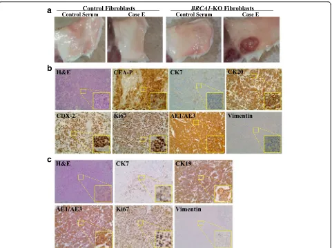
To determine whether cancer sera promote tumor for-mation in vivo, NOD/SCID mice were injected subcuta-neously with BRCA1-KO fibroblasts exposed to healthy control or cancer patients’sera. Cells were injected fol-lowing 2 weeks of treatment. Mice were followed up for tumor growth, and they were euthanatized 3 to 4 weeks after cell inoculation due to the massive growth of can-cerous masses, which had compromised the ability to move of the mice. Independently of the cancer serum used, all mice injected with cancer sera-treatedBRCA1 -KO fibroblasts developed visible tumors as early as one week following inoculation (Fig. 2a and Additional file 4: Figure S3). In contrast, none of the mice injected with
control fibroblasts, treated with healthy control or can-cer patient sera, developed tumors during the course of the experiments (4 weeks latency) (Fig. 2a).
Histopathological analyses of excised tumors showed solid growth of tumor cells sheets with high proliferation index (over 85–90 % Ki67 positivity) (Fig. 2b, c and Add-itional file 5: Figure S4). We further characterized these tumors for differentiation patterns based on the primary tumor of the blood donors. The histology of tumors ob-tained with cells treated with sera derived from five dif-ferent patients (Cases A, B, C, D and F) confirmed that they were poorly differentiated carcinomas (Fig. 3 and Additional file 4: Figure S3). Notably, vimentin staining of all the cancerous masses was negative indicating that cancer factors carried in the sera of all tested patients had integrated in the genome of the BRCA1-KO fibro-blasts changing their fate permanently. Even more strik-ing was the discovery thatBRCA-KO fibroblasts treated with sera taken from four patients with colorectal cancer liver metastases (Cases E, G, H and I), and one patient with pancreatic cancer (Case J) displayed epithelial differ-entiating features typical of colorectal adenocarcinomas and pancreatic ductal adenocarcinoma, respectively (Table 3 and Fig. 2b, c and Additional file 5: Figure S4). The tumors generated with cells treated with cases E, G, H and I (colorectal cancer) were negative for CK7 but they all stained positive for CEA, CK20, CDX-2 and AE1/AE3, which are universal markers of colorectal cancer. The tu-mors generated with BRCA-KO fibroblasts treated with
a
b
Fig. 1Cancer patient sera increasedBRCA1-KO fibroblasts growth.
case J (pancreatic cancer), stained strongly positive for CK7/CK19 which is a typical feature of pancreatic adeno-carcinoma differentiation. Interestingly, CK7 expression was not uniform in the cancer specimen but it was strongly expressed in the areas with pancreatic tumor morphology indicating an evolving differentiation from undifferentiated morphology to pancreatic cancer pheno-type. Attempts to characterize immunohistochemically the other tumors failed to show any differentiating features re-sembling the tumors of blood donors as they all stained negatively for tumor-specific markers (Table 3 and Fig. 3).
BRCA1-KO fibroblasts treated with healthy control sera failed to acquire any malignant transformation
To rule out the possibility that BRCA1-KO fibroblasts might have an innate tendency to turn malignant due to the impaired function of the BRCA1 gene, BRCA1-KO
fibroblasts were cultured with healthy patients’serum for 2 weeks. Inoculation of these cells in NOD/SCID mice failed to form any mass even at longer latency period (12 weeks post-transplantation), confirming the known notion that a single oncosuppressor mutation is not enough to trigger cancerogenesis and activate the cascade of events that eventually lead to cancer transformation (Fig. 2a). Furthermore, the results of this test confirmed also that the putative factors responsible for the malignant transformation of theBRCA1-KO fibroblasts were exclu-sively present in the serum of patients with metastatic cancer and were absent in the serum of healthy patients.
Malignant transformation of theBRCA1-KO fibroblasts is permanent and not transient
Further, we wanted to test whether the malignant trans-formation of the BRCA1-KO fibroblasts was permanent
Fig. 2Cancer patient sera induced the transformation ofBRCA1-KO fibroblasts.aNOD/SCID mice were injected with either control orBRCA1-KO
and not secondary to a short-term and transient effect. Therefore,BRCA1-KO fibroblasts were treated with can-cer serum for 2 weeks. Subsequently, they were exposed to DMEM-F12 media supplemented with 10 % FBS and 1 % Pen-Strep, for two more weeks to allow them to re-cover. Interestingly we found that cells had kept their tumorigenic potential and still formed tumors when they were injected into NOD/SCID mice, indicating thus that the transformation was indeed permanent and not tran-sient (Fig. 4).
BRCA1-KO fibroblasts demonstrate a significant increased uptake of cancer-derived exosomes
A number of recent evidences indicate that exosomes are involved in cancer invasion and metastasis [4, 8, 18, 32]. We had noticed in previous unpublished experi-ments that cancer exosomes had the tendency to accu-mulate in the cytoplasm of HEK293 cells in larger number than what was observed in normal cells. Follow-ing this observation, we hypothesized that perhaps one of the methods implemented by the oncosuppressor genes to protect the genome of the cells was to prevent the uptake of genetic material potentially harmful to the cell. As a consequence of that, any cell with a single oncosuppressor mutation would show a superior uptake of cancer exosomes. In order to test our hypothesis,
exosomes were isolated from cancer patients’sera. The size of the isolated particles was between 50 and 120 nm as visualized by electron microscopy (Fig. 5a), and Nano-sight analyses (size = 103 +/−7; Fig. 5b). This is in the range of the expected size for exosomes. The isolated exosomes were labeled with PKH-26 and added to BRCA1-KO and wild type fibroblasts, and HEK293 cell cultures to assess their internalization. Exosomes uptake was assessed by measuring the number of positive intra-cellular spots (Fig. 5c and d). As hypothesized, BRCA1 -KO fibroblasts and HEK293 cells showed an uptake of cancer exosomes 3 to 13 times higher (6.6 +/−0.6 times for BRCA1-KO fibroblasts and 7.7 +/− 2.0 for HEK293 cells) than that measured in control wild type fibroblasts. This suggests that one of the ways oncosuppressor genes might use to protect the integrity of the genome would be to act as gatekeepers at the membrane level and block the integration of dangerous genetic material.
Discussion
In the present study, we confirmed that sera of patients with metastatic cancer contain tumorigenesis-signaling factors that, once delivered to recipient target cells, are capable to complete the cascade of events that eventu-ally lead the cells to acquire malignant traits. In our pre-vious research we had used the HEK293 cells as a model
Fig. 3Tumors generated with cancer patient sera-treatedBRCA1-KO did not all display differentiation characteristics.BRCA1-KO fibroblasts were
of “initiated” cell to demonstrate that the horizontal transfer of blood-circulating cancer factors is also applic-able to human cells [26], confirming thus, the validity of the“genometastatic” theory in humans [23–25]. Accord-ing to this theory, carcinogenetic steps such as initiation, promotion and progression might not represent events limited to the cells forming the primary tumor, but may actually be a process reproducible through cancer fac-tors, shed by primary tumors and carried through the blood, to susceptible cells, located at metastatic sites [5, 7, 10–16]. In order to strengthen the validity of this al-ternative metastatic pathway in humans, we used fibro-blasts, which are among the most represented cells in
human body and central players at metastatic sites [33, 34]. In addition, these cells display a high level of plasti-city [35]. We induced a BRCA1 oncosuppressor muta-tion to reproduce an in vitro model that would be as close as possible to what it is encountered in real clinical scenarios. We hypothesized that not only immortal cells, as previously demonstrated [23–26], but also any human cell carrying a single oncosuppressor mutation might represent a target cell susceptible of malignant trans-formation if exposed to blood-circulating cancer factors. The observation that BRCA1-KO fibroblasts cultured in cancer patients’sera displayed oncogenic properties such as increased proliferation potential and ability to form tumors following subcutaneous injection into NOD/ SCID mice supports the belief that truly a horizontal transfer of cancer material might have been overlooked when trying to understand metastatic disease. Moreover, the discovery that BRCA1-KO fibroblasts change their fate completely and turn into colon cancer cells and pancreatic cancer cells, when exposed to serum derived from patients affected by metastatic colon and pancre-atic cancer, strengthen the belief of the authors, that similarity doesn’t imply sameness and therefore meta-static cells might not necessarily be only cells detaching from primary tumors. In line with this concept, the mo-lecular profiles of primary and metastatic lesions are not usually identical and therefore, at least theoretically, the possibility that metastases might not be deriving from the same cells is still open [36–39]. The data gathered in our study prove that any cell as long as it is “initiated”
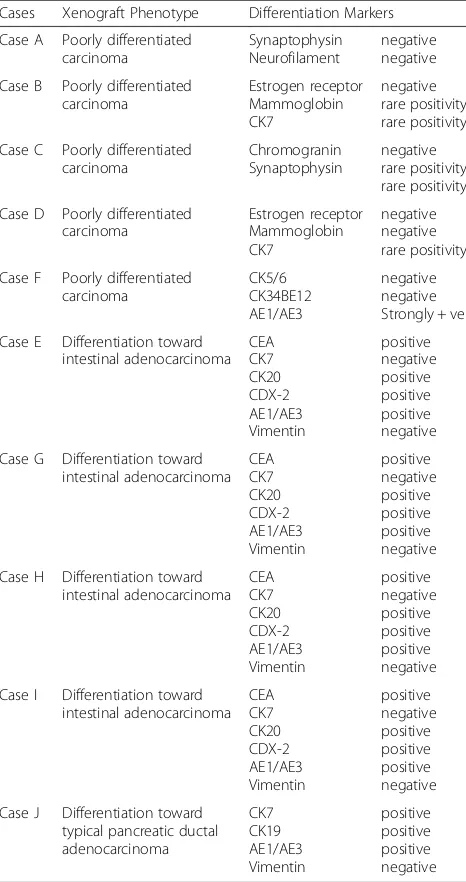
Table 3Summary of the immunohistochemistry analyses of the xenografts obtained with cancer patients’serum-treatedBRCA1 -KO fibroblasts
Cases Xenograft Phenotype Differentiation Markers
Case A Poorly differentiated carcinoma
Synaptophysin Neurofilament
negative negative
Case B Poorly differentiated carcinoma Estrogen receptor Mammoglobin CK7 negative rare positivity rare positivity
Case C Poorly differentiated carcinoma Chromogranin Synaptophysin negative rare positivity rare positivity
Case D Poorly differentiated carcinoma Estrogen receptor Mammoglobin CK7 negative negative rare positivity
Case F Poorly differentiated carcinoma CK5/6 CK34BE12 AE1/AE3 negative negative Strongly + ve
Case E Differentiation toward intestinal adenocarcinoma CEA CK7 CK20 CDX-2 AE1/AE3 Vimentin positive negative positive positive positive negative
Case G Differentiation toward intestinal adenocarcinoma CEA CK7 CK20 CDX-2 AE1/AE3 Vimentin positive negative positive positive positive negative
Case H Differentiation toward intestinal adenocarcinoma CEA CK7 CK20 CDX-2 AE1/AE3 Vimentin positive negative positive positive positive negative
Case I Differentiation toward intestinal adenocarcinoma CEA CK7 CK20 CDX-2 AE1/AE3 Vimentin positive negative positive positive positive negative
Case J Differentiation toward typical pancreatic ductal adenocarcinoma CK7 CK19 AE1/AE3 Vimentin positive positive positive negative
Fig. 4Cancer patient sera permanently transformedBRCA1-KO
has the potentials to incorporate in its genome “signal-ing”factors, change its fate and acquire aberrant pheno-typical traits identical or similar to the source of the signaling factors.
To our knowledge, this is the first study to demon-strate the transformation of human fibroblasts carrying a single oncosuppressor mutation (i.e., BRCA1) into colon cancer or pancreatic cancer cells after exposure to serum of patients with metastatic colon or metastatic pancre-atic cancer. This result becomes even more striking and fascinating when considering that the recovery test indi-cated that the phenotypical modifications were indeed permanent and not related to a transient alteration of the genetic asset of the cells. In other words, a short ex-posure to cancer factors present in the serum seems to have been strong enough to overcome the repair
mechanisms of the treated cells. The main implication of this stable modification of the genome of human cells is that at least in vitro, metastatic transformation, through horizontal transfer, is not a theory anymore but a fascinating reality. The scientific soundness of this data is corroborated by the evidence brought about in our ex-periments that the malignant transformation would be unlikely to be secondary to an innate instability of the fi-broblasts caused by theBRCA1mutation. In fact, the ex-posure of the BRCA1-KO cells to healthy serum never made them susceptible of malignant transformation, con-firming the known notion that a single mutation is not able to trigger the cascade of events that leads to tumori-genesis [40].
In our study, we documented, for the first time, a sig-nificant increased uptake of cancer-derived exosomes by
a
c
d
b
Fig. 5BRCA1-KO fibroblasts internalized exosomes more efficiently than control cells.aExosomes were isolated as described under Methods.
Representative micrographs of transmission electron microscopy on cancer patient sera exosome preparations. The image showed small vesicles of approximately 50–120 nm in diameter. Scale bars 100 nm.bNanoSight analysis of samples prepared as in (a). The size was centered around 94 nm in diameter.cConfocal microscopy monitoring of PKH-26-labeled exosome uptake in vitro intoBRCA1-KO fibroblasts, control fibroblasts, and HEK293 cells. Note that exosomes internalized more inBRCA1-KO cells. They were dispersed in the cytoplasm and tended to form aggregates in the perinuclear regions.dThe number of PKH-26-labeled exosomes (dots) was counted. Data are expressed as relative number (Rel. Num.) of exosomes per cell compared to that in control fibroblasts. In the insert, data are expressed as mean +/−SD (n= 6 exosome preparations;P= 0.032 for HEK293 cells compared to control fibroblasts,P= 0.028 forBRCA1-KO fibroblasts compared to control fibroblasts, and
BRCA1-KO fibroblasts and HEK293 cells when com-pared to wild type fibroblasts. This finding noted only in cells carrying a single oncosuppressor mutation suggests a potential role that oncosuppressor genes might have in exosomes trafficking. Perhaps, they might protect the in-tegrity of the cellular genome not only by repairing DNA damages and controlling cell cycle checkpoints [27, 41], but also by blocking the uptake of extracellular mutating material and thus preventing their integration in the genome with subsequent DNA alterations. Al-though we reported an increased uptake of cancer de-rived exosomes in oncosuppressor mutated cells, their putative role in the documented malignant transform-ation of the cells yet has to be determined as well as the true nature of all the factors involved in the transfer of malignant traits. While cancer cells-derived circulating factors (i.e., DNA, mRNA, miRNA, proteins) were de-tected in the blood of cancer patients, and their regula-tory role in cancer progression and development were reported in many studies [16, 22, 32, 42, 43] still their respective roles have to be fully defined. If on one hand, it has been reported that exosomes originating from the primary tumor paves the way for organ-specific metasta-sis by preparing a niche for the engraftment of circulat-ing cancer cells, [5–7, 9], on the other hand, the results of our study might offer a different prospective, which do not exclude, but might integrate the conventional view on metastasis.
The inability of theBRCA1-KO fibroblasts to fully dif-ferentiate in all cancer phenotypes is a limitation of our study. In our view, this failure is not perceived necessar-ily as a weakness of the theory but it is perceived as awareness that the genometastatic process is not under-stood in its entirety. When we exposed HEK293 to can-cer patients’ sera the cells turned only into poorly differentiated carcinomas regardless of the type of can-cer studied [26]. The exposure ofBRCA1-KO fibroblasts has added value to the genometastatic theory since we have observed a full differentiation into at least two can-cer cell lineages (colon and pancreas). We are planning to repeat the experiments with different cells and differ-ent single oncosuppressor mutations to see if the hori-zontal transformation targets different types of cells in different organs and at different stages of their physio-logical differentiation according to the type of cancer.
Conclusion
The data presented in this study support the hypothesis that any human cells carrying a single oncosuppressor mutation is capable of integrating cancer factors carried in the blood of cancer patients and undergo complete malignant transformation. The evidence shown in our experiments that uptake of cancer-derived exosomes is significantly increased in these cells suggests a possible
role that oncosuppressor genes might have in exosomes trafficking.
The reported findings support the notion of the pos-sible role of a non-classical pathway to explain cancer traits exchange between malignant and non-malignant cells that may have implications during cancer progres-sion and metastasis. Based on these results, the hypoth-esis that dissemination and migration of cancer cells from primary tumors might not be the only mechanism to explain metastases seems rational and merits further study.
Additional files
Additional file 1: Figure S1.Methodology design followed to knock-out
BRCA1in fibroblasts. pX-458 plasmid was used as a vector for CRISPR-Cas9 system forBRCA1knocking down. Human fibroblasts were transfected using lipofectamine 3000 (A). Transfected fibroblasts with sgBRCA1-pX458 or empty pX-458 vectors were sorted using a FACSAria cell sorter based on their GFP positivity. Only GFP positive cells were obtained and expanded in culture (B). (i) Naïve fibroblasts were gated as negative fraction (GFP negative fibroblasts). (ii) Fraction of cells sorted as control fibroblasts (empty pX-458 vector-transfected cells). (iii) Fraction of cells sorted as sgBRCA1-pX458 transfected fibroblasts (BRCA1-KO). Transfection efficiency showed percentage of 4–6 %. Fibroblasts were treated with cancer patients’ serum or healthy individual serum. Treated cells were analyzed for their in vitro proliferation or injected into NOD/SCID mice for tumor growth potential. (PPT 524 kb)
Additional file 2: Figure S2.Validation ofBRCA1Knockout in human
fibroblasts. (A) Surveyor nuclease assay was performed as described under Materials and Methods. DNA was extracted from sorted fibroblasts. After amplification, DNA was denatured, reannealed and subjected to endonuclease digestion. Digestion products were run on 1 % agarose gel. Only with DNA extracted from sgBRCA1-transfected fibroblasts, we detected 3 bands: the full-length PCR product (i) and the digestion products (ii and iii). Note that the cumulative size of bands ii and iii equals the size of band i. (B) Western blot analysis of proteins extracted from control fibroblasts (empty vector transfected) andBRCA1-KO fibroblasts. Note that BRCA1 signal is absent in BRCA1-KO fibroblasts. (*) points to the BRCA1 signal. (PPT 265 kb)
Additional file 3: Table S1.List of antibodies used in this study. Table
S2. BRCA1 guide sequence as designed by the CRISPR Design Tool. Table S3. Analyses of off-target sequences of the BRCA1 guide. (DOC 58 kb)
Additional file 4: Figure S3.Cancer patient sera induced the
transformation ofBRCA1-KO fibroblasts.BRCA1-KO fibroblasts were treated with cancer patient sera for 2 weeks (6 Cases). Treated cells were injected into NOD/SCID mice that were followed for 4 weeks for tumors growth. Developing tumors were excised and photographed. (PPT 189 kb)
Additional file 5: Figure S4.Cancer patient sera changed the fate of
BRCA1-KO fibroblasts.BRCA1-KO fibroblasts were treated with CRC-LM patient sera for 2 weeks (Cases G, H and I). Treated cells were injected into NOD/SCID mice that were followed for 4 weeks for tumors growth. Generated tumors were processed for H&E staining, or immunolabeled with antibodies against tumor specific markers. (PPT 36551 kb)
Abbreviations
ANOVA:analysis of variance;BRCA1: breast cancer susceptibility gene 1; BSA: bovine serum albumin; DAB: diaminobenzidine; FBS: fetal bovine serum; H&E: hematoxylin and eosin; HEK293: human embryonic kidney cells; KO: knockout; PDL: Population doubling level.
Competing interests
Authors' contributions
GA supervised the study. MA, VA, MA and GA conceived and designed the study. MA, DH and GA developed the methodology. MA, DH, ZHG and GA acquired and analyze data, and managed patients. MA, DH and GA drafted the manuscript. All authors read and approved the final draft of the manuscript.
Acknowledgement
We are grateful to Ayat Salman for her assistance with the Ethical Committee approvals, Laura Montermini for her assistance with Nanosight data acquisition, and Diane Gingras for her assistance with electron microscopy data acquisitions. This work was financially supported by Giuseppe Monticciolo. The funder had no role in study design, data collection and analysis, decision to publish, or preparation of the manuscript.
Author details
1Cancer Research Program, McGill University Health Centre-Research Institute,
1001 Decarie Boulevard, Montreal H4A 3J1, QC, Canada.2Department of
Experimental Surgery, Faculty of Medicine, McGill University, 845 Rue Sherbrooke O, Montreal H3A 0G4, QC, Canada.3Department of Pathology, McGill University Health Centre-Research Institute, 1001 Decarie Boulevard, H4A 3J1 Montreal, QC, Canada.4Department of Obstetrics and Gynecology,
Santo Bambino Hospital, via Torre del Vescovo 4, Catania, Italy.5Department
of Surgical Sciences, Organ Transplantation and Advances Technologies, University of Catania, via Santa Sofia 84, Catania, Italy.6Department of
Surgery, McGill University, St. Mary Hospital, 3830 Lacombe Avenue, Montreal H3T 1M5, QC, Canada.
Received: 4 February 2016 Accepted: 9 May 2016
References
1. Eccles SA, Welch DR. Metastasis: recent discoveries and novel treatment strategies. Lancet. 2007;369(9574):1742–57.
2. Jemal A, Bray F, Center MM, Ferlay J, Ward E, Forman D. Global cancer statistics. CA Cancer J Clin. 2011;61:69–90.
3. Nguyen DX, Bos PD, Massagué J. Metastasis: from dissemination to organ specific colonization. Nat Rev Cancer. 2009;9:274–84.
4. Skog J, Wurdinger T, van Rijn S. Glioblastoma microvesicles transport RNA and proteins that promote tumour growth and provide diagnostic biomarkers. Nat Cell Biol. 2008;10:1470–81.
5. Hood JL, San RS, Wickline SA. Exosomes released by melanoma cells prepare sentinel lymph nodes for tumor metastasis. Cancer Res. 2011;71: 3792–801.
6. Grange C, Tapparo M, Collino F, Vitillo L, Damasco C, Deregibus MC, Tetta C, Bussolati B, Camussi G. Microvesicles released from human renal cancer stem cells stimulate angiogenesis and formation of lung premetastatic niche. Cancer Res. 2011;71(15):5346–56.
7. Peinado H, AlečkovićM, Lavotshkin S, Matei I, Costa-Silva B, Moreno-Bueno G, Hergueta-Redondo M, Williams C, García-Santos G, Ghajar C, Nitadori-Hoshino A, Hoffman C, Badal K, Garcia BA, Callahan MK, Yuan J, Martins VR, Skog J, Kaplan RN, Brady MS, Wolchok JD, Chapman PB, Kang Y, Bromberg J, Lyden D. Melanoma exosomes educate bone marrow progenitor cells toward a prometastatic phenotype through MET. Nat Med. 2012;18:883–91.
8. Abdel-Mageed ZY, Yang Y, Thomas R, Ranjan M, Mondal D, Moraz K, Fang Z, Rezk BM, Moparty K, Sikka SC, Sartor O, Abdel-Mageed AB. Neoplastic reprogramming of patient-derived adipose stem cells by prostate cancer cell-associated exosomes. Stem Cells. 2014;32:983–97.
9. Fujita Y, Yoshioka Y, Ochiya T. Extracellular vesicle transfer of cancer pathogenic components. Cancer Sci. 2016. [DOI: 10.1111Epub ahead of print]. 10. Valadi H, Ekström K, Bossios A, Sjöstrand M, Lee JJ, Lötvall JO.
Exosome-mediated transfer of mRNAs and microRNAs is a novel mechanism of genetic exchange between cells. Nat Cell Biol. 2007;9:654–9.
11. Runz S, Keller S, Rupp C, Stoeck A, Issa Y, Koensgen D. Malignant ascites-derived exosomes of ovarian carcinoma patients contain CD24 and EpCAM. Gynecol Oncol. 2007;107:563–71.
12. Pisetsky DS, Gauley J, Ullal AJ. Microparticles as a source of extracellular DNA. Immunol Res. 2010;49:227–34.
13. Subra C, Grand D, Laulagnier K, Stella A, Lambeau G, Paillasse M, De Medina P, Monsarrat B, Perret B, Silvente-Poirot S, Poirot M, Record M. Exosomes account for vesicle mediated transcellular transport of
activatable phospholipases and prostaglandins. J Lipid Res. 2010;51: 2105–20.
14. Gaiffe E, Pretet JL, Launay S, Jacquin L, Saunier M, Hetzel G. Apoptotic HPV positive cancer cells exhibit transforming properties. PLoS One. 2012;7: e36766.
15. Balaj L, Lessard R, Dai L, Cho YJ, Pomeroy SL, Breakefield XO. Tumour microvesicles contain retrotransposon elements and amplified oncogene sequences. Nat Commun. 2011;2:180.
16. Fleischhacker M, Schmidt B. Circulating nucleic acids (CNAs) and cancer–a survey. Biochim Biophys Acta. 2007;1775(1):181–232.
17. Thery C, Zitvogel L, Amigorena S. Exosomes: composition, biogenesis and function. Nat Rev Immunol. 2002;2:569–79.
18. Ogorevc E, Kralj-Iglic V, Veranic P. The role of extracellular vesicles in phenotypic cancer transformation. Radiol Oncol. 2013;47(3):197–205. 19. Candelario KM, Steindler DA. The role of extracellular vesicles in the
progression of neurodegenerative disease and cancer. Trends Mol Med. 2014;20(7):368–74.
20. Pant S, Hilton H, Burczynski ME. The multifaceted exosome: biogenesis, role in normal and aberrant cellular function, and frontiers for
pharmacological and biomarker oppertunities. Biochem Pharmacol. 2012; 83(11):1484–94.
21. Colombo M, Raposo G, Théry C. Biogenesis, secretion, and intercellular interactions of exosomes and other extracellular vesicles. Annu Rev Cell Dev Biol. 2014;30:255–89.
22. Falcone G, Felsani A, D’Agnano I. Signaling by exosomal microRNAs in cancer. J Exp Clin Cancer Res. 2015;34:32.
23. Garcıa-Olmo D, Garcıa-Olmo DC, Ontanon J, Martinez E, Vallejo M. Tumor DNA circulating in the plasma might play a role in metastasis. The hypothesis of the genometastasis. Histol Histopathol. 1999;14:1159–64. 24. Olmo DC, Domínguez C, Arranz M, Anker P, Stroun M,
García-Verdugo JM, García-Olmo D. Cell-free nucleic acids circulating in the plasma of colorectal cancer patients induce the oncogenic transformation of susceptible cultured cells. Cancer Res. 2010;70:560–7.
25. Trejo-Becerril C, Perez-Cardenas E, Taja-Chayeb L, Anker P, Herrera-Goepfert R, Medina-Velazquez LA, Hidalgo-Miranda A, Perez-Montiel D, Chavez-Blanco A, Cruz-Velazquez J, Dıaz-Chavez J, Gaxiola M, as-Gonzalez AD. Cancer Progression Mediated by Horizontal Gene Transfer in an In Vivo Model. PLoS One. 2012;7(12):e52754.
26. Abdouh M, Zhou S, Arena V, Arena M, Lazaris A, Onerheim R, Metrakos P, Arena G. Transfer of malignant trait to immortalized human cells following exposure to human cancer serum. J Exp Clin Cancer Res. 2014;33(1):86. 27. Gudmundsdottir K, Ashworth A. The roles of BRCA1 and BRCA2 and
associated proteins in the maintenance of genomic stability. Oncogene. 2006;25(43):5864–74.
28. Welcsh PL, King MC. BRCA1 and BRCA2 and the genetics of breast and ovarian cancer. Hum Mol Genet. 2001;10(7):31–42.
29. Campeau PM, Foulkes WD, Tischkowitz MD. Hereditary breast cancer: New genetic developments, new therapeutic avenues. Hum Genet. 2008; 124(1):31–42.
30. Pal T, Permuth-Wey J, Betts JA. BRCA1 and BRCA2 mutations account for a large proportion of ovarian carcinoma cases. Cancer. 2005;104(12): 2807–16.
31. Ran FA, Hsu PD, Wright J, Agarwala V, Scott DA, Zhang F. Genome engineering using the CRISPR-Cas9 system. Nat Protoc. 2013;8(11): 2281–308.
32. Hoshino A, Costa-Silva B, Shen TL, Rodrigues G, Hashimoto A, Tesic MM. Tumour exosome integrins determine organotropic metastasis. Nature. 2015;527(7578):329–35.
33. Polanska UM, Orimo A. Carcinoma-associated fibroblasts: non-neoplastic tumour-promoting mesenchymal cells. J Cell Physiol. 2013;228(8):1651–7. 34. Paulsson J, Micke P. Prognostic relevance of cancer-associated fibroblasts in
human cancer. Semin Cancer Biol. 2014;25:61–8.
35. Takahashi K, Tanabe K, Ohnuki M, Narita M, Ichisaka T, Tomoda K, Yamanaka S. Induction of pluripotent stem cells from adult human fibroblasts by defined factors. Cell. 2007;131(5):861–72.
36. Suzuki M, Tanin D. Gene expression profiling of human lynph node metastases and matched primary breast carcinomas: clinical implications. Mol Oncol. 2007;1(2):172–80.
38. Liu X, Zhang M, Go VLW, Hu S. Membrane proteomic analysis of pancreatic cancer cells. J Biomed Sci. 2010;17(1):74.
39. Yoshida A, Okamoto N, Tozawa-Ono A, KoizUmi H, Kiguchi K, Ishizuka B, Kumai T, Suzuki N. Proteomic analysis of differential protein expression by brain metastases of gynecological malignancies. Hum Cell. 2013;26(2):56–66. 40. Boehm JS, Hession MT, Bulmer SE, Hahn WC. Transformation of human and
murine fibroblasts without viral oncoproteins. Mol Cell Biol. 2005;25: 6464–74.
41. Jiang Q, Greenberg RA. Deciphering the BRCA1 tumor suppressor network. J Biol Chem. 2015;290(29):17724–32.
42. Diehl F, Li M, Dressman D, He Y, Shen D, Szabo S, Diaz Jr LA, Goodman SN, David KA, Juhl H, Kinzler KW, Vogelstein B. Detection and quantification of mutations in the plasma of patients with colorectal tumors. Proc Natl Acad Sci U S A. 2005;102(45):16368–73.
43. Kahlert C, Melo SA, Protopopov A, Tang J, Seth S, Koch M, Zhang J, Weitz J, Chin L, Futreal A, Kalluri R. Identification of double-stranded genomic DNA spanning all chromosomes with mutated KRAS and p53 DNA in the serum exosomes of patients with pancreatic cancer. J Biol Chem. 2014;289(7): 3869–75.
• We accept pre-submission inquiries
• Our selector tool helps you to find the most relevant journal
• We provide round the clock customer support
• Convenient online submission
• Thorough peer review
• Inclusion in PubMed and all major indexing services
• Maximum visibility for your research
Submit your manuscript at www.biomedcentral.com/submit