R E S E A R C H A R T I C L E
Open Access
Quantitative detection of
DNMT3A
R882H mutation
in acute myeloid leukemia
Rimma Berenstein, Igor Wolfgang Blau, Nikola Suckert, Claudia Baldus, Antonio Pezzutto, Bernd Dörken
and Olga Blau
*Abstract
Background:DNMT3Amutations represent one of the most frequent gene alterations detectable in acute myeloid leukemia (AML) with normal karyotype. Although various recurrent somatic mutations ofDNMT3Ahave been described, the most common mutation is located at R882 in the methyltransferase domain of the gene. Because of their prognostic significance and high stability during disease evolution,DNMT3Amutations might represent highly informative biomarkers for prognosis and outcome of disease.
Methods:We describe an allele-specific PCR with a Blocking reagent for the quantitative detection ofDNMT3AR882H mutation providing the possibility to analyze the quantitative amount of mutation during the course of disease. Next, we analyzed 62 follow-up samples from 6 AML patients after therapy and allogeneic stem cell transplantation (alloSCT).
Results:We developed an ASB-PCR assay for quantitative analysis of R882HDNMT3Amutation. After optimization of blocker concentration, a R882H-positive plasmid was constructed to enhance the accuracy of the sensitivity of quantitative detection. The assay displayed a high efficiency and sensitivity up to 10−3. The reproducibility of assay analyzed using follow-up samples showed the standard deviation less than 3.1 %. This assay displayed a complete concordance with sequencing and endonuclease restriction analysis. We have found persistence ofDNMT3A R882H mutations in complete remission (CR) after standard cytoreduction therapy that could be indicating presence of DNMT3Amutation in early pre-leukemic stem cells that resist chemotherapy. The loss of correlation betweenNPM1and DNMT3Ain CR could be associated with evolution of pre-leukemic and leukemic clones. In patients with CR with complete donor chimerism after alloSCT, we have found no DNMT3A R882H. In relapsed patients, all samples showed an increasing of bothNPM1andDNMT3Amutated alleles. This suggests at least in part the presence ofNPM1and DNMT3Amutations in the same cell clone.
Conclusion:We developed a rapid and reliable method for quantitative detection ofDNMT3AR882H mutations in AML patients. Quantitative detection ofDNMT3AR882H mutations at different time points of AML disease enables screening of follow-up samples. This could provide additional information about the role ofDNMT3Amutations in development and progression of AML.
Keywords:AML,DNMT3Amutations, Quantitative PCR
* Correspondence:olga.blau@charite.de
Department of Hematology, Oncology and Tumorimmunology, Charité University School of Medicine, Hindenburgdamm 30, 12200 Berlin, Germany
Introduction
Somatic mutations in the DNA nucleotide methyltrans-ferase 3A gene (DNMT3A)have been reported approxi-mately in 22 % of de novo acute myeloid leukemia (AML) and 36 % of cytogenetically normal AML [1]. Mutations inDNMT3Awere first described by Leyet al.
using whole genome sequencing [2]. DNMT3A belongs to the mammalian methyltransferase gene family which is responsible for tissue-specific gene expression [3]. DNA methyltransferases are the key enzymes for gen-ome methylation, which plays an important role in epi-genetically regulated gene expression and repression.
DNMT3A together with other methyltransferases con-ducts de novo methylation of cytosine residues in CpG islands by the enzymatic addition of methyl residues from S-adenosyl-L-methionine to the 5-carbon atom of the cytosine ring. CpG islands are often located proximate to gene promoters thereby regulating their activation. Actively transcribed genes display unmethylated CpG islands which supports the euchromatin structure whereas methylated CpG islands are associated with untranscribed genes stabilizing the heterochromatin structure [4, 5]. Cancer genomes are most commonly characterized by global DNA hypomethylation. However, cancer cells also typically exhibit distinct regions of DNA hypermethyla-tion, which are particularly well characterized in the CpG islands of promoter regions of tumor-suppressor genes. Although various recurrent DNMT3A mutations have been described, the most common mutation affects resi-due R882 within the methyltransferase domain.DNMT3A
mutations are typically heterozygous [2, 6].
The biology ofDNMT3Ais not fully understood. Holz-Schietinger et al. reported that mutations in DNMT3A
could retard its function by multiple mechanisms as changes in the catalytic properties, its processivity and the disruption of interaction with binding partners [7]. Fur-thermore, Russler-Germainet al.found that mutations in the position R882 inhibit the formation of active tetramers of DNMT3A [8]. The impaired function of mutated
DNMT3A leads to a hypomethylated genome of myeloid cells possibly promoting leukemogenesis and influencing disease outcome [9].
Since theDNMT3Amutations are present in the early pre-leukemic cells, this alteration seems to be a“founder” mutation, which can be implicated as functional compo-nents of AML evolution [10, 11].DNMT3Amutations are highly associated with mutations in the nucleophosmin 1 gene (NPM1), fms-related tyrosine kinase 3 gene (FLT3), and isocitrate dehydrogenase 1 gene (IDH1) [12, 13].
Several studies reported a negative prognostic impact of DNMT3A mutations [12–16]. Prognostic effect is known to depend on certain biological factors as well as a combination of cytogenetics and other mutations such as those inFLT3andNPM1.
Some authors have found stability ofDNMT3A muta-tions during the course of disease; therefore those aber-rations could be potential marker for minimal residual disease (MRD). Furthermore, the presence of DNMT3A
mutations seems to be associated with the incidence of
FLT3-ITD-positive clones at relapse possibly influencing the responsiveness ofFLT3-positive cases to chemother-apy [17, 18]. Last published data have demonstrated that
DNMT3A mutations are also detectable in AML patients in long-term complete remission (CR) and can occur in pre-leukemic stem cells [10, 11, 19]. The identification of pre-leukemic cells with genetic mutations in CR has im-portant implications for the MRD monitoring. Moreover, the persistence of DNMT3A mutations in CR may have important implications for the management of AML.
Recent discoveries utilizing whole-exome sequencing in a large cohort of persons, unselected for cancer or hematologic phenotypes have demonstrated somatic mutations in significant proportion of persons particu-larly which older than 65 years. Moreover, DNMT3A
gene together withTET2, ASXL1, and PPM1D had dis-proportionately high numbers of somatic mutations [20, 21]. The data suggest that mutations in pre-leukemic cells could precede leukemia. Furthermore, DNMT3A
mutations could drive clonal expansions. Based on these data,DNMT3Amutation might represent highly inform-ative biomarkers for AML. Thus, a neginform-ative prognostic impact and, in addition, conflicting reports on the po-tential role of DNMT3A mutations for the evolution of leukemic stem cell, require fast, reliable, quantitative and available methods for detection of mutation.
Sanger sequencing is well-established but not very sensi-tive as well as time-consuming and cost-intensive method. HRM analysis provides the possibility of high throughput screening of mutations, but data interpretation occasion-ally can be difficult. Therefore, good validated controls and standards are needed. Previously we reported a rapid and reliable restriction fragment length polymorphism based method for the qualitative detection of DNMT3A
R882H mutation [22]. Current quantitative established assays have low sensitivity and therefore cannot be used as reliable methods for MRD diagnostic. Here, we describe an Allele-Specific PCR with a Blocking reagent (ASB-PCR) for the quantitative detection of DNMT3A R882H mutation with sensitivity up to 10−3providing the possibil-ity to analyze the quantitative amount of this mutation for routine diagnostic during the course of disease.
Methods
Patient and control materials
patients after chemotherapy and allogeneic stem cell transplantation (alloSCT). All patients were treated at the University Clinic Charité from September 2009 to May 2013. Diagnoses were established according to the WHO criteria [23]. Written informed consent was ob-tained from all patients in accordance with the Declar-ation of Helsinki and the ethical guidelines of the Charite University School of Medicine, which approved this study. In 6 AML patients included in the follow up study, induc-tion therapy consisted of “7 + 3” therapy with Cytosine Arabinoside and Daunorubicine. Second part of induction with “7 + 3” started at day 22, if at day 15 count of BM blasts were reduced. Once complete remission (CR) was achieved, 2–4 courses of consolidation chemotherapy with high-dose Cytosine Arabinoside were administered. At 1-st CR or at 2-nd CR patients were allocated to alloSCT.
Control material for DNMT3A mutation included DNA from K562 cell line (wt) and constructed plasmid which contains the R882H mutation.
DNA extraction
Mononuclear cells (MNCs) from BM aspirates were iso-lated using Ficoll density centrifugation as described [24]. DNA was extracted using Allprep DNA/RNA mini kit (Qiagen) from 1*107MNCs as recommended by the manufacturer. DNA yields ranged from 50 to 300 ng/μl.
Allele-Specific PCR with a Blocking reagent (ASB-PCR)
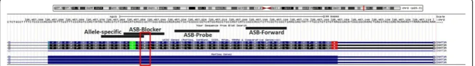
The ASB-PCR assay was designed using Primer3, Oligocalc and UCSC software. We used an allele-specific reverse primer containing the mutational spot (R882H G > A) at its 3′-end. The blocking sequence was developed comple-mentary to the wt allele. The discriminating base was located in the middle of the blocker. To prevent elongation by Taq polymerase, a phosphate group was added to the 3′-end of the blocker. For fluorescence detection, a Taq-Man probe was used (Fig. 1). All primer sequences are listed in Table 1. Further properties, such as melting temperature were developed in accordance with Morlan
et al.[25]. The reaction mixture contained 12.5μl 2 × Ab-solute qPCR Mix (Applied Biosystems), 10 pmol of each forward and reverse allele-specific primer, 40 pmol of ASB-Blocker, 5 pmol of probe, and 30 ng of DNA in a final
reaction volume of 25 μl. Reaction was run at 95 °C for 10 min followed by 40 cycles of denaturation at 95 °C for 20 s and annealing/elongation at 67 °C for 45 s on a Rotor Gene 6000 Real-Time PCR Cycler (Qiagen).
Plasmid preparation
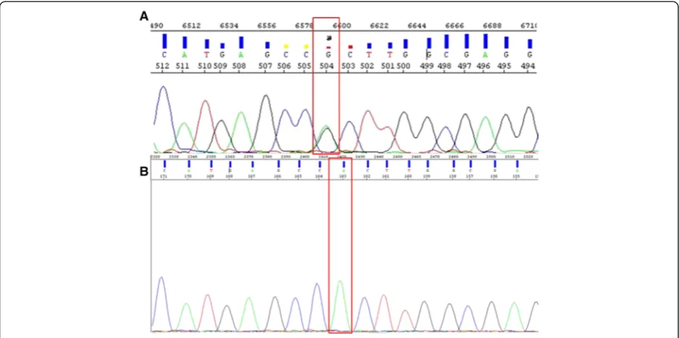
For absolute quantification, a plasmid containing the
DNMT3AR882H G > A mutations was constructed. The template was amplified using ASB-PCR primers, and the fragment size (94 bp) was analyzed on a 1.5 % agarose gel. The plasmid was prepared using the TOPO TA cloning Kit (Invitrogen) and chemically competentE.coli
as per the manufacturer’s instructions. Fragment inser-tion was checked using DNA sequencing with provided M13 forward and reverse primers.
DNA sequencing and endonuclease restriction analysis of
DNMT3Amutations
PCR sequencing reaction was performed as previously described [22]. Amplified products were purified using the PCR Purification Kit (Qiagen) according to the man-ufactures instruction. Sequencing was performed using ABI310 Genetic Analyzer (Applied Biosystems), and data were analyzed using DNA Sequencing Analysis Software v.5.2.0. Endonuclease restriction analysis of DNMT3A
R882H mutation was performed using Fnu4HI (New England Biolabs) as previously reported [22].
Qualitative and quantitative evaluation of mutations
Qualitative evaluation for presence ofNPM1,DNMT3A,
IDH1 and IDH2 mutations were performed by Sanger sequencing using ABI310 Genetic Analyzer (Applied Biosystems) as previously described [22]. FLT3-ITD and
FLT3 D835Y were quantified by fluorescence fragment analysis on a 310 Genetic Analyzer (Applied Biosystems) as previously described [26]. Quantitative analysis of pres-ence NPM1 mutations was performed by real-time PCR using Rotor Gene 6000 Real-Time PCR Cycler (Qiagen). The reaction mixture contained 12.5 μl 2 × Absolute qPCR Mix (Applied Biosystems), 30 pmol of each forward and reverse allele-specific primer, 10 pmol of forward and 6 pmol of reverse HCK probes, 25 pmol of each FAM-MGB and HEX probes, and 50 ng of DNA in a reaction
final volume of 25 μl. Reaction was run at 95 °C for 15 min followed by 45 cycles of denaturation at 95 °C for 15 s and annealing/elongation at 60 °C for 1 min.
Results
ASB-PCR assay performance
At first, we have tested the performance of ASB-PCR by standard PCR amplification and agarose gel electrophoresis.
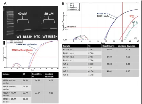
Specificity of ASB-PCR was verified using Sanger sequencing. To determine the optimal blocker concen-tration, PCR was performed with 40μM and 80 μM of blocker, successively. A specific amplification of mu-tated DNA was determined producing a PCR fragment of approximately 94 bp. DNA of the wt sample was also amplified, but with a considerably lower efficiency com-pared to R882H-positive DNA (Fig. 2.ai). No significant
Table 1Oligonucleotides used in this study
Name Sequence Application Tm [°C]
DNTM3A-Ex23F 5′-GTGTGGTTAGACGGCTTCC Sequencing 59.5
DNMT3A-Ex23R 5′-CTCTCCCACCTTTCCTCTG Sequencing 59.5
ASB-F 5′- CAGCGGAGCGAAGAGGTG ASB-PCR 60.8
Allele-specific 5′- CGTCTCCAACATGAGCCA ASB-PCR 56.3
ASB-Blocker 5′- CATGAGCCGCTTGGCGAG-PH ASB-PCR 60.8
ASB-Probe 5′- FAM-CTCCATGACCGGCCCAGCAGTC-BBQ ASB-PCR 69.5
The mutational spot in the allele-specific primer and blocker sequence is bold and punctuated. ASB, indicates Allele-Specific PCR with a Blocking reagent;DNMT3A, indicates DNA methyltransferase 3 gene
difference was seen for PCR amplification with 40 μM and 80μM of blocker. However, the presence of 40μM blocker in the reaction led to detection of R882H around 2 Ct’s earlier compared to the assay without blocker (22.64vs. 24.38; Fig. 2.aii). Therefore, 40μM of blocker were used for further applications. Next, we de-termined the quantitative performance of the assay (Fig. 2.b). The fluorescence signal of mutated samples exceeded the threshold 11–14 cycles earlier as com-pared with wt samples (27.64/27.83 vs. 38.28/41.41). Based on this analysis, we chose a Ct-value of 35 as the
cut-off limit.
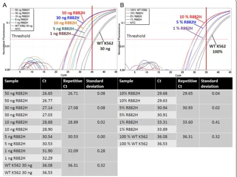
Subsequently, the sensitivity of ASB-PCR was studied. First, a dilution series of genomic DNA was prepared ranging from 50 ng to 1 ng (Fig. 3.a). Differentiation be-tween wt and R882H samples was possible for all dilu-tion steps. One ng of R882H DNA was detected 4 cycles earlier compared with 30 ng of wt DNA (32.09 vs.
36.31). In addition, a dilution series of R882H-positive DNA with wt DNA was analyzed (Fig. 3.b). Discri-mination of 1 % of R882H-positive DNA was possible
exceeding the threshold approximately 3 cycles earlier as the wt DNA (33.60 vs. 36.31). The cut-off limit of Ct=
35 was constant for these analyses. To enhance the ac-curacy of the sensitivity determination of quantitative detection of the mutation, a R882H-positive plasmid was constructed.
Plasmid performance and absolute quantification
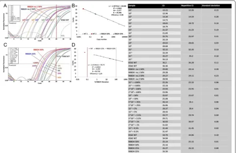
We have generated a plasmid which contains a homozy-gous R882H mutation (Fig. 4). Significant differentiation to the wt DNA was possible up to a dilution of 10−1copies. A copy number of 10−2 exceeded the threshold approxi-mately at the cut-off value of 35 cycles (Fig. 5.a). However, the assay effectiveness was distinct when the plasmid con-taining a homozygous R882H mutation was compared with heterozygous patient samples. Two samples obtained from AML patients at diagnosis containing approximately 50 % ofDNMT3AR882H mutations exceeded the thresh-old at a similar cycle as the 10° dilution of the plasmid (29.14/29.11 vs. 28.65; Fig. 5.b). The initial mutation portion was determined by Sanger sequencing. Since
the absolute quantification was not possible in this set-ting, we performed an absolute quantification by the percentage of mutated allele. Based on the previous measurement, the 10° plasmid dilution was defined to contain 50 % of DNMT3A R882H mutation. Fig. 5.c demonstrates a possibility of reliable quantification of patient samples using this setting. The sample containing 50 % ofDNMT3AR882H was detected at the same cycle as the 100plasmid dilution (25.15 vs.25.07). In addition, the sample with 25 % R882H mutation exceeded the threshold at a similar cycle compared to the 5*10−1 plas-mid controls (26.33 vs. 26.1). The assay displayed a high efficiency between 0.98 and 1.03 (Fig. 5.b and d). The sen-sitivity of assay ranged up to 10−3.
In addition, follow-up samples of one patient were an-alyzed three times to examine the reproducibility of assay results. As shown in Table 2 the standard deviation was≤3.1 %.
Assays concordance
Next, the concordance of different methods for detec-tion ofDNMT3Amutation was examined.
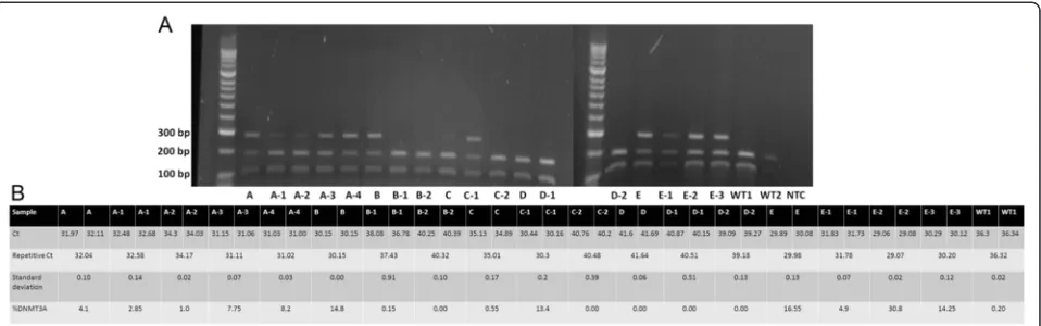
Initially, 25 samples from AML patients were analyzed using DNA sequencing. Next, endonuclease restriction analysis and ASB-PCR were used (Fig. 6). We have pre-viously described that endonuclease restriction analysis has a perfect concordance with Sanger sequencing and is characterized by high sensitivity [22]. Qualitative assessment of band sizes (Fig. 6.a) corresponded to quantitative detection of R882H mutation using
ASB-PCR (Fig. 6.b) in follow up samples from patient A, B, C, und E. Patient D showed noDNMT3AR882H muta-tion using both methods. For example, increasing of
DNMT3A R882H mutation in samples from patient C from 0.55 % to 13.4 % by ASB-PCR assay (Fig. 6.b) matched with a gain of band by endonuclease restriction analysis (Fig. 6.a). A cut-off of Ct = 35 was also applic-able for analysis of follow-up samples. No false negative or positive signals were detected using the endonuclease restriction and ASB-PCR.
Analysis of follow-up samples from AML patients
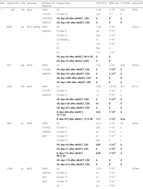
We analyzed 62 follow up constitutive samples obtained from 6 AML patients at diagnosis, after induction, consoli-dation therapy, and after alloSCT. All patients included in the follow-up analysis harbored aNPM1mutation at diag-nosis. This enabled comparison ofDNMT3Astability dur-ing both CR and complete molecular remission (molCR) using the well-established marker for detection of MRD. The main characteristics, karyotype, concurrent muta-tions, percent of DNMT3A mutation, and MRD data of these patients are shown in Table 3. Median age was 55 years (range 43–60 years), and the median survival time by the end of this study was 30 months (range 9–52 months).
from 0.05 % to 4.05 % during therapy. Furthermore, even in CR and molCR according to analysis ofNPM1, a signifi-cant proportion of DNMT3A R882H mutation could be detected. In patients 1, 2, 3, 4, and 5 (Table 3),DNMT3A
mutation was present at diagnosis and in CR after stand-ard therapy. The amounts of mutation were decreased, for example, from 21.45 % to 16.5 % in patient 1 and from 14.95 % to 1.2 % in patient 4. In patient 2 (Table 3), amount of mutated allele was later increased despite the
patients was in CR with slight positive or negative MRD. The percentage of mutated allele was higher (30.8 %) as compared to diagnostic sample (11.95 %). At this time point, the patient exhibited thrombocytopenia in periph-eral blood with normal blasts count in BM. In samples obtained in CR after alloSCT (Patients 1, 2, and 3, Table 3) with complete donor chimerism (CDC),DNMT3A muta-tion was not found. Interestingly, NPM1 mutation was found in very small amount. Overall, in CR no significant Fig. 5Absolute quantification ofDNMT3AR882H mutations. Values of ASB-PCR are listed in the corresponding table.aAnalysis of copy number dilutions of the R882H homozygote plasmid. Patient samples with approximately 50 % of R882H mutated allele are indicated by dotted lines; the 10° plasmid dilutions are pink line.bThe standard curve (copy numbers over cycle). Patient samples with 50 % R882H are shown as a blue diamond and a green triangle. Wt samples are shown as red quadrats. R2= correlation coefficient. B = Intercept with the ordinate. M = Slope of the standard curve.cAbsolute quantification was performed by percentage of mutated allele in a sample. Plasmid concentrations were adjusted to 50 % and 25 % of mutated R882H allele according to the measurement of heterozygote patient samples (dotted lines).dStandard curve (%R882H over cycle). The dotted red line shows the limit of 100 % R882H mutated allele in a sample. Patient samples are shown as a blue diamond and a green triangle. wt samples are shown as red quadrats. R2= correlation coefficienct. B = Intercept with the ordinate. M = Slope of the standard curve
Table 2Reproducibility of ASB-PCR
Sample Run 1 (%R882H) Run 2 (%R882H) Run 3 (%R882H) Standard deviation (%)
187 23.05 21.6 17.19 3.052
187-1 8.2 8.65 8.4 0.225
187-2 3.5 3.5 2.82 0.392
187-3 2.1 1.4 1.07 0.525
187-4 2.35 3.35 1.70 0.831
correlation between NPM1 and DNMT3A was detected.
DNMT3Amutation was not detectable in patients 1, 2, and 3 after alloSCT, in CR with CDC. After alloSCT no
DNMT3AR882H mutation was detected in cases when the patient displayed a complete or high percent of donor cells.
In relapse of AML, significant proportion ofDNMT3A
R882H mutation was detected in all relapsed patients (Table 3). For instance, percentage of R882-positive allele increased significantly in patient 4 at relapse from 0 % to 9.25 % correlating with the molecular relapse detected by the percentage of NPM1 and reduced amount of donor cells (10 %). In patient 6 the decreasing of donor cells from 92 % to 60 % associated with a growing of R882-positive allele from 0 % to 0.45 %. However, per-centage ofNPM1did not increase and a relapse was not diagnosed. This patient underwent match related alloSCT after reduced intensity conditioning, and demonstrated delayed engraftment with slowly growing of donor cells. Notably, in the CR, the majority of patients showed a high level of DNMT3A mutations despite the low or negative rates ofNPM1mutation. In relapse, levels of both muta-tions have been increased.
Discussion
Last years, potential role of DNMT3A mutation for prognosis and outcome of AML has been extensively studied. Several authors have shown a negative impact of DNMT3A mutations on outcomes of AML patient [12, 13, 15, 18, 27–29]. DNMT3A mutation has been shown an independent poor prognostic factor for overall survival and relapse-free survival [15]. Currently, only few methods for the quantitative detection of DNMT3A
R882H mutations are available [19, 30]. Here, we devel-oped an ASB-PCR assay that allows discrimination be-tween wt and mutated DNMT3A and quantification of R882H allele in one reaction. The main feature of this assay is the combination of an allele-specific primer with
a competitive blocker as described by Morlan et al.[25]. Features ofDNMT3AR882H sequence allowed introduc-tion of a purin-purin-mismatch (G-A) to the allele-specific primer leading to its high selectivity. Thus, a significant amplification difference for the wt and mutant allele was seen also without the use of the competitive blocker. Add-ing blocker enhanced selective amplification of R882H-mutated allele that lead to earlier detection of mutation. Furthermore, the maximum Ctdifference between wt and
mutated allele was 14 cycles. Thereby, effectiveness of the blocker is caused by intersection with the allele-specific primer and location of the discriminating base in the mid-dle of the blocker sequence. In contrast, the allele-specific primer includes the discriminating base at its 3′-end.
In addition to the enabling of discrimination between wt and mutant allele, the assay had to facilitate analysis of follow-up samples that contain a low percentage of mutated allele. The high sensitivity of 10−3and the pos-sibility to use low DNA amounts, up to 1 ng, make this approach feasible.
Here, absolute quantification was performed by the percentage of mutated R882H allele in a sample. Since the generated plasmid contained a homozygous R882H mutation the amplification characteristics using ASB-PCR were different as compared to patient samples containing the heterozygous mutation. Possibly, the missing competition between wt and mutant allele dur-ing the amplification of plasmid DNA leads to a more efficient enrichment of mutant R882H generating higher Ct values. Modification of plasmid concentrations
ac-cording to the percentage of mutated allele in the patient samples determined by sequencing enabled a reliable absolute quantification. Alternatively, the generation of an additional plasmid containing the wt allele is possible. Ab-solute quantification can be performed by combining the wt plasmid with different proportions of the plasmid con-taining the mutated allele [31].
Table 3Quantitative analysis ofDNMT3AR882H in follow-up samples from 6 AML patients
Patient Age/Gender FAB Karyotype Mutations at diagnosis
Disease status DNMT3A% NPM1log FLT3Ratio Outcome/OS
1 52/F M4 46,XX NPM1 DS 21,45 1^106 0,72 CR/9+
FLT3-ITD CR after CT 16,4 1^-5 0
FLT3-TKD2 CR, day+28 after alloSCT , CDC 0 0 0 DNMT3A CR, day+195 after alloSCT, CDC 0 0 0
2 60/M M2 46,XY, del(9q) NPM1 DS 11,95 1^105 CR/31+
DNMT3A CR after IT 5,8 1^10-2
CR after CT 0,05 1^10-4
CR, Platelet↓ 0,1 1^10-4
CR 16,5 1^10-5
CR 30,8 0
CR 11,3 1^10-4
CR, day+26 after alloSCT, 98 % DC 0 0
CR, day+73 after alloSCT (CDC) 0 0
3 43/F M4 46,XX NPM1 DS 17,9 1^105 0,78 CR/52+
FLT3-ITD CR, day+563 after alloSCT, CDC 0 1^10-5 0
DNMT3A CR, day+731 after alloSCT, CDC 0 1^10-4 0
CR, day+1082 after alloSCT, CDC 0 0 0
CR, day+1445 after alloSCT, CDC 0 0 0
4 50/M M4 46,XY NPM1 DS 14,95 1,0^105 0,41 R/17+
FLT3-ITD CR after IT 1,6 1^10-4 0
DNMT3A CR after CT 1,2 1^10-5 0
CR, day+40 after alloSCT, CDC) 0 1^10-4 0
CR, day+130 after alloSCT, CDC 0,1 0 0
CR, day+217 after alloSCT, CDC 0 0 0
R, day+306 after alloSCT, 10 % DC
9,25 1^105 0
R, day+373 after alloSCT, 15 % DC 11,7 1^104 0,25
5 50/F 5a 46,XX NPM1 DS 22,15 1^105 0,5 CR/26
FLT3-ITD CR after IT 0,35 1^10-4 0
DNMT3A CR after IT 0,2 1^10-4 0
IDH1 CR after CT 0,1 1^10-4 0
CR after CT 0,05 0 0
CR, day+29 after alloSCT, CDC 0,05 1^10-4 0 CR, day+71 after alloSCT, CDC 0 1^10-5 0 R, day+114 after alloSCT,
90 % DC
0,05 1^10-4 0 CR, day+174 after alloSCT, CDC 0 0 0
CR, day+215 after alloSCT, CDC 0 0 0
6 71/M 5a 46,XY NPM1 DS 12,75 1^104 CR/46+
DNMT3A CR after IT 0,1 1^10-2
IDH1 CR after CT 0,1 1^10-4
IDH2 CR after CT 1,65 1^10-4
For application of ASB-PCR in diagnostic routine the concordance with other analysis methods is important. ASB-PCR displayed a perfect concordance with sequen-cing and endonuclease restriction analysis [22]. This ac-counts for a high specificity of the assay. However, because low amplification of wt samples is present a wt control should be run together with patient samples. Here, the cut-off value of Ct= 35 was applicable in every run but this
could depend on the PCR reagents and machine used for Real-Time quantification.
To demonstrate the need and effectiveness of new established method, we analyzed 62 follow-up samples from 6 AML patients after therapy and alloSCT. More-over, data about the stability ofDNMT3Amutation dur-ing the course of disease are restricted and controversial. Previously published studies have demonstrated that
DNMT3A mutations are stable at relapse [12, 15]. Thol
et al. found that the mutations disappeared at CR and recurred at relapse in one patient [12]. Later, Houet al.
reported persistence ofDNMT3A mutations at CR in 5 patients, which later achieved relapse and died of disease progression. These data could relate the persistence of
DNMT3A mutations and high risk of relapse [18]. Re-cently, Pløenet al.have identified persistence ofDNMT3A
mutations in long-term remission of patients with AML that received cytoreduction or palliative therapy [19]. Using cell-sorting, the authors showed thatDNMT3A mu-tationswere present in T-cells and B-cells at diagnosis in some patients, and also in T-cells several years after diagnosis. The presence of DNMT3Ain both B-cells and T-cells could lead to the assumption that mutation had occurred in an early pre-leukemic stem cell prior to the acquisition of other genetic events, and could be resistant
to chemotherapy [19]. Therefore, further exploration of the role of DNMT3A R882H mutations for the progres-sion of AML disease is needed.
In accordance with previous studies, we found persist-ence of DNMT3A R882H mutations after standard cytoreduction therapy. In most cases the amount of mu-tated allele was lower as compared with the diagnostic sample. But in one case (Patient 2, Table 3) we have found an increasing of DNMT3A R882H allele in CR without relapse signs. Furthermore, NPM1 remained negative indicating potential persistence of a different clone. The permanent presence of DNMT3A R882H after therapy in deep CR could be indicating presence of
DNMT3Amutation in early pre-leukemic stem cells that resist chemotherapy. This could also explain the loss of correlation between NPM1 and DNMT3A because the clonal expansion of pre-leukemic stem cells could ori-ginate independent ofNPM1positive clones [10, 11]. In relapse, all samples showed an increasing of bothNPM1
and DNMT3A mutated alleles. This suggests at least in part the presence of NPM1and DNMT3A mutations in the same cell clone. After alloSCT in patients in CR with complete donor chimerism we have no foundDNMT3A
R882H. This data suggests the removal of leukemic stem cells after alloSCT and indicates the importance of alloSCT for high risk AML patients.
In summary, we developed a rapid, sensitive and specific method for quantitative detection of DNMT3A R882H mutations in AML patients. This assay could be easily ap-plicable for routine screening ofDNMT3AR882 mutation not only at time of diagnosis but also after the treatment. Quantitative detection of DNMT3A R882H mutations at different time points of AML disease enables screening of
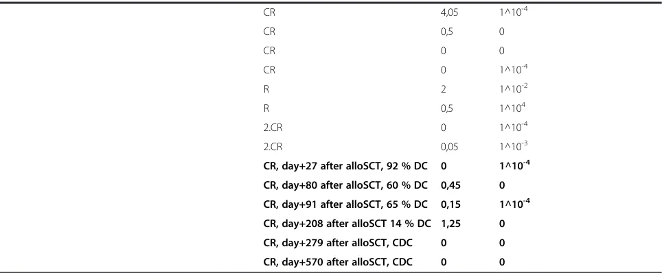
Table 3Quantitative analysis ofDNMT3AR882H in follow-up samples from 6 AML patients(Continued)
CR 4,05 1^10-4
CR 0,5 0
CR 0 0
CR 0 1^10-4
R 2 1^10-2
R 0,5 1^104
2.CR 0 1^10-4
2.CR 0,05 1^10-3
CR, day+27 after alloSCT, 92 % DC 0 1^10-4
CR, day+80 after alloSCT, 60 % DC 0,45 0
CR, day+91 after alloSCT, 65 % DC 0,15 1^10-4
CR, day+208 after alloSCT 14 % DC 1,25 0
CR, day+279 after alloSCT, CDC 0 0
CR, day+570 after alloSCT, CDC 0 0
FABFrench–American–British classification systems,DSprimary diagnosis,ITinduction therapy,CTconsolidation therapy,CRcomplete remission,RRelapse, alloSCTallogeneic stem cell transplantation,CDCcomplete donor chimerism,DCdonor cells
follow-up samples. This could assist to evaluate response to therapy and provide additional information about the role ofDNMT3Amutations in development and progres-sion of AML.
Competing interests
The authors declare that they have no competing interests.
Authors’contributions
RB designed and carried out the molecular genetic studies, drafted the manuscript. IWB designed research and revised the manuscript. NS carried out the molecular genetic studies. CB, AP, and BD participated in the design of study and revised the manuscript. OB designed research, carried out the sequencing analyses and revised the manuscript. All authors read and approved the final manuscript.
Acknowledgment
This work was supported by the Stefan-Morsch-Stiftung for Leukemia Tumour Patients.
Received: 25 February 2015 Accepted: 12 May 2015
References
1. Marcucci G, Metzeler KH, Schwind S, Becker H, Maharry K, Mrozek K, et al. Age-related prognostic impact of different types of DNMT3A mutations in adults with primary cytogenetically normal acute myeloid leukemia. J Clin Oncol. 2012;30(7):742–50.
2. Ley TJ, Ding L, Walter MJ, McLellan MD, Lamprecht T, Larson DE, et al. DNMT3A mutations in acute myeloid leukemia. N Engl J Med. 2010;363(25):2424–33.
3. Zhang Y, Chen FQ, Sun YH, Zhou SY, Li TY, Chen R. Effects of DNMT1 silencing on malignant phenotype and methylated gene expression in cervical cancer cells. J Exp Clin Cancer Res. 2011;30:98.
4. Jasielec J, Saloura V, Godley LA. The mechanistic role of DNA methylation in myeloid leukemogenesis. Leukemia. 2014;28(9):1765–73.
5. Li KK, Luo LF, Shen Y, Xu J, Chen Z, Chen SJ. DNA methyltransferases in hematologic malignancies. Semin Hematol. 2013;50(1):48–60.
6. O’Brien EC, Brewin J, Chevassut T. DNMT3A: the DioNysian MonsTer of acute myeloid leukaemia. Ther Adv Hematol. 2014;5(6):187–96.
7. Holz-Schietinger C, Matje DM, Reich NO. Mutations in DNA
methyltransferase (DNMT3A) observed in acute myeloid leukemia patients disrupt processive methylation. J Biol Chem. 2012;287(37):30941–51. 8. Russler-Germain DA, Spencer DH, Young MA, Lamprecht TL, Miller CA,
Fulton R, et al. The R882H DNMT3A mutation associated with AML dominantly inhibits wild-type DNMT3A by blocking its ability to form active tetramers. Cancer Cell. 2014;25(4):442–54.
9. McDevitt MA. Clinical applications of epigenetic markers and epigenetic profiling in myeloid malignancies. Semin Oncol. 2012;39(1):109–22. 10. Shlush LI, Zandi S, Mitchell A, Chen WC, Brandwein JM, Gupta V, et al.
Identification of pre-leukaemic haematopoietic stem cells in acute leukaemia. Nature. 2014;506(7488):328–33.
11. Corces-Zimmerman MR, Hong WJ, Weissman IL, Medeiros BC, Majeti R. Preleukemic mutations in human acute myeloid leukemia affect epigenetic regulators and persist in remission. Proc Natl Acad Sci U S A. 2014;111(7):2548–53. 12. Thol F, Damm F, Ludeking A, Winschel C, Wagner K, Morgan M, et al.
Incidence and prognostic influence of DNMT3A mutations in acute myeloid leukemia. J Clin Oncol. 2011;29(21):2889–96.
13. Ribeiro AF, Pratcorona M, Erpelinck-Verschueren C, Rockova V, Sanders M, Abbas S, et al. Mutant DNMT3A: a marker of poor prognosis in acute myeloid leukemia. Blood. 2012;119(24):5824–31.
14. Ibrahem L, Mahfouz R, Elhelw L, Abdsalam EM, Soliman R. Prognostic significance of DNMT3A mutations in patients with acute myeloid leukemia. Blood Cells Mol Dis. 2015;54(1):84–9.
15. Im AP, Sehgal AR, Carroll MP, Smith BD, Tefferi A, Johnson DE, et al. DNMT3A and IDH mutations in acute myeloid leukemia and other myeloid malignancies: associations with prognosis and potential treatment strategies. Leukemia. 2014;28(9):1774–83.
16. Shivarov V, Gueorguieva R, Stoimenov A, Tiu R. DNMT3A mutation is a poor prognosis biomarker in AML: results of a meta-analysis of 4500 AML patients. Leuk Res. 2013;37(11):1445–50.
17. Wakita S, Yamaguchi H, Omori I, Terada K, Ueda T, Manabe E, et al. Mutations of the epigenetics-modifying gene (DNMT3a, TET2, IDH1/2) at diagnosis may induce FLT3-ITD at relapse in de novo acute myeloid leukemia. Leukemia. 2013;27(5):1044–52.
18. Hou HA, Kuo YY, Liu CY, Chou WC, Lee MC, Chen CY, et al. DNMT3A mutations in acute myeloid leukemia: stability during disease evolution and clinical implications. Blood. 2012;119(2):559–68.
19. Ploen GG, Nederby L, Guldberg P, Hansen M, Ebbesen LH, Jensen UB, et al. Persistence of DNMT3A mutations at long-term remission in adult patients with AML. Br J Haematol. 2014;167(4):478–86.
20. Genovese G, Kahler AK, Handsaker RE, Lindberg J, Rose SA, Bakhoum SF, et al. Clonal hematopoiesis and blood-cancer risk inferred from blood DNA sequence. N Engl J Med. 2014;371(26):2477–87.
21. Jaiswal S, Fontanillas P, Flannick J, Manning A, Grauman PV, Mar BG, et al. Age-related clonal hematopoiesis associated with adverse outcomes. N Engl J Med. 2014;371(26):2488–98.
22. Berenstein R, Blau IW, Kar A, Cay R, Sindram A, Seide C, et al. Comparative examination of various PCR-based methods for DNMT3A and IDH1/2 mutations identification in acute myeloid leukemia. J Exp Clin Cancer Res. 2014;33:44. 23. Vardiman JW, Thiele J, Arber DA, Brunning RD, Borowitz MJ, Porwit A, et al.
The 2008 revision of the World Health Organization (WHO) classification of myeloid neoplasms and acute leukemia: rationale and important changes. Blood. 2009;114(5):937–51.
24. Jaatinen T, Laine J. Isolation of mononuclear cells from human cord blood by Ficoll-Paque density gradient. Curr Protoc Stem Cell Biol. 2007;Chapter 2:Unit 2A.1. doi:10.1002/9780470151808.sc02a01s1.
25. Morlan J, Baker J, Sinicropi D. Mutation detection by real-time PCR: a simple, robust and highly selective method. PLoS ONE. 2009;4(2), e4584.
26. Blau O, Berenstein R, Sindram A, Blau IW. Molecular analysis of different FLT3-ITD mutations in acute myeloid leukemia. Leuk Lymphoma. 2013;54(1):145–52. 27. Lu Q, Chen Y, Wang H, Li Z. DNMT3A mutations and clinical features in
Chinese patients with acute myeloid leukemia. Cancer Cell Int. 2013;13(1):1. 28. Markova J, Michkova P, Burckova K, Brezinova J, Michalova K, Dohnalova A,
et al. Prognostic impact of DNMT3A mutations in patients with intermediate cytogenetic risk profile acute myeloid leukemia. Eur J Haematol. 2012;88(2):128–35.
29. Schwarz J, Markova J. DNMT3A mutations in AML: a new prognostic factor? Leuk Res. 2013;37(11):1432–3.
30. Shivarov V, Ivanova M, Naumova E. Rapid detection of DNMT3A R882 mutations in hematologic malignancies using a novel bead-based suspension assay with BNA(NC) probes. PLoS ONE. 2014;9(6), e99769.
31. Szankasi P, Reading NS, Vaughn CP, Prchal JT, Bahler DW, Kelley TW. A quantitative allele-specific PCR test for the BRAF V600E mutation using a single heterozygous control plasmid for quantitation: a model for qPCR testing without standard curves. J Mol Diagn. 2013;15(2):248–54.
Submit your next manuscript to BioMed Central and take full advantage of:
• Convenient online submission
• Thorough peer review
• No space constraints or color figure charges
• Immediate publication on acceptance
• Inclusion in PubMed, CAS, Scopus and Google Scholar
• Research which is freely available for redistribution