Tanaffos (2011) 10(1), 12-18
©2011 NRITLD, National Research Institute of Tuberculosis and Lung Disease, Iran
Evaluation of Polymerase Chain Reaction for Diagnosis of
“Tuberculous Pleurisy”
Amir M.H. Asnaashari 1, Mohammad Towhidi 1, Reza Farid 2, Mohammad R.Abbaszadegan 2,3, Davood Attaran 1, Seiedeh Sedigheh Fatemi 1, Atefeh Amiri Darban 1
1 Research Center of Pulmonary Diseases, Department of Pulmonology, 2 Research Center of Immunology, Ghaem Hospital, Mashhad University of Medical Sciences (MUMS), 3 Pardis Clinical Laboratory Center, MASHHAD-IRAN.
ABSTRACT
Background: Differential diagnosis between tuberculous pleurisy (TBP) and non- tuberculosis pleural effusion represents a
critically important clinical problem. In recent years, several noninvasive methods have been found for diagnosis of
tuberculous pleurisy. This study aimed to evaluate the value of detection of the genome of Mycobacterium tuberculosis
(MTB) by polymerase chain reaction (PCR) method for the diagnosis of tuberculous pleurisy and compare the results with
those of conventional methods.
Materials and Methods: In this cross-sectional study, we studied 62 patients (42 men and 20 women) with pleural effusion
in Ghaem Hospital, affiliated to Mashhad University of Medical Sciences from January 2006 to June 2007.
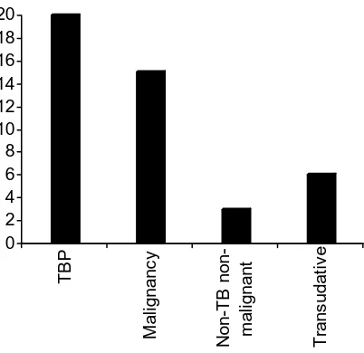
Results: A total of 20 patients had tuberculous pleurisy (45.4%), 15 patients had malignant pleural effusion (34%), 3 patients
had pleural effusion with various “non-tuberculosis non-malignant” etiologies (6.8%) and 6 patients had transudative pleural
effusion (13.6). The sensitivity, specificity, positive predictive value and negative predictive value of PCR in tuberculous
pleurisy were 85%, 100%, 100% and 88.8%, respectively.
Conclusion: The value of PCR test and pleural biopsy was similar in the diagnosis of TBP. However, PCR detected MTB in
pleural effusion when conventional pleural biopsy failed to do so. (Tanaffos2011; 10(1): 12-18)
Key words: Tuberculosis, Polymerase chain reaction, Pleurisy, Diagnosis
INTRODUCTION
Tuberculosis (TB) is a major cause of morbidity and mortality worldwide, infecting approximately one third of the world's population (1). WHO
Correspondence to: Asnaashari A MH
Address: Research center of pulmonary diseases, Department of pulmonology, Ghaem hospital, Mashhad University of Medical Sciences (MUMS), Mashhad, Iran.
Email address: asnaashariam@mums.ac.ir Received: 3 July 2010
Accepted: 1 December 2010
TBP occurs not only as a result of a delayed hypersensitivity reaction to Mycobacterium tuberculosis (MTB) antigens but also due to MTB entering the pleural space. Increased vascular permeability and decreased protein clearance cause pleural effusion (7). TBP is almost always exudative and lymphocyte- predominant and it is important to differentiate it from other causes of exudative and lymphocyte- predominant pleural effusions.
The diagnosis of TBP is not always easy:
• PPD test is negative in 30% of patients during the first visit, although a positive test does not support the diagnosis due to the high prevalence of tuberculosis in Iran and unknown results of BCG vaccination in newborns.
• Direct smear of pleural fluid has limited diagnostic value because of the need for at least 10,000 / ml of organisms (8). Therefore, in the best case scenario, it is positive in less than 10% of cases (9-14). Culture takes 6 weeks but only needs 10-100 live tubercle bacilli. As a result, pleural fluid culture is a more sensitive method, but the diagnostic value of culture has reported to be 12-70% in the most precise studies (10). • Granuloma is found in 50 to 97% of patients and
culture of tissue samples can be diagnostic in 30 - 80% of cases (8, 10-13, 15-17). Thus, the diagnostic value is not 100% and we should keep it in mind that needle biopsy is an invasive procedure occasionally associated with complications such as pneumothorax and hemothorax.
• Sputum smear and culture have limited diagnostic values for TBP (18).
Therefore, we need a non-invasive diagnostic method that only requires pleural fluid to detect TBP. This study aimed to evaluate the diagnostic value of detecting the genome of MTB by polymerase chain reaction (PCR) in pleural fluid for the diagnosis of TBP.
MATERIALS AND METHODS
This cross-sectional study was performed from January 2006 to June 2007 aiming to assess the value of PCR in the diagnosis of TBP. A total of 62 patients with clinical and radiological diagnosis of pleural effusion referred to the respiratory clinic or admitted to the respiratory ward of Ghaem University Hospital, affiliated to Mashhad University of Medical Sciences, enrolled in this study. An informed consent was obtained from all patients and the Ethics Committee of Ghaem Hospital approved this study. Clinical signs and symptoms, demographic data, and radiological results were all recorded.
Study method:
• Exudative pleural effusion
Plus one or more of the following findings:
• Granuloma in pleural biopsy and ruling out other causes of granulomatous pleuritis by clinical examination (for Rheumatoid Arthritis) and radiological findings (for Sarcoidosis)
• Finding acid-fast positive bacilli in pleural fluid ,sputum, or bronchoalveolar lavage(BAL)
• Positive culture for MTB in “pleural fluid”, “sputum”, or “BAL”
Plus
• Appropriate and acceptable clinical response to anti-TB treatment
DNA Extraction and PCR
Enzymatic DNA extraction was performed for pleural effusion as previously reported (19-21). Briefly, 5 ml of pleural sample was collected in 500 µl of 10% EDTA (1 mg/ml). For samples with high viscosity, equal volume of NaOH 4% was added and vortex for homogenizing. Samples were incubated for 30 min in a 37 ˚C water bath and centrifuged for 30 min at 1,500 rpm. The supernatant was completely discarded. Then the pH was adjusted to 7.5. Equal volumes of concentrated pleural fluid samples were lysed in tris-ethylenediaminetetraacetic acid buffer (Tris-Cl, 1 M; ethylenediaminetetraacetic acid, 0.5mM; pH, 8.0) and 10% sodium dodecyl sulfate, in the presence of 5µl of proteinase K and 1µl of glycogen and then incubated in a 55 °C water bath for 2 h. The DNA was purified in phenol-chloroform and then precipitated with ethanol at -20°C overnight.
For non-viscous pleural samples, the sample was centrifuged at 3000 rpm for 20 min. Supernatant was transferred into a clean microfuge tube and centrifuged to collect the pellet.
For increased sensitivity in molecular testing of
TB, a nested PCR reaction was performed (22). A PCR reaction solution was prepared by mixing 15ul of a previously mixed reaction mixture (1 × PCR buffer, 1.5 mM MgCl2, 200µM each of deoxynucleoside triphosphates, 0.5 mM of each forward primer and reverse primer, 0.5 U of Taq DNA polymerase enzyme and 10µl of the target DNA to create a total of 25µl). Then, the thermal cycler (Techne, England) was programmed for 30 cycles with initial denaturation at 94°C for 5 min, denaturation at 94°C for 45 sec, annealing at 56°C for 45 sec, extension at 72°C for 45 sec, and final extension at 72°C for 5 min.
For the second PCR reaction, 1µl of the amplified products from the first PCR was used as template and mixed with 24µ1 of a freshly prepared reaction mixture. This was followed by the same procedures used to obtain the first PCR product, but annealing was performed at 50°C for 30 sec.
RESULTS
We studied 62 patients, 42 men and 20 women, with a mean age of 58.6 years (range 16 to 100 years). Fifty six patients had exudative (90.3%) and 6 cases had transudative pleural effusion (%9.7).
Fifteen patients with exudative pleural effusion were excluded from the study because we did not find any specific diagnosis or we were unable to follow them.
development of cancer in the lungs or other organs during the 12 months follow-up after the completion of anti TB treatment.
Therefore, these 3 patients were considered as having TBP. However, we excluded them from the statistical analysis because they did not match the inclusion criteria. PCR was negative in all 3 patients.
Accordingly, patients were divided into 4 categories (Figure 1):
1) Tuberculosis pleural effusion (TBP): 20 cases (45.4%)
2) Malignant pleural effusion: 15 cases (34%) 3) Non-malignant non-tuberculosis, exudative pleural effusion with definite diagnosis: 3 cases (6.8%)
4) Transudative pleural effusion: 6 cases (13.6%)
Figure 1. Distribution of patients in 4 categories
Group 1: Patients with TBP:
The diagnosis of PTB was confirmed in 20 patients (45.4% of patients). The mean age of patients was 60 years (range 18 to 70 years). There were 16 males and 7 females.
• Granuloma was found in the pleural biopsy of 17 patients (74%) which in one case was associated
with caseous necrosis. In one of these patients, MTB was found by Zheil Neelson staining and culture of pleural fluid (4% of patients with TBP). Sputum smear was positive in 2 cases in this group. Therefore, only 15 patients were diagnosed solely by pleural biopsy.
• In 5 patients in this group, sputum or BAL was reported positive for MTB. As mentioned above, pleural biopsy was diagnostic in only 2 of these patients. In the other 2, the diagnosis of TBP was only based on sputum smear and culture. In the fifth patient sputum was negative, pleural biopsy was not diagnostic and the diagnosis of TBP was based on bronchoalveolar lavage (BAL). Culture was positive for MTB in all 5 patients.
PCR test results in patients with TBP:
In this study, 20 patients had TBP based on standard criteria:
• The PCR test was positive in 17 patients (% 85) and negative in 3 patients.
• In all 5 patients with positive sputum or BAL, pleural fluid PCR was also positive.
• PCR was positive in only one patient whose pleural fluid smear and culture were positive as well.
• Among 15 patients who were considered as having TBP only based on finding granuloma in their pleural biopsy, pleural fluid PCR was reported positive in 12 of them (82.3%)
The total number of patients with TBP was 20 people (Table 1).
− Pleural biopsy showed granuloma in 17 cases (85%)
− PCR test was positive in pleural fluid of 17cases (% 85)
− Positive sputum smear or BAL in 5 cases (%21.7)
− Positive smear and culture of pleural fluid in 1 patient (4%)
0 2 4 6 8 10 12 14 16 18 20
TB
P
M
a
li
gnanc
y
N
on-T
B
non-m
a
lig
nant
T
rans
udat
iv
Table 1. Evaluation of the value of different diagnostic methods for the diagnosis of pleural tuberculosis
Diagnostic Total Percent
Granuloma in pleural biopsy 17 Sensitivity = 85%, Specificity = 100% PPV = 100%, NPV = 88.8%
Positive PCR of pleural fluid 17 Sensitivity = 85%, Specificity = 100%, PPV = 100%, NPV = 88.8%
BK in sputum and/or BAL 5 Sensitivity = 25%, Specificity = 100%, PPV = 100%, NPV = 61.5%
BK in pleural fluid + positive culture 1 Sensitivity = 5%, Specificity = 100%, PPV =100%, NPV = 55.8%
Group 2: Patients with malignant pleural effusion: Definitive diagnosis of malignant pleural effusion was made in 15 patients. The mean age of these patients was 64.7 years (range 42 to 100 years). They were 60% males and 40% females.
In 11 patients the diagnosis was solely based on finding malignant cells in pleural tissue biopsy (73%).
In 1 patient the diagnosis was solely based on finding malignant cells (6%) in pleural fluid.
In 2 patients, the diagnosis of malignancy was based on lymph node biopsy
In 1 patient the diagnosis was made based on finding malignant cells in bronchial biopsy during bronchoscopy (6%).
In 2 patients despite the malignant pleural biopsy, sputum study was positive for BK.
Pleural fluid study showed that all these patients except one had exudative pleural effusion. Pleural biopsy in this patient was normal but the diagnosis of malignancy was based on bronchial biopsy. Cytopathologic examination of pleural fluid showed malignant cells only in 26% of patients.
Pleural fluid PCR in patients with malignant pleural effusion:
In both patients with biopsy proven malignant pleural effusion, BK was also found in their sputum and PCR of their pleural fluid was positive. Therefore, presence of both cancer and tuberculosis was confirmed in these two patients.
In other patients with malignant pleural effusion, PCR was negative.
Group 3: Patients with transudative pleural effusion:
Pleural effusion was transudative in 6 patients. The mean age of these patients was 55.2 years (range 41 to 80 years).
Heart failure, chronic renal failure, nephrotic syndrome and acute eosinophilic pleurisy due to nitrofurantoin were considered as the causes of pleural effusion in these patients. PCR was negative in all of them.
Group 4: Patients with exudative, non-TB, non malignant pleural effusion with specific diagnosis: There were 3 patients in this group:
− Pleural effusion after esophageal surgery
− Acute polyserositis
− Post CABG (coronary artery bypass graft) pleural effusion
The study did not prove tuberculosis or malignancy in these patients.
PCR test was negative in all these patients.
DISCUSSION
In our Study, the mean age of patients with TBP was 60 years which was more than that of similar studies like 56 years in a study by Epstein et al. (11) and 28 years in a study by Aho et al.(23).
is probably due to different causes of TBP in the areas with high prevalence of TB like Iran, which is hypersensitivity to the organism rather than BK entering the pleural space. The other etiology may be the use of conventional methods for culture. We know that inoculation of pleural fluid and using liquid culture media or BACTEC system can improve the sensitivity of mycobacterial culture (24).
Diagnostic value of pleural biopsy is not 100% .In other studies granuloma was found in 50-97% of cases (8,10-13,15-17). In our study, pleural biopsy showed granuloma in 85% of patients. In 3 patients, despite the absence of granuloma in pleural biopsy, definite diagnosis of TBP was made by sputum and BAL which shows the inadequate value of pleural biopsy in the diagnosis of TBP. Traditionally, sputum smear is done only in people with abnormal chest radiography, but recently the sputum examination of patients with TBP even with normal radiography was performed and interesting results were obtained (18). In our study, sputum examination was done only in those with abnormal chest x-ray. In 4 patients sputum was reported positive for Bk. Interestingly, pleural biopsy showed granuloma in only 2 of these patients. In the other 2 patients, biopsy showed malignant pleural effusion. PCR was positive in all these 4 patients; thus, we ruled out the probability of a false positive test.
The value of PCR test and pleural biopsy was completely similar in the diagnosis of TBP and both tests were associated with a significant percentage of false negative results.
PCR test was positive in 14 out of 17 patients who had granuloma in biopsy (82.3%); probably PCR would have been positive for all patients if we had also used other sequences rather than IS1160.A review article published in 2007 showed that studies that used IS6110 insertion sequence had sensitivity
from 31% to 81%. The sensitivity of PCR in our study was 85% (24).
We did not culture the pleural biopsy specimens. If we had, it might have increased the diagnostic yield of pleural biopsy (24).
Positive PCR test in the presence of BK in the lungs (BK positive sputum) and in the absence of granuloma, confirmed that the PCR test in cases with non-diagnostic biopsy can lead to TB diagnosis.
In 5 of our study patients, sputum, BAL or pleural fluid was positive for MTB. As mentioned above, pleural biopsy was diagnostic only in 2 of these patients, but PCR was positive in all of them.
Positive PCR test in the presence of a pleural biopsy that confirmed malignancy showed that PCR test can help in the diagnosis of accompanying TB in these patients. Similar to our study, Czystowska recently reported a patient with carcinomatous cells and positive PCR of pleural effusion (25).
CONCLUSION
The diagnostic value of PCR test and pleural biopsy was similar in the diagnosis of TBP but PCR detected MTB in pleural effusion when conventional pleural biopsy failed to do so. We recommend using PCR in all patients suspected of having tuberculous pleurisy if the pleural biopsy is not diagnostic in an endemic area for tuberculosis like Iran.
REFERENCES
1. Song CH, Lee JS, Nam HH, Kim JM, Suhr JW, Jung SS, Na MJ, Paik TH, Kim HJ, Park JK, Jo EK. IL-18 production in human pulmonary and pleural tuberculosis. Scand J
Immunol 2002; 56 (6): 611- 8.
2. Tuberculosis control: Surveillance, planning and financing. Geneva, Switzerland: World Health Organization, 2006; 242 3. Dye C. Global epidemiology of tuberculosis. Lancet 2006;
367 (9514): 938- 40.
5. Ferrer Sancho J. Pleural tuberculosis: incidence, pathogenesis, diagnosis, and treatment. Curr Opin Pulm
Med 1996; 2 (4): 327- 34.
6. Aoe K, Hiraki A, Murakami T, Eda R, Maeda T, Sugi K, et al. Diagnostic significance of interferon-gamma in tuberculous pleural effusions. Chest 2003; 123 (3): 740- 4. 7. Stead WW, Eichenholz A, Stauss HK. Operative and
pathologic findings in twenty-four patients with syndrome of idiopathic pleurisy with effusion, presumably tuberculous.
Am Rev Tuberc 1955; 71 (4): 473- 502.
8. Escudero Bueno C, García Clemente M, Cuesta Castro B, Molinos Martín L, Rodríguez Ramos S, González Panizo A, et al. Cytologic and bacteriologic analysis of fluid and pleural biopsy specimens with Cope's needle. Study of 414 patients. Arch Intern Med 1990; 150 (6): 1190- 4.
9. Sharma SK, Mohan A. Extrapulmonary tuberculosis. Indian
J Med Res 2004; 120 (4): 316- 53.
10.Berger HW, Mejia E. Tuberculous pleurisy. Chest 1973; 63 (1): 88- 92.
11.Epstein DM, Kline LR, Albelda SM, Miller WT. Tuberculous pleural effusions. Chest 1987; 91 (1): 106- 9. 12.Valdés L, Alvarez D, San José E, Penela P, Valle JM,
García-Pazos JM, et al. Tuberculous pleurisy: a study of 254 patients. Arch Intern Med 1998; 158 (18): 2017- 21.
13..Aggarwal AN, Gupta D, Jindal SK. Diagnosis of tuberculosis pleural effusion. Indian J Chest Dis 1999; 41: 89- 100.
14.Sibley JC. A study of 200 cases of tuberculous pleurisy with effusion. Am Rev Tuberc 1950; 62 (3): 314- 23.
15..Seibert AF, Haynes J Jr, Middleton R, Bass JB Jr. Tuberculous pleural effusion. Twenty-year experience. Chest 1991; 99 (4): 883- 6.
16.Kumar S, Seshadri MS, Koshi G, John TJ. Diagnosing tuberculous pleural effusion: comparative sensitivity of mycobacterial culture and histopathology. Br Med J (Clin
Res Ed) 1981; 283 (6283): 20.
17.Prakash UB, Reiman HM. Comparison of needle biopsy with cytologic analysis for the evaluation of pleural effusion: analysis of 414 cases. Mayo Clin Proc 1985; 60 (3): 158- 64.
18.Rossi GA, Balbi B, Manca F. Tuberculous pleural effusions. Evidence for selective presence of PPD-specific T-lymphocytes at site of inflammation in the early phase of the
infection. Am Rev Respir Dis 1987; 136 (3): 575- 9.
19.Villegas MV, Labrada LA, Saravia NG. Evaluation of polymerase chain reaction, adenosine deaminase, and
interferon-gamma in pleural fluid for the differential diagnosis of pleural tuberculosis. Chest 2000; 118 (5): 1355- 64.
20.Nagesh BS, Sehgal S, Jindal SK, Arora SK. Evaluation of polymerase chain reaction for detection of Mycobacterium tuberculosis in pleural fluid. Chest 2001; 119 (6): 1737- 41.
21.Folgueira L, Delgado R, Palenque E, Noriega AR. Detection of Mycobacterium tuberculosis DNA in clinical samples by using a simple lysis method and polymerase chain reaction. J
Clin Microbiol 1993; 31 (4): 1019- 21.
22.Yuen KY, Yam WC, Wong LP, Seto WH. Comparison of two automated DNA amplification systems with a manual
one-tube nested PCR assay for diagnosis of pulmonary tuberculosis. J Clin Microbiol 1997; 35 (6): 1385- 9.
23.Aho K, Brander E, Pätiälä J. Studies of primary drug
resistance in tuberculous pleurisy. Scand J Respir Dis Suppl 1968; 63: 111- 4.
24..Gopi A, Madhavan SM, Sharma SK, Sahn SA. Diagnosis
and treatment of tuberculous pleural effusion in 2006. Chest 2007; 131 (3): 880- 9.
25.Czystowska M, Stokłosa A, Szczepulska-Wójcik E,
Maszkowska-Kopij K, Opoka L, Skoczylas A, et al. Simultaneous detection of tumor cells and the positive result of genetic test for Mycobacterium tuberculosis in pleural