P R E L I M I N A R Y C O M M U N I C A T I O N
Open Access
Filming a live cell by scanning electrochemical
microscopy: label-free imaging of the dynamic
morphology in real time
Michelle Meng-Ni Zhang
1,2, Yi-Tao Long
1*and Zhifeng Ding
2*Abstract
The morphology of a live cell reflects the organization of the cytoskeleton and the healthy status of the cell. We established a label-free platform for monitoring the changing morphology of live cells in real time based on scanning electrochemical microscopy (SECM). The dynamic morphology of a live human bladder cancer cell (T24) was revealed by time-lapse SECM with dissolved oxygen in the medium solution as the redox mediator. Detailed local movements of cell membrane were presented by time-lapse cross section lines extracted from time-lapse SECM. Vivid dynamic morphology is presented by a movie made of time-lapse SECM images. The morphological change of the T24 cell by non-physiological temperature is in consistence with the morphological feature of early apoptosis. To obtain dynamic cellular morphology with other methods is difficult. The non-invasive nature of SECM combined with high resolution realized filming the movements of live cells.
Keywords:Label-free, Scanning electrochemical microscopy, Dynamic morphology, Human bladder cancer cell
Findings
The morphology of single live cells reflects the organiza-tion of the cytoskeleton and the healthy status of the cell [1]. Monitoring the morphological changes of live cells may provide dynamic information of the cell attachment or cytoskeleton organization [1]. The most commonly used cell imaging method is fluorescent microscopy, which is extremely sensitive and allows the visualization of particular structure or compounds inside the cells [2,3]. On the other hand, the preparation of the specimen is relatively time-consuming, and the live cells are sensitive to photo-damage [3]. As an imaging method with high spatial resolution, atomic force micro-scopy (AFM) avoids staining process and photo-damage. However, its cantilever tip mechanically damages the soft cells thus is not suitable for time-lapse experiments [4]. As a non-invasive single-cell analysis method, scan-ning electrochemical microscopy (SECM) has been
successfully applied to live cell-imaging due to its high temporal and spatial resolution [5-13]. This technique is based on the measurement of the electrochemical cur-rent flowing through the SECM tip, which is usually an ultramicroelectrode (UME). The current detected at the tip is dependent on the separation space between the tip and the cell, therefore morphological information of the live cell can be revealed by the electrochemically mapped images [7-9,11]. Compared to fluorescent microscopy, sample preparation of SECM is simple without any staining or labeling procedure. Unlike AFM, the SECM probe does not need to touch the cell, thus it can carry on time-lapse measurement without mechani-cally scratching the cell. Nevertheless, most SECM ima-ging experiments were conducted with the addition of a certain redox mediator, which is usually non- physiolo-gic and undesired [7-9,11,12]. In our previous study [14], we found that dissolved oxygen in the medium solution could be detected by SECM, which provides an opportunity of label-free imaging cellular morphology using dissolved oxygen as the redox mediator. Bladder cancer is the fourth most common cancer of men and the eighth most common cancer of women [15]. Like most of cancers, bladder cancer begins with the * Correspondence: ytlong@ecust.edu.cn; zfding@uwo.ca
1State Key Laboratory of Bioreactor Engineering and Department of
Chemistry, East China University of Science and Technology, 130 Meilong Road, Shanghai, China 200237
2
Department of Chemistry, The University of Western Ontario, 1151 Richmond Street, London, ON, Canada N6A 5B7
Full list of author information is available at the end of the article
mutation of one single cell [16,17]. Investigations of bladder cancer, especially the interaction with anti-can-cer drugs, at the single cell level can provide new insight into its physiology, pathology and pharmacology, and promote the development of chemotherapy in response to single-cell behaviours [18]. Herein, the real-time mor-phological changes of single live T24 cells under non-physiological temperature are revealed by time-lapse SECM with dissolved oxygen as the indicator. While the reactive oxygen species (ROS) released by live cells may interfere with the detection of dissolved oxygen [12,14,19], we determined that under physiological con-ditions oxygen can be reduced at -0.455 V and hydrogen peroxide can be reduced at -0.745 V, while superoxide is oxidized at +0.055 V. Thus in this research the potential was set at -0.500 V to reduce the dissolved oxygen. We also found that the resting status when the T24 cells do not release ROS can last for up to 5 h [20], which sus-tains imaging T24 cells with only dissolved oxygen.
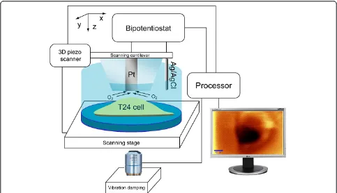
The typical SECM setup is illustrated in Figure 1. The changing morphology of a T24 cell under room temperature was investigated by time-lapse SECM images scanned at about 0.8 μm above the nucleus of the cell with a 5 μm diameter Pt UME biased at -0.500 V vs. Ag/AgCl. (Figure 2b, d and 3) for oxygen reduc-tion [14,21]. The distance between the UME and the
cell was calibrated with ferrocenemethanol after the time-lapse SECM experiment [12].
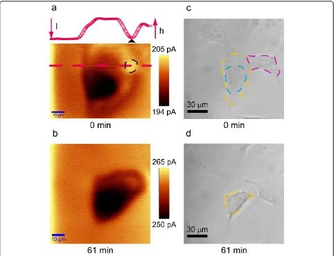
At -0.500 V vs. Ag/AgCl, the dissolved oxygen can be reduced at the diffusion controlled rate at the Pt surface of the UME [14,21]. The time-lapse SECM images in Figure 2 present negative feedback as the current over the T24 cell is lower than the background current, which is the current measured over the Petri dish bot-tom. This indicates the cell is not releasing anything redox active, and the current flowing through the UME only results from the reduction of dissolved oxygen in medium solution [20]. The T24 cell blocks the dissolved oxygen from diffusing to the Pt surface of the UME (Figure 1), resulting in decreased current over the T24 cell, which is called negative feedback [22,23] in SECM (Figure 2b, d and 3).
In comparison of the SECM images and the 50 × opti-cal micrographs obtained with the same T24 cell (Figure 2), the SECM images present higher spatial resolution than the optical microscopic images. The nucleus (black area) could be clearly distinguished from the membrane (brown area) in the SECM images (Figure 2a and 2b). Conversely, in the first optical microscopic images, the great nuclear area (blue dashed area) and membrane (orange dashed area) can be vaguely distinguished (Fig-ure 2c); 1 h later, only an outline of the cell (blue
dashed area) can be seen (Figure 2d). The time-lapse SECM images in Figure 2 demonstrate that the cellular morphology has been changed within 1 h. The mem-brane has been shrunk and the nucleus has been rounded and compacted (Figure 2b). The morphological change observed in the time-lapse SECM images is in consistence with the time-lapse optical micrographs. It is plausible that in the SECM image in Figure 2a, the current over the black dashed area on the cell mem-brane is higher than the background current. However, the cross section line drawn from the red dashed tion presenting the cellular topography along this posi-tion (inset in Figure 2a) shows that this area corresponds to the edge of the cellular membrane, and the current over this area (black triangle pointed current in the cross section line) is no higher than the back-ground current. The color in this area is relatively
brighter since the adjacent cell (purple dashed area in Figure 2c) has elevated membrane, and the color scale of a SECM image is adjusted laterally in WiTec software with which the SECM experiments were conducted.
Figure 3 clearly demonstrates the real-time morpholo-gical change of the T24 cells over 156 min under room temperature. We could easily distinguish the black con-densing nucleus in the center and the significantly shrinking cellular membrane around the nucleus in each image, and observe the dynamic morphological changes of each part. The time-lapse cross section lines drawn from the color dashes in each image of Figure 3 reveal the real-time topographical change of the T24 cell. At the beginning (Figure 3a), the great nuclear area is ele-vated from the extended cellular membrane; the non-nucleus part of the great nuclear area (green triangles) is higher than the nucleus part (yellow triangles); the
whole cell is flat but the surface is uneven and wavy (Figure 3a-d). Then the non-nucleus part of the great nuclear area gradually merges to the nucleus (Figure 3a-f), and cannot be distinguished from the nucleus 30 min later (Figure 3f). The extended cellular membrane slowly shrinks in a worm-like manner, that is, partially elevates and pushes forward into the great nuclear area, for 93
min (red arrows in Figure 3a-k). 93 min later, the cellu-lar topography becomes steady and no obvious change was observed in the next 63 min; the total cell height is raised but the cellular surface is smooth (Figure 3l-p). As can be seen from the lowest current value in each cross section line, the cell height was found to fluctuate periodically in a small range during the 156 min. The
real-time morphological changes observed here with time-lapse SECM are consistent with the morphological features of early apoptosis [24], which suggests that the prolonged exposure to room temperature may induce apoptosis of T24 cells. An additional movie file shows the dynamic morphology of the T24 cell in more detail (see Additional file 1).
Experimental
T24 cells were supplied by American Type Culture Collection (ATCC, Manassas, VA, USA). The T24 cells were cultured in DMEM (Dulbecco’s Modified Eagle’s Medium) supplemented with 4 mM L-gluta-mine, 100 units/ml of penicillin, 100 μg/ml of strepto-mycin, and 10% fetal bovine serum (FBS). All the culture media and supplement were obtained from Gibco (Invitrogen, Burlington, ON, Canada). The cells were incubated at 37°C and 5% CO2 over night before SECM experiments. Cell culture was performed with plastic tissue culture tools (Becton, Dickinson and Company. Mississauga, ON, Canada). The cells were washed with Opti-MEM (no phenol red, Invitrogen, Burlington, Canada) for 3 times, then refilled with 4 mL fresh Opti-MEM prior to SECM experiments. Optical images were taken with an inverted micro-scopic lens (50×, Nikon, Japan). The resolution of SECM was about 5 μm because a 5.0 μm diameter Pt ultramicroelectrodes (UME) was used in the experi-ment. The SECM principle, instrumentation, operating procedures, and fabrication of 5 μm Pt UME were described in previous publications [10,12].
Conclusions
Time-lapse SECM is an ideal platform for monitoring real-time morphological change of live cells. With dis-solve oxygen as the probing molecule, undesired arti-facts caused by additive redox mediators are avoided, and the morphological change reflects actual natural response to the stimulation of interest (e.g. tempera-ture stress). 5μm diameter Pt UMEs provide adequate resolution to follow the dynamic morphological changes. The time-lapse SECM images presented in this paper possess remarkably high spatial resolution compared to 50 × optical microscopic images. Time-lapse cross section lines extracted from the time-Time-lapse SECM images reveal specific local details of the dynamic topographical change. The cross section lines can be drawn from any position thus can be utilized to monitor the real-time topographical change of any interested local spot of a live cell. The acquisition time of each SECM image is only 3 min, which make it simple and convenient to film the movements of live cells.
Additional material
Additional file 1: Dynamic morphology of a T24 cell under room temperature. temperature.
Abbreviations
SECM: Scanning electrochemical microscopy; UME: Ultramicroelectrode.
Acknowledgements
This research was supported by NSERC Grant (Canada), the National Science Fund for Distinguished Young Scholars (21125522, China), the National Natural Science Foundation of China (91027035),the Fundamental Research Funds for the Central Universities (WK1013002, China) and the Open Project Program of the State Key Laboratory of Chemical Engineering (ECUST, SKL-ChE-11 C01, China). MMNZ was supported by the China Scholarship Council for her research in Canada.
Author details
1
State Key Laboratory of Bioreactor Engineering and Department of Chemistry, East China University of Science and Technology, 130 Meilong Road, Shanghai, China 200237.2Department of Chemistry, The University of Western Ontario, 1151 Richmond Street, London, ON, Canada N6A 5B7.
Authors’contributions
MMNZ carried out the experiment and drafted the manuscript. YTL prepared the manuscript for submission and coordinated final formulation, ZD formulated the research idea and prepared the manuscript draft version. All authors read and approved the final manuscript.
Competing interests
The authors declare that they have no competing interests.
Received: 20 December 2011 Accepted: 21 March 2012 Published: 21 March 2012
References
1. Lepekhin EA, Walmod PS, Berezin A, Berezin V, Bock E:Evaluation of Cell Morphology.Cytoskel Meth Protoc2000,161:85-100.
2. Lippincott-Schwartz J, Patterson GH:Development and Use of Fluorescent Protein Markers in Living Cells.Science2003,300:87-91.
3. Stephens DJ, Allan VJ:Light Microscopy Techniques for Live Cell Imaging.
Science2003,300:82-86.
4. You HX, Lau JM, Zhang S, Yu L:Atomic force microscopy imaging of living cells: a preliminary study of the disruptive effect of the cantilever tip on cell morphology.Ultramicroscopy2000,82:297-305.
5. Beaulieu I, Kuss S, Mauzeroll J, Geissler M:Biological Scanning
Electrochemical Microscopy and Its Application to Live Cell Studies.Anal Chem2011,83:1485-1492.
6. Zhao X, Diakowski PM, Ding Z:Deconvoluting Topography and Spatial Physiological Activity of Live Macrophage Cells by Scanning
Electrochemical Microscopy in Constant-Distance Mode.Anal Chem2010,
82:8371-8373.
7. Bard AJ, Li X, Zhan W:Chemically imaging living cells by scanning electrochemical microscopy.Biosens Bioelectron2006,22:461-472. 8. Shigeru Amemiya AJB, Fan Fu-Ren F, Mirkin Michael V, Unwin Patrick R:
Scanning Electrochemical Microscopy.Annu Rev Anal Chem2008,
1:95-131.
9. Bard AJ, Mirkin MV:Scanning Electrochemical MicroscopyNew York: Mercel Dekker; 2001.
10. Renkang Zhu ZD:Enhancing image quality of scanning electrochemical microscopy by improved probe fabrication and displacement.Can J Chem2005,83:1779-1791.
11. Amemiya S, Guo J, Xiong H, Gross D:Biological applications of scanning electrochemical microscopy: chemical imaging of single living cells and beyond.Anal Bioanal Chem2006,386:458-471.
13. Diakowski PM, Ding Z:Interrogation of living cells using alternating current scanning electrochemical microscopy (AC-SECM).Phys Chem Chem Phys2007,9:5966-5974.
14. Zhao X-C, Zhang M-N, Long Y-T, Ding Z-F:Redox reactions of reactive oxygen species in aqueous solutions as the probe for scanning electrochemical microscopy of single live T24 cells.Can J Chem2010,
88:569-576.
15. Kamat AM, Lamm DL:Chemoprevention of urological cancer.J Urology
1999,161:1748-1760.
16. What you need to know about bladder cancer.[http://www.cancer.gov/ pdf/WYNTK/WYNTK_bladder.pdf].
17. Weinberg R:One Renegade Cell: The Quest For The Origin Of Cancer (Science Masters). Basic Books1999.
18. Templer RH, Ces O:New frontiers in single-cell analysis.J Roy Soc Interface
2008,5:S111-S112.
19. Zhao X, Lam S, Jass J, Ding Z:Scanning electrochemical microscopy of single human urinary bladder cells using reactive oxygen species as probe of inflammatory response.Electrochem Comm2010,12:773-776. 20. Zhang MMN, Long Y-T, Ding Z:Cisplatin effects on evolution of reactive
oxygen species from single human bladder cancer cells investigated by scanning electrochemical microscopy.J Inorg Biochem2011,108:115-122. 21. Mancuso S, Papeschi G, Marras AM:A polarographic, oxygen-selective,
vibrating-microelectrode system for the spatial and temporal characterisation of transmembrane oxygen fluxes in plants.Planta2000,
211:384-389.
22. Bard AJ, Fan FRF, Kwak J, Lev O:Scanning electrochemical microscopy. Introduction and principles.Anal Chem1989,61:132-138.
23. Kwak J, Bard AJ:Scanning electrochemical microscopy. Theory of the feedback mode.Anal Chem1989,61:1221-1227.
24. Ndozangue-Touriguine O, Hamelin J, Bréard J:Cytoskeleton and apoptosis.
Biochem pharmacol2008,76:11-18.
doi:10.1186/1752-153X-6-20
Cite this article as:Zhanget al.:Filming a live cell by scanning electrochemical microscopy: label-free imaging of the dynamic morphology in real time.Chemistry Central Journal20126:20.
Open access provides opportunities to our colleagues in other parts of the globe, by allowing
anyone to view the content free of charge.
Publish with
Chemistry
Central and every
scientist can read your work free of charge
W. Jeffery Hurst, The Hershey Company.
available free of charge to the entire scientific community peer reviewed and published immediately upon acceptance cited in PubMed and archived on PubMed Central yours you keep the copyright
Submit your manuscript here: