Gestation
(or pregnancy)
is the carrying of
developing young within the female reproductive tract
Human pregnancy averages:
280 days or
40 weeks or
9 months
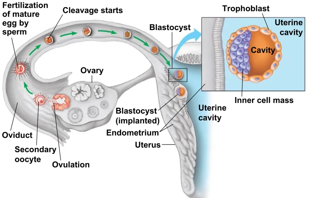
The embryo and placenta take
shape during the first month of
pregnancy
•
Human development begins with
fertilization
in the oviduct creating a
zygote
•
Cleavage continues producing a solid ball of cells called a
morula
which then becomes a hollow ball of cells called a
blastocyst
whose
inner cell mass becomes the embryo.
•
The outer cell layer of the blastocyst called the
trophoblast or
blastoderm
will become implanted in the uterine wall & form part of
the placenta
•
Gastrulation
occurs and organs develop from the three embryonic
Figure 27.15ab
Trophoblast
Cavity
Uterine
cavity
Blastocyst
Cleavage starts
Fertilization
of mature
egg by
sperm
Oviduct
Secondary
oocyte
Ovulation
Ovary
Blastocyst
(implanted)
Endometrium
Uterus
Uterine
cavity
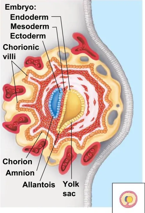
4 extraembryonic membranes
develop
amnion
•
surrounds the embryo and
•
forms a fluid-filled amniotic cavity that protects the embryo.
yolk sac
•
in reptiles, stores yolk, and
•
in humans, source of blood cells and initial circulation
allantois
•
contributes to the umbilical cord,
•
forms part of the urinary bladder, and in reptiles, stores embryonic waste.
chorion
•
contributes to the placenta and
Figure 27.15e
Embryo:
Endoderm
Mesoderm
Ectoderm
Chorionic
villi
Amnion
Chorion
Figure 27.15f
Placenta
Amnion
Amniotic
cavity
Mother’s
blood
vessels
Allantois
Yolk
sac
Chorionic
villi
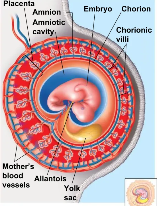
Placenta – contains both
embryonic & maternal parts
•
Formed from chorion and trophoblast
•
mediates exchange of nutrients, gases, and the products of excretion
between the embryo and the mother.
•
consists of
chorionic villi
closely associated with the blood vessels of
Hormonal changes during
pregnancy
1
st
10 Weeks
HCG or human chorionic gonadotropin –
•
Secreted by developing embryo about
8-10 days after fertilization
•
thought to be cause of morning
sickness
•
detected in urine during pregnancy
tests
Progesterone-
•
secreted by corpus luteum
After 10 weeks
HCG
•
production stops (eliminating
morning sickness)
Progesterone
Figure 27.16-0
TRIMESTER 1
TRIMESTER 2
TRIMESTER 3
Gill pouches (primitive
gill-like structures) Placenta
Limb buds Tail Umbilical cord Amnion Umbilical cord Placenta
Most radical changes for mother and embryo; embryo particularly susceptible to radiation, drugs, alcohol.
The fetus continues to grow; its eyes can open, teeth form, and bones begin to harden. The placenta begins to secrete progesterone and stops secreting hCG. The corpus luteum degenerates.
The fetus grows rapidly and gains strength. The mother’s abdominal organs become squeezed, causing frequent urination, digestive troubles, and backaches. Babies born prematurely—as early as 24 weeks—may survive, but they require special medical care.
Timeline of Human Development Conception
Week 5: Highly organized multicellular embryo about 7 mm long, with notochord and coelom. Brain and spinal cord taking shape. Gill pouches will develop into parts of throat and ear.
Week 8: All major structures present in rudimentary form. The embryo is about 4 cm long. Fetus can move its limbs and head and make facial expressions.
Week 14: Fetus is about 6 cm long. Features have been refined, and the fetus now appears more human.
Week 20: The fetus is about 19 cm long, weighs about 0.5 kg, and has eyebrows, eyelashes, fingernails, and toenails and is covered with fine hair. It may be quite active and “kick.” The mother’s abdomen is markedly enlarged. With limited space, the fetus bends into the fetal position.
Week 40 (newborn): The circulatory system and
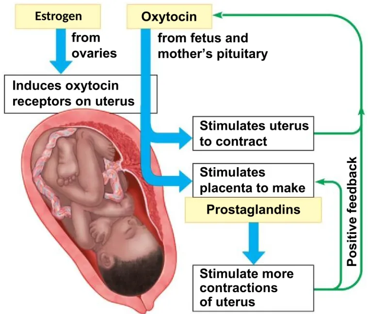
The induction of labor involves
positive feedback
•
Oxytocin and prostaglandins cause uterine
contractions
•
that in turn stimulate the release of
more
oxytocin
and prostaglandins.
•
The result is a steady increase in contraction
intensity
•
climaxing in forceful muscle contractions that
Figure 27.17a
from
ovaries
Induces oxytocin
receptors on uterus
Oxytocin
from fetus and
mother’s pituitary
Stimulates uterus
to contract
Stimulates
Childbirth is induced by hormones
and other chemical signals
•
The series of events that expel an infant from the uterus is called
labor
.
•
Hormonal changes induce birth.
•
Estrogen makes the uterus more sensitive to oxytocin.
•
Oxytocin acts with prostaglandins to initiate labor.
•
The cervix dilates to about 10 cm.
Labor occurs in three stages:
Latent
- dilation of the cervix,
Active
- expulsion, delivery of the
infant, and