Progress in Biological Sciences
Vol. 5, Number 2, Summer / Autumn 2015/207-221Isolation and identification of native
sulfur-oxidizing bacterium capable of uranium
extraction
Received: 8 March, 2015; Accepted: 10 July, 2015
Faezeh Fatemi1*, Abbas Rashidi2,Samaneh Jahani1
1. Nuclear Fuel Cycle Research School, Nuclear Science and Technology Research Institute, Tehran, Iran
2. Department of Chemical Engineering, Faculty of Engineering, University of Mazandaran, Babolsar, Iran
Bioleaching is the extraction of metals from their ores through the use of microorganisms. In this process, the use of native bacteria leads to achieve more yields of metals. So, in the present study, native sulfur-oxidizing bacterium in potentiality of uranium extraction was isolated from Ghachin mine in Iran and identified by partial gene sequencing. For this purpose, the water samples were collected from Ghachin mine and cultivated in Starkey medium. In following, the isolate was inoculated into individual Starkey plates and incubated until the colonies indicating the purified bacterium appeared. Then, the identification was carried out based on phenotypic characteristics and 16s rDNA sequencing. After that, bioleaching of uranium experiments carried out using uranium ore at 2.5 and 5% pulp densities. The result showed that after 15 days of incubation, the bacteria in the fresh samples was grown. Following 5-7 days of the plate's incubation, we obtained the single purified colonies of the bacteria. On the basis of 16s rDNA nucleotide sequencing, the bacteria showed 99.71% similarity to Acidithiobacillus ATCC 19377. Besides, the bioleaching experiments indicated that the bacterium is capable of uranium extraction in 2.5 and 5% pulp densities during 3 and 5 days. In conclusion, in this study, for the first time, we isolated the native sulfur-oxidizing bacterium capable of uranium extraction,from uranium mine of Gachin in Bandar Abbas, Iran.
ammonium salts
Introduction
Bioleaching is the extraction of metals from the ores or soils by biological processes, mostly by microorganisms. In this process, by using the bacteria, the metals such as uranium, copper, zinc, cobalt, gold were dissolved. Bioleaching processes are commonly more eco-friendly than physico-chemical processes. They do not use big amount of energy as compared to roasting and smelting and do not produce sulfur dioxide or other harmful gases. This process can be considered to be compatible with antipollution rules (1). In the recent years, recovery of metals from low grade ores using bacteria was developed (2).
Acidithiobacillus is a sulfur-oxidizing bacterium used in industrial process of bioleaching. It is a genus of proteobacteria, gram-negative, acidophilus and tolerant to a range of metal ions. This bacterium is able to perform oxidation and reduction of sulfur which can change ferrous sulphate to ferric sulphate (3) leading to sulfuric acid production under aerobic conditions (4). This process can cause the water in their areas acidic (5). Acidithiobacillus grow chemoautotrophically
by fixing CO2, obtaintheirenergyby using
reduced inorganic sulfur compounds as an electron donor and use oxygen as the electron acceptor. In addition to sulfur, these bacteria can use thiosulfate or tetrathionate as the sources of energy, but growth in a liquid medium on thiosulfate is slow (6). This bacterium is obligatory aerobic because it uses atmospheric oxygen for the oxidation of sulfur to sulfuric acid. Acidithiobacillus requires only little amounts of nitrogen due to its small quantity of growth but the best
sources are of inorganic
acids, especially sulfate, followed by the
ammonium salts of organic acids, nitrates, asparagines and amino acids (7). In the bioleaching process, A. thiooxidans adhere to the surface of a mineral, typically form an exopolysaccharide layer (EPS) which serve as the reaction space (8-9).
Acidithiobacillus appear to be present in a wide range of environments, especially in sites where pH is low, such as mining areas, hot sulfur springs and bioleaching operations (10). In addition, this has been found in acid mine drainage. These mines contain of sulfur materials necessary for the growth of sulfur-oxidizing bacteria (11).
There is variation within and between species of sulfur-oxidizing bacteria. However, since their natural habitats are ecologically extremely diverse, different strains of the same species developing in various ecological niches are characterized by differences in growth rate, tolerance to heavy metal ions and activities of sulfide mineral oxidation (12). Analysis of 16s rDNA fragment has been successfully used to identify bacteria in the environmental samples (10) as it is a good tool for inferring intergeneric and intrageneric relationships. 16s rDNA gene sequence intergenic spacer was compared to sequences in the GenBank database by using BLAST program (13). In this study, for the first time, we isolated and identified a native sulfur-oxidizing bacterium from uranium mine of Gachin in Bandar Abbas, Iran. In addition, we considered the capability of this bacterium in bioleaching of uranium.
Materials and Methods
Sampling from uranium mineTen water samples were collected from the
sterilized falcons which one-third of spaces were left blank. The samples were transferred to the laboratory at 4°C.
Isolation of bacterium in liquid medium The collected samples cultured in Starkey liquid medium (14-15) that the compositions is as follows: 1.0 g (NH4)2SO4, 0.14 g
CaCl2.2H2O, 3.0 g KH2PO4, 0.1 g
Mgcl2·6H2O and 10 g Sulfur, in 1000 ml of
distilled water. In order to isolate the sulfur-oxidizing bacterium, 10 ml of the water samples were inoculated into 90 ml of the Starkey medium in 250 ml Erlenmeyer flasks. Initial pH adjusted to 4 by addition of 10N H2SO4. These flasks were incubated at 30°C
at 180 rpm in shaker incubator for several days until the bacterium growth.
Purification of bacterium on solid medium After the bacteria was grown, the cells were cultured on solid medium plates. The solid medium, containing of 0.1g NH4Cl, 3g
KH2PO4, 0.1g MgCl2, 0.25g CaCl2, 5g
Na2S2O3.5H2O, 20g agar and 1000 ml of
distilled water (16). Initial pH adjusted to 4 using 10N H2SO4 and then autoclaved for 15
minutes in 121°C. The isolates were then streaked on solid Starkey medium (streak culture) and incubated at 30°C. The isolated bacterium was purified by repeated single colony purification three times.
Morphological characterization of isolated bacterium
The characterization of isolated bacterium was carried out on colony morphology characterization such as size, shape, color and gram’s staining (17, 18). Afterwards, for total bacterial count, a single colony was picked and inoculated into 20 ml liquid medium in 50 ml Erlenmeyer flask. The flask was incubated at 30°C at 180 rpm in shaker
incubator. Total bacterial count was done daily by using neubauer haemocytometer. In addition, variation of pH was measured with a pH meter (Metrohm 827).
DNA Extraction
Purified bacterium was set up for identification process. In the first step, DNA extraction was performed using 200 ml fresh liquid medium of bacterium. The bacterium filtered by 0.22 µm filter following washing twice with 1×PBS (8g NaCl, 0.2g KCl, 44.1g Na2HpO4.2H2O, 0.24g KH2PO4, 1000 ml
distilled water and pH of 1× PBS adjusted to 1.2). Afterwards, the filter washed with washing buffer (50 ml A2X, 25 ml Glycerol and 25 ml distilled water (buffer A2X containing 200 mM Trise-HCl, 50 mM EDTA, 200 mM NaC6H5O7 and 10 mM
CaCl2)). The filter transferred to a falcon and
PCR amplification of 16s rDNA gene The amplification was performed by using
universal primers, 27F (5/
-AGAGTTTGACCTGGCTCAG-3/), 1492R
(5/-TACGGCTACCTTGTTACGACTT-3/). PCR reactions including 10 µl of 10xPCR buffer, 0.5 µM of each primer, 200 µM dNTPs, extracted DNA and 2 unit of taq polymerase in volume of 100 µl (20). In following, initial denaturation carried out in 95°C for 5 min. The replication cycle was included 30 iterative cycle with denaturation at 94°C for 1 min, primer annealing at 49.3°C for 1 min and primer extension at 72°C for 1 min (21). After the cycles were completed, the amplified DNA preserved at 72°C for 5 min (20). To assess the quality of amplified DNA, these were electrophoresed on 1% agarose gel.
DNA fragments analysis through agarose gel electrophoresis
Agarose gel electrophoresis is the benchmark technique for separation and purification of nucleic acids as well as PCR-analysis (22). By using 1% agarose concentration as standard, smaller fragments were able to migrate faster than the larger fragments. 5 µl of the DNA mixed with loading buffer poured into the slots of the submerged gel using a disposable micropipette. Also, load size standards (ladder) were poured into slots. The agarose gel was placed into electrophoresis tank including TBE buffer and SYBER SAFE at voltage 100v for 45 min. Then,the gel was
placed in the transilluminator machine that reflected to the gel UV light and the gel image was taken.
Sequencing of amplified gene and phylogenetic tree assembly
The PCR product was purified and sequenced by Seqlab Company in Germany. 16s rDNA sequence obtained were arranged by using Bioedit software and identified close phylogenetic of isolated bacteria carried out by EzTaxon database (24). Finally, the phylogenetic tree was drawn with using ClustalX and Mega version 3 software’s.
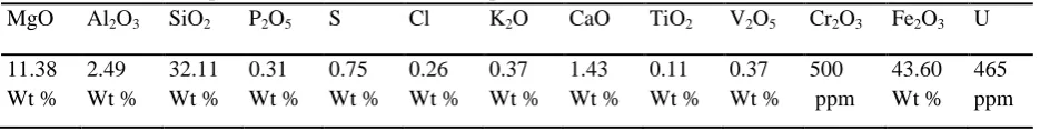
Ore sample and analysis
In the present study, the Iranian uranium mine was selected for bioleaching propose. The mine is located in the central of Iran in Yazd province. The samples included the rocks of the mine and concentrate in the form of soil, were collected from different sites of Saghand mine in Yazd. The rocks were crushed into a size of 106 µm (optimum liberation degree of uranium mineral according to a report of Processing Technology Developed for Uranium Ores from Saghand Deposit Islamic Republic of Iran). After crushing, the chemical analysis of the ore was carried out by XRF (Table 1). According to the XRF analysis of the ore, the main minerals are pyrite, quartz and also uraninite as the main uranium-bearing mineral in the ore.
Table 1. Chemical compositions of the uranium ore sample
MgO Al2O3 SiO2 P2O5 S Cl K2O CaO TiO2 V2O5 Cr2O3 Fe2O3 U
Bioleaching experiments
Bioleaching process was conducted in 250mL Erlenmeyer flask containing 90 ml APH medium and 2.5% uranium ore powder. Also, 10% (v/v) of sulfur-oxidizing bacteria isolated from Ghachin mine was used as inoculum. APH medium containing 2g (NH4)2SO4,0.5gK2HPO4,0.5gMgSO4.7H2O,
0.1g KCl, 0.01g Ca(NO3)2.4H2O, 10g S as
energy source and 1000 ml of distilled water (25). Initial pH was adjusted to 4 with H2SO4
(10N). The flask was maintained in a shaker incubator at 180rpm and 30◦C. When uranium extraction rate reached 100%, 10 ml of the supernatant was removed and added to a flask containing 90 ml APH medium and 5% uranium ore powder. The flask incubated in shaker incubator at 180rpm and 30◦C. The soluble uranium, variation of pH and Eh measured daily as a determined parameters.
Analysis
Sample was taken at regular intervals to determine total soluble uranium. It was measured by o-phenanthroline method (using UV–Vis spectrophotometer) (26, 27). The Eh and pH of the supernatant at room temperature was measured with a Metrohm pH meter, model 827.
Results
Isolation and purification of sulfur-oxidizing bacterium
The growth of sulfur-oxidizing bacterium sampling from acidic mine water was observed after 15 days incubation in Starkey medium. The presence of sulfur-oxidizing bacterium in liquid medium was confirmed by the sulfur precipitation and decrease of the medium pH. Following isolation of the bacterium, purification carried out on the Starkey solid medium. After 5-7 days of the plate incubation
at 30°C, the single colonies of sulfur-oxidizing bacterium was observed (Fig. 1).
Figure 1. The single colonies of the isolated bacterium on Starkey solid medium
Characterization and colony morphology The morphology observation indicated that the colonies of isolated bacterium were circular, relatively small in size and yellow in color. Gram’s staining revealed that this strain was gram-negative. In following, the strain was grown after 5 days incubation of a single colony into 20 ml fresh Starkey liquid medium to obtain pure cultures. The microscopical observation revealed that the isolated bacterium was motile and rod shaped. In addition, the isolated bacterium showed chemolithotrophic properties using carbon dioxide as a sole source of carbon (33) and oxidized sulfur and reduced sulfur compounds to sulfuric acid.
Figure 2. pH variations of isolated bacteria into liquid medium. In any 24-hour period, variations of pHregistered and graph were drawn
Figure 3. Growth curve of the isolated bacteria into liquid medium. In any 24-hour period, the total bacterial counted using neubauer haemocytometer
Identification of sulfur-oxidizing
bacterium
Amplification conditions were optimized using genomic DNA from pure culture. The electrophoretical analysis of the PCR products showed that the size of the fragment amplified from isolated bacterium matched the expected size of 1465 bp for primers 27F and 1492R (Fig. 4).
Sequencing and phylogenetic tree assembly Follwing 16s rDNA nucleotide sequence of
the isolated bacterium, a comparative phylogenetic analysis of the 16s rDNA sequence was performed with respect to the large group of A. thiooxidans strains which 16s rDNA nucleotide sequences available in EzTaxon database. The phylogenetic tree of the bacterium is shown in Figure 5. As, it is clear from the results, the isolated strain has 99.71% similarity to A. thiooxidans ATCC19377(accession number: PRJNA157459)
based on 16s rDNA sequence. 3.2
3.4 3.6 3.8 4 4.2 4.4
pH
Time (h)
0 0.5 1 1.5 2 2.5 3
cell/ml
(×
10
7
)
Time (h)
0 24 48 72 96 120 168 192 216 240 264 288 336 360 408 432 504
Figure 4. Agarose gel electrophoresis of the amplified product. The 16S rDNA fragment was amplified using the primer pair 27F, 1492R
Bioleaching experiment
The result showed that the sulfur-oxidizing bacterium isolated from Ghachin is capable of uranium extraction (100%) during 3 days (72h) in 2.5% pulp density (Fig. 6a). Variation of pH and Eh in time intervals in the bioleaching experiment is presented in Figure 6b and c. The pH of the medium decreased from 4 to 1.67 during total uranium extraction. The Eh of the medium increased from 203 mV to 440 mV showing bacterium activity.
Figure 6. Bioleaching experiment in 2.5% pulp density of uranium ore. a) Uranium concentrations in 2.5% pulp density. Variation of b) pH c) Eh during the experiments
In the 5% pulp density, the result showed the total uranium extraction (100%) during 5 days (120h) (Fig. 7a). The curve variation (Fig. 7b) showed that the pH of the medium decreased of 4 to 1.46. Figure 7c shows the variation of Eh during 5 days of uranium extraction. The Eh were increased of 142 mV to 425 mV.
Discussion
Our study indicated that the native isolated bacterium from uranium mineof Ghachin in Bandar Abbas is capable of uranium extraction from low grade uranium ore.
The results indicated that the sulfur-oxidizing bacterium was isolated from water sample after 15 days incubation in Starkey medium. Vidyalakshmi and Sridar were also succeeded to isolate sulfur-oxidizing bacterium taking from mine acidic soil within 15 days in Starkey medium (28). Bergamo et al. isolated sulfur-oxidizing bacterium obtained from different sites in the State of São Paulo, Brazil using modified Postgate medium during 7-10 days (10). The difference in the days of isolation is influenced by several factors such as sample type and location, taxonomy of bacteria, bacterial strain, type of used liquid medium and culture conditions (29). Based on the research conducted by Rojas-Avelizapa et al., the Starkey medium is the best medium for the isolation of sulfur-oxidizing bacteria compared with five other medium (ATCC medium 125, thiosulfate mineral medium, Starkey medium-dextrose, Starkey thiosulfate medium and 9K medium) (30). Our results confirm these finding indicated the isolation of sulfur-oxidizing bacterium from the water sample using Starkey medium. As indicated in other studies, sulfur was the only energy source in the Starkey medium (15). According to Qiang et al., as this bacteria gain their energy from the oxidation of only sulfur compounds, no more than sulfur-oxidizing bacteria could survive in these conditions (31). As mentioned in the results, precipitation of sulfur and decrease in pH is the first signs of bacterial growth. Wetting sulfur and its attachment to the cells are resulted into sulfur precipitation (32). Production of sulfuric acid during bacterium
growth in liquid Starkey medium leaded to decrease of the medium pH. The sulfuric acid is produced by oxidizing elemental sulfur bacteria contained in the culture medium (33).
As shown in Figure 1, the colonies of the bacterium observed after 5-7 days incubation, while Shahroz et al. observed some colonies after 12-15 days of incubation at 30oC using thiosulphate solid medium for purification of sulfur-oxidizing bacterium (17). Furthermore, Selman and Waksman observed the colonies of sulfur-oxidizing bacterium during 5-6 days using thiosulphate solid medium (34). The significant differences in the days number of purification have several reasons such as bacterial strain, the type of used solid medium, the density of the bacterium and growing conditions (29).
The results of morphological
characterization (Fig. 1) in this study are in agreement with the general observation according to Ryu et al. which indicated that the colonies of sulfur-oxidizing bacterium were circular, small, yellow in color on solid medium and gram-negative (35). Also, Vishniac indicated that the colonies of sulfur-oxidizing bacterium were circular on solid medium and gram-negative (36). In addition, the result was similar with isolated bacteria by Ryu et al. indicated the isolated bacterium was motile and rod shaped (35).
As shown in Figure 2, decrease in pH was observed in medium that caused by sulfur-oxidizing bacteria with producing H2SO4.
group of microorganisms decreases the environmental pH from 7 to 2 through the oxidation of elemental sulfur to sulfuric acid or oxidation of metal sulfides to metal sulfates (33, 38). On the basis of Carmen’s studies, decreasing the pH value was influenced by the taxonomic and physiologic diversity of the acidophilic chemolithotrophic bacteria belonging to Acidithiobacillus genus (33).
As shown in Figure 3, the isolate bacterium in the growth curve showed no lag phase. On the other hand, the maximum bacterial growth was shown in 4 to 5 days of incubation. Ryu et al. also showed that the isolated sulfur-oxidizing bacterium was without lag phase following maximum bacterial growth in 8 to 10 days of incubation at pH 2 and 4 (35). Lag time is distinct as the initial period in the growth number of a bacterial when cells are changing to a new environment before starting log phase growth. Several reasons affect the duration of lag time, containing inoculums mass, the physiological of bacteria, and the physiochemical environment of new growth medium (39). The log phase of growth is a design of stable growth where all the cells are dividing frequently by double division. The cells division at a continuous rate depended upon the structure of the growth medium and the conditions of incubation. The rate of log growth of a bacterial culture is expressed as the doubling time of the bacterial population (40). From this description, it can be concluded that the difference in the number of bacteria in different phases of bacterial growth can be due to the variation in composition and supply of bacterial biochemical structure including cellular proteins and enzymes, nucleic acids, and polysaccharides.
In this study, the universal primers i.e. 27F and 1492R were used in 16s rDNA
sequencing technique done for bacterium identification. Bergamo et al. concluded that the 16s rDNA region is a useful target for developing molecular methods that focus on the detection, rapid differentiation and identification of Acidithiobacilli (10).
Tsukasa et al. identified a
chemolithoautotrophic sulfur-oxidizing bacterium using universal 27F and 1492R primers (41). Also, Paulino,s research indicated that identification based on 16s rDNA sequence for this bacteria was successful (42). Xia et al. isolated the sulfur-oxidizing bacterium from acid mine drainage of copper ore in Baiyin area, Gansu province. The 16s rDNA gene sequence of this isolate was analyzed and phylogenetic analysis showed that the strain has 99.8% sequence similarity with that of the known strain A. thiooxidans ATCC 19377 in the Genebank, respectively (43). Moreover, Yongqing et al. identified sulfur-oxidizing bacterium by using 16s rDNA sequence. They showed that their isolated bacterium has 96-97% similarity with
A. thiooxidans ATCC 19377 (44).
bioleaching process. Moreover, the properties of the leached ores are main importance (46). In addition, the results indicated that Eh and pH value decreased by increasing pulp density (Figs. 6b, c; 7b,c). The studies indicated that decreasing of the bacterium activity can be due to toxicity of heavy metal (47, 48). The toxicity of a metal primarily depends on its interaction with the organism or reactions with biomolecules. Thus, the chemical and physical properties of the metal can help to predict its toxicity (48). It is not only the reactivity with biomolecules but also determines the toxicity of a metal. Before the metal can exhibit its effects, it needs to enter
the cell, therefore, several other aspects need to be considered including bioavailability of the metal, interactions with other ions at the binding site and transport into the cell (48).
Conclusion
1. Borisovich, U.A., Mihaylovich, K.A. (2013) Bioleaching of low grade uranium ore containing pyrite using A. ferrooxidans and A. thiooxidans. J. Radioanal. Nucl. Chem., 295, 151-156.
2. Rawlings, E. (1997) Biomining: theory, microbes and industrial processes. Bioscience, Georgetown, Tex.
3. Glazer, A.N., Nikaido, H. (1995) Application of biotechnology for mineral processing. Microb.
Biotechnol., 268-287.
4. Steudel, R. (1989) On the nature of the "elemental sulfur" (S0) produced by sulfuroxidizing bacteria. In: Schegel, H.G., Bowien, B. (Eds.), A model for S0 globules. Biology of Autotrophic Bacteria. Science Tech. Publication., 289-303.
5. Bond, P.L., Druschel, G.K., Banfield, J.F. (2000) Comparison of acid mine drainage microbial communities in physically and geochemicaly distinct ecosystems. Appl. Environ.Microbiol., 66, 4962-4971.
6. Dopson, M., Craig, B.A., Koppineedi, P., Philip, L. (2003) Growth in sulfidic mineral environments, metal resistance mechanisms in acidophilic micro-organisms. Microbiology., 149, 1959-1970.
7. Waksman, S.A., Joffe, J.S. (1922) The chemistry of the oxidation of sulfurby microorganisms to sulfuric acid and transformation of insoluble phosphates into soluble forms. J. Biol. Chem., 50,
35-45.
8. Rohwerder, T., Gehrke, T., Kinzler, K., Sand, W. (2003) Progress in bioleaching: fundamentals andmechanisms of bacterial metal sulphide oxidation. Appl. Microbiol. Biotechnology., 63, 239-248.
9. Sand, W., Gehrke, T. (2006) Extracellular polymeric substances mediate bioleaching/ biocorrosionvia interfacial processes involving iron(III) ions and acidophilic bacteria. Res. Microbiol., 157, 49-56.
10. Bergamo, R.F., Novo, M., Verissimo, R., Paulino, L., Stoppe, N., Sato, M., Manfio, G., Prado, P., Garsia, O., Ottoboni, L. (2004) Differentiation of Acidithiobacillus ferrooxidans and
Acidithiobacillus thiooxidans strains based on 16s-23s rDNA spacer polyphormism analysis. Res. Microbiol., 155, 559-567.
11. Rawlings, E. (2005) Characteristics and adaptability of iron- and sulfur-oxidizing
microorganismsused for the recovery of metals from minerals and their concentrates. Microb. Cell Fact.,4-13.
12. Ageeva, S.N., Kondrat'eva, T.F., Karavaiko, G.I. (2001) Phenotypic characteristics of Thiobacillus ferrooxidans strains. Mikrobiologiia., 70, 226-234.
13. Altschul, S.F., Gish, W,. Miller, W., Myers, E.W., Lipman, D.J. (1990) Basic local alignment search tool. J. Mol. Biol., 215, 403-410.
14. DSMZ. List of media. (2002) Deutsche Sammlung von Mikroorganismen und Zellkulturen Gmb Germany.
16. Waksman, S.A. (1922) Microorganisms concerned in oxidation of sulur in the soil. J. Bacteriol., 7(6), 605-608.
17. Shahroz, K., Faizul, H., Fariha, H., Kausar, S., Rahat, U. (2012) Growth and biochemical
activities of Acidithiobacillus thiooxidans collected from black shale. Microbiol. Res.,2, 78-83.
18. Gram, H.C. (1884) "Über die isolierte Färbung der Schizomyceten in Schnitt- und Trockenpräparaten" (in German). Fortsch. Medizin., 2, 185-189.
19. Escobar, B., Bustos, K., Morales, G., Salazar, O. (2008) Rapid and specific detection of
Acidithiobacillus ferrooxidans and leptospirillum ferrooxidans by PCR. Hydrometallurgy., 92,
102-106.
20. Ai, O., Satoshi, W., Tadayoshi, K., Tsuyoshi, S., Kazuo, K. (2005) Diversity of 16s ribosomal DNA-defined bacterial population in acid rock drainage from japanese pyrite mine. J. Biosci. Bioeng., 100, 644–652.
21. Leloup, J., Loy, A., Knab, N.J., Borowski ,C., Wagner, M., Jorgensen, B.B. (2007) Diversity andabundance of sulfate-reducing microorganisms in the sulfate and methane zones of a marinesediment, Black Sea. Environ. Microbiol., 9, 131-142.
22. Sambrook, J., Russell, D.W. (2001) Molecular cloning: A laboratory manual. New York: Cold Spring Harbor Laboratory Press.
23. Altschul, S.F., Madden, T.L., Schaeffer, A.A., Zhang, J., Zhang, Z., Miller, W., Lipman, D.J. (1997) Gapped BLAST and PSI-BLAST: a new generation of protein database search programs.
Nucleic Acids Res., 25, 33-89.
24. Chun, J., Lee, J.H., Jung, Y., Kim, M., Kim, S., Kim, B.K., Lim, Y.W. (2007) EzTaxon: a web-based tool for the identification of prokaryotes web-based on 16S ribosomal RNA gene sequences. Int. J.Syst. Evol. Microbiol., 57, 2259-2261.
25. Ronald, M.A. (1997) Handbook of microbiological media. Second Ed. Robert Stern Publisher, New York.
26. Underwood, A.L., Day, R.A. (1991) Quantitative Analysis. Prentice Hall, London., pp. 645-646.
27. Ryu, H.W., Cho, K.S., Chang, Y.K., Kim, S.D., Mori, T. (1995) Refinement of low-grade clay by microbial removal of sulfur and iron compounds using Thiobacillus ferrooxidans. J.Ferment. Bioeng., 80, 46.
28. Vidyalakshmi, R., Srida, R. (2006) Isolation and characterization of sulphur oxidizing bacteria.
Microbiol. Cult. Collect., 5, 73-75.
29. Ward, G. (1916) Laboratory manual in general microbiology, 1st edn. New York.
30. Rojas-avelizapa, N.G., Gómez-ramírez, M., Hernández-gama, R., Aburto, J., García, D.E. (2013) Isolation and selection of sulfur-oxidizing bacteria for the treatment of sulfur-containing
hazardous wastes. Chem. Biochem. Eng. Q., 27(1), 109-117.
31. Qiang, L.I., Cong, W., Baobin, L., Cunmin, S., Fei, D., Cunjiang, S., Shufang, W. (2012) Isolation of Thiobacillus spp. and its application in the removal of heavy metals from activated sludge.
32. Sivaji, R., Leslie, R. (1971) Berger G. The requirement of low pH for growth of Thiobacillus thiooxidans . Archiv für Mikrobiologie., 79, 338-344.
33. Carmen, M. (2010) The taxonomic and physiologic diversity of the acidophilic chemolithotrophic bacteria of the genus thiobacillus used in ores solubilization processes. Trav. Inst. Spéol. «Émile Racovitza»,97-112.
34. Selman, A., Waksman, S.A. (1922) Microorganisms concerned in the oxidation of sulfur in the soil. New Jersey Agricultural Experiment Station, Department of Soil Chemistry and Bacteriology., 84, 605-608.
35. Ryu, H.W., Moon, H.S., Lee, E.Y., Cho, K.S., Choi, H. (2003) Leaching characteristics of
heavy metals from sewage sludge by Acidithiobacillus thiooxidans MET. J. Environ. Qual., 32, 751-759.
36. Vishniac, W.V. (1974) The genus Thiobacillus. Bergey's manual of determinative bacteriology, 8th edn.
37. Kempner, E. (1966) Acid Production by Thiobacillus thiooxidans. Journal of Bacteriology., 92, 1842-1843.
38. Rao, T. (2005) Advances in water and wastewater treatment. American Society of Civil Engineers.
39. Swinnen, I.A.M., Bernaert, S, K., Dens, E.J.J., Geeraerd, A.H., Vanimpe, J.F. (2004) Predictivemodelling of the microbial lag phase: a review. Int. J. Food Microbiol., 94, 137-159.
40. Al-qadiri, H., Al-alami, N., Lin, M., Al-holy, M., Cavinato, A., Rasco, B. (2008) Studying of the bacterial growth phases using fourier transform infrared spectroscopy and multivariate analysis.
J. Rapid Meth. & Aut. Mic., 16, 73-89.
41. Tsukasa, I., Kenichi, S., Satoshi, O. (2004) Isolation, characterization, and in situ detection of a
novel chemolithoautotrophic sulfur-oxidizing bacterium in wastewater biofilms growing under
microaerophilic conditions. Appl Environ Microbiol., 70(5), 3122-3129.
42. Paulino, L., Rog´erio, F., Maricilda, P., Oswaldo, G., Gilson, P., Laura, M. (2001) Molecular characterization of Acidithiobacillus ferrooxidans and A. thiooxidans strains isolated from mine wastes in Brazil. Antonie van Leeuwenhoek., 80, 65–75.
43. Xia, J., Peng, A., He, H., Yang, Y., Liu, X., Qiu, G. (2004) Acidithiobacillus albertensis BY-05, a new strain for bioleaching of metal sulfides ores. Project (50321402) supported by Nature Science Foundation of China for innovation research group; Project (2004CB619204) supported by National Major Basic Research Item, China.
44. Yongqing, N., Dongshi, W., Kaiyu, H. (2008) 16s rDNA and 16s–23s internal transcribed spacer sequence analyses reveal inter- and intraspecific Acidithiobacillus phylogeny. Microbiology., 154, 2397-2407.
45. Scott, P. (2013) What is Biomining. Wise Geek.
46. Acevedo, F., Gentina, J. (1989) Process engineering aspects of the bioleaching of copper ores.
Biopr. Engin., 4, 223-229.
47. Newman, L.A., Doty, S.L., Gery, K.L., Heilman, P.E., Muiiznieks, I., Shang, T.Q., Siemieniec,
contaminants: a review of phytoremediation research at the University of Washington. J. Soil Commun., 7, 531-542.