Published online 2016 February 20. Review Article
Non-Union Current Treatment Concept
Arash Moghaddam,
1,*Claudia Ermisch,
1and Gerhard Schmidmaier
11Center for Orthopedics, Trauma and Spinal Cord Injury, University Hospital Heidelberg, Heidelberg, Germany
*Corresponding author: Arash Moghaddam, Center for Orthopedics, Trauma and Spinal Cord Injury, University Hospital Heidelberg, Heidelberg, Germany. Tel: +49-62215635394, Fax: +49-62215629123, E-mail: [email protected]
Received 2015 October 31 ; Accepted 2015 December 25.
Abstract
Context: This article wants to give a current concept for the challenging decision for conservative or operative treatment strategies of non-unions according to the principles of ‘diamond concept’ and aspects that have to be attended.
Evidence Acquisition: Between February 2010 and March 2014, 424 patients with non-unions were treated at Heidelberg university hospital. This database has been analyzed at least one year after the treatment. The analysis and the experience in surgery and treatment of non-unions as well as present literature were prepared for this review as a current concept.
Results: If an atrophic non-union is suggested, reosteosynthesis and biological stimulation is required. A revision surgery of autologous transplantation of cancellous bone from the iliac crest is often enough. Alternatively, reamer-irrigator-aspirator (RIA) can be taken out of the femur with lower complications and pain in the extraction area and be combined with growth factors like bone morphogenetic proteins (BMPs), if consolidation after cancellous bone is still absent. In complex cases, consequential and radical removal of the infection often improved circulation through interventional angiography and use of the two-step procedure (the Masquelet technique) as well as a tissue covering are required.
Conclusions: By using the ‘diamond concept’ as a complex concept, non-unions can be treated in different stages in a targeted manner.
Keywords: Diamond Concept, Reamer-Irrigator-Aspirator, Non-Union, Masquelet Technique
Copyright © 2016, Iran University of Medical Sciences. This is an open-access article distributed under the terms of the Creative Commons Attribution-NonCommer-cial 4.0 International License (http://creativecommons.org/licenses/by-nc/4.0/) which permits copy and redistribute the material just in noncommerAttribution-NonCommer-cial usages, provided the original work is properly cited.
1. Context
The percentage of delayed or non-union after fractures of long bones is approximately 10%, but depends on patient’s risk profile. The current definition states that a non-union is a fracture that will not consolidate without any further intervention, independent of the treatment time. With adequate stability, conservative treatment in the early stages of a non-union is possible. The operative treatment depends on the type of non-union. There are one-step or two-step procedures according to the principles of the ‘di-amond concept’. This involves the improvement of the me-chanical situation (in most cases with a reosteosynthesis) and vascularization, local application of osteoconductive carriers e.g. tricalciumphosphate, vital cells from autolo-gous bone, and osteoinductive substances like bone mor-phogenetic proteins (BMP-2 or BMP-7).
Hypertrophic and atrophic non-unions without large defect gaps or signs of infection can be treated with a one-step procedure. For treating infected non-unions or non-unions with large defect gaps, the Masquelet tech-nique is recommended.
2. Evidence Acquisition
University of Heidelberg hospital is the major center for orthopaedic treatments in Germany. This database has been analyzed at least one year after treatment. Our
results were on one hand based on a collective of 424 patients with non-unions who were treated in our cen-ter between February 2010 and March 2014, and on the other hand they were based on the current literature on e.g. PubMed. The analysis and the experience in surgery and treatment of non-unions as well as present literature were prepared for this review as a current concept.
3. Results
3.1. Epidemiology
In general, non-union as a complication of fracture healing occurs in 5% - 10% of cases (1) and can be as high as 30% in high-risk groups (2-5).
The high incidence on the tibia can be explained by poor tissue covering and therefore poor blood circulation. In ad-dition, there is more often 2° - 3° of open injuries with com-plex damage of the bone and the surrounding soft tissue (5). The incidence of non-unions in bones of the extremi-ties is as follows: the tibia has on average an occurrence of 8.7% (6, 7), the femur only slightly less at 6.1% (8-10), the humerus 3% - 5%, and the lower arm 5% (11-18).
3.2. Risk Factors
Besides the type of fracture and the soft tissue damage, there are other risk factors for the development of non-unions (19). Smoking increases the risk of delayed heal-ing or non-unions (5, 20). In addition, advanced age has a negative effect on physiological fracture healing (21, 22). Other factors such as diabetes mellitus, the use of non-steroidal anti-inflammatory drugs (NSAIDs) and previous fractures of the same extremity also have negative effects (23, 24). In the risk score according to Moghaddam et al. all of these risk factors have their relative importance for measuring the individual risk of a patient for developing a non-union (5) (Table 1).
3.3. Diagnosis
The diagnosis begins with extensive history taking and includes the individual risk profile of the patient, taking into account previous illnesses and medications as well as all previous conservative and operative treatments. History of previous infections should be taken into ac-count as well as the analysis of previous clinical and ra-diological findings. Blood circulation and soft tissue at the site of injury should also be evaluated.
Clinical indications of non-union include pain on weight bearing, limitations to the mobility of an extremity, or in-stability. In addition, there are clinical signs of infection to be aware of, such as redness, swelling and warmth or de-velopment of a fistula. One should especially look out for local, systemic or anamnestic signs of osteitis.
Conventional radiological imaging of the affected ex-tremity in two levels with inclusion of joints is standard for diagnosing faulty positioning or instability. One should note that defects cannot be completely observed with native X-rays due to summation and covering. An additional computed tomography (CT) can be used to evaluate whether there is partial or entire bridging of the defect gap. In addition, a contrast magnetic resonance imaging (MRI) can be used to evaluate vascularization of the bone and separate vital and non-vital areas (25) (A CT scan is considered the gold standard for evaluating non-unions and providing information for treatment).
3.4. Classification
According to the current definition from European society of tissue regeneration in orthopedics and traumatology (ES-TROT), a non-union is a fracture that does not heal without further intervention, independent of the length of treatment.
Non-unions are classically divided into four types: hy-pertrophic due to mechanical causes, atrophic due to biological causes, defect, and infection non-unions.
Hypertrophic non-unions develop due to insufficient mechanical stability and can lead to formation of a callus in the area of fracture. Bone consolidation does not occur. If therapy is delayed, atrophic non-unions can develop (26). Atrophic non-unions often involve reduced vascu-larity of the defect gap and surrounding bone, which can
lead to atrophy of the fracture ends. Defect non-unions usually occur due to high-speed trauma and higher grade open fractures, which can cause loss of bone through nu-merous fragments. Furthermore, infected non-unions develop primarily in open fractures after traumas in which germs can get into the wound.
Besides the purely morphological classification, there is a new classification system according to the non-union scoring system (NUSS) which incorporates bone quality, soft tissue damage, and the individual patient risk (e.g. smoking) into a score (27). From this NUSS-score, there are therapy considerations that can be adapted to the pa-tient (28-30). The higher the score, the more specialized and custom the therapy concept must be to offer the pos-sibility of consolidation (Table 2).
3.5. Therapy
The goal of non-union therapy is the consolidation of bone defects with correction of the axis and leg length as well as reaching weight bearing stability. A sufficient tissue covering as well as removal of the infection are the basic prerequisites. Independent of the type of non-union and localiza-tion, therapy can differ in conservative and operative ap-proaches.
3.5.1. Conservative
Conservative approaches are especially useful in the early phase of non-union treatment and require sufficient me-chanical stability as well as bone regeneration potential. The newest methods in conservative treatment in delayed fracture healing of the lower extremity are consequential weight bearing until full weight bearing can be mustered.
The most common methods of conservative treatment in delayed fracture healing in the area of the lower extremity are consequential weight bearing to full weight bearing.
Additionally, fractures are treated with daily lower ener-gy ultrasound (a possibility of conservative therapy is the application of low energy ultrasound to the defect gap), over a defined interval of three to six months.
The deciding criteria for successful treatment with low energy ultrasound are sufficient stability as well as a de-fect gap of under 10 mm, free of inde-fection in the previous history, start of therapy in less than five months after the fracture, and a NUSS-score less than 35 (31).
3.5.2. Operative Treatment Diamond Concept
The multi-factorial causes of delayed fracture healing make an individual patient’s specific therapy noteworthy. In this way, the so-called diamond concept (Figure 1) has become evermore established (3, 32). Therapy consists of an optimized combination of biological and biomechani-cal factors (29, 33-38) (The diamond concept has five differ-ent factors that must be analyzed for therapy) (39).
- Osteogenic cells: in form of mesenchymal stem cells (MSCs), autologous cancellous bone (iliac crest or reamer irrigator aspirator (RIA)).
- Osteoconductive structures; example: autologous can-cellous bone, synthetic bone replacement material (ex-ample: tricalcium phosphate).
- Growth hormones; examples: bone morphogenetic protein (BMP)-2 and BMP-7.
- Vascularization: through improving the macro-circu-lation and local induction of a Masquelet membrane re-newal of the fracture ends.
In looking at individual clinical and radiological param-eters, there are a number of factors lacking in a combi-nation treatment and therefore, systemic optimization of the biological and biomechanical treatment of non-unions should be looked at (29, 34, 40, 41).
3.5.3. Isolation of Bone Replacement Material
The iliac crest often serves as the source of autologous cancellous bone. The method involved is widely used and is considered the gold standard in the surgical treatment of non-unions. It has the optimal consistency and can es-tablish good primary stability as a tricortical graft. Prob-lematic is the limited availability and differing quality and quantity as well as a high extraction morbidity (25, 42-46).
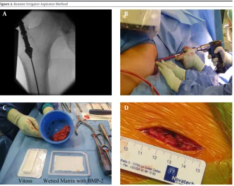
An alternative to autologous cancellous bone extraction is the use of RIA (Depuy-Synthes, USA) (Figure 2). In this procedure, the percutaneous opening of the medullary cavity is performed with a combination of drilling, clean-ing and vacuum extraction of MSCs out of long bones. This method is primarily performed on the femur, but can also be done on the tibia. In this way, it is possible to extract 80 cm of the autologous bone material for transplantation (25, 35, 43-48) (an alternative to extraction of cancellous bone from the iliac crest is the RIA method, which involves winning cancellous bone out of the femur).
There is a larger amount of growth factors (e.g. BMP-2) in the RIA material as in autologous cancellous bone from the iliac crest. RIA material shows a higher osteo-conductive potential in comparison to autologous can-cellous bone (25, 46, 49, 50).
3.5.4. Complications
The main complications of removal of cancellous bone from the iliac crest are infections, development of hema-tomas, fractures, hypertrophic scars, and chronic pain from the extraction area (51).
The complications associated with RIA to win autolo-gous bone material are substantially less. In our patient collective, we observed a perforation of the femoral shaft (1/200), which could have been avoided by correctly posi-tioning the guide wire in both levels.
3.5.5. Growth Factors
Growth factors play a deciding roll in physiological
frac-ture healing and bone regeneration. Currently, there are two growth hormones permitted for use in orthopedics and trauma surgery. These include BMP 2 for open lower leg fractures and BMP-7 for non-unions of the tibia.
Studies over the past years have shown that the use of BMP-7 is at least equal to a single cancellous bone graft (52). Furthermore, there are adequate indications in everyday clinical routine to suggest increases in the healing rate of non-unions (18). The combination of autologous cancellous bone with BMPs is clearly superior to a single cancellous bone graft (53).
BMP-7 is not currently commercially available; so, increased BMP-2 (Medtronic) can be used in non-union therapy.
The authors recommend the additional use of BMPs in a failed therapy with autologous cancellous bone. In addi-tion, when the effect is clearly proven by clinical studies, we recommend receiving approval of costs from the insurance company and approval of its use from a panel of experts.
3.6. Bone Marrow Drilling in Hypertrophic
Non-Unions
According to the diamond concept, optimizing mechani-cal stability is indicated in hypertrophic non-unions. In the case of an instable osteosynthesis, a reosteosynthesis is nec-essary. In simple cases, the dynamization of an adjoining nail with full weight bearing is possible. In complex cases, a reosteosynthesis with medullary cavity drilling and medul-lary nail osteosynthesis with a thick nail is necessary (54) (in hypertrophic non-unions in the tibia shaft area, medullary cavity drilling and nail osteosynthesis has been indicated).
3.6.1. Positioning
A patient is positioned on the back and the x-ray beam is po-sitioned on the contralateral side. The placement of a blood tourniquet is not necessary for intramedullary osteosynthe-sis, but can be necessary for other intervention techniques.
3.6.2. Execution: Tibia Non-Union
The knee was bent maximally with the help of orienta-tion points, the tuberositas tibiae and middle of the pa-tella, and the skin was cut. The patellar ligament was cut lengthwise. After the opening of the medullary cavity with an awl, a guide wire was inserted. The correct positioning was tested with the aid of an x-ray intensifier in two levels.
The drilling began with the smallest drill and was in-creased incrementally thereafter. The largest drill was used under intensified image control and was complete only when the drill head had entirely filled the smallest shaft area of the medullary cavity. As a result, the medul-lary nail can be inserted. This should be 1 mm less in di-ameter than maximal drilling.
3.6.3. Follow-Up
After wound healing is over, full weight bearing is pos-sible. Up until this point, the patient needs low-dose heparin. For improving the functional outcome, physio-therapy of the bordering joints plays an important role.
3.7. Atrophic Non-Unions
Atrophic non-unions are described as insufficient and stagnated bone regeneration. According to the diamond concept, an additional biological activation of bone re-generation is often necessary (In atrophic non-unions, biological activation of bone regeneration is necessary).
3.7.1. Surgery According to the Diamond Concept
In the first step, previously implanted osteosynthesis material is completely removed and the proximal and distal ends of the non-union gaps are debrided down to healthy bone. In long bones, debridement should always be expanded to the entire medullary cavity (29, 33, 35, 46). Often a complete implant removal and reosteosynthesis are needed to ensure optimal biomechanical stability and correction of faulty positioning.
After radical debridement of the non-union and reosteo-synthesis, defect gaps are filled with autologous cancel-lous bone in combination with synthetic resorbable ma-terial (e.g. tricalciumphosphate) as an osteoconductive scaffold. This can occur in the form of autologous cancel-lous bone from the iliac crest or through the RIA System from the contralateral femur or tibia. An optimal scaffold should not only be osteoconductive, but also have good cell adhesion capabilities and adequate stability as well as flexibility. The criteria for a good scaffold can be summa-rized under the 4 F’s: form, function, fixation and forma-tion. The currently available material only fulfills these criteria partially (56).
In bone defects under 2 cm (or 5 cm) with no signs of infection, defect gap filling is the only treatment concept used. In larger defects and in the presence of an infection, bone formation involves a two-step method, the Masque-let technique (Figure 3).
The one-step method is contraindicated when there were signs of infection. Furthermore, the age of the patients plays a role as a possible contraindication. The morbidity as-sociated with the procedure should be considered. In some cases, cancellous bone from a donor can be used. Patients with a too narrow medullary cavity of the femur of less than 10 mm in the isthmus are not right for RIA removal (46).
3.7.2. Further Bone Replacement Materials
In addition to allogeneic bone grafts, bone replacement material tricalciumphosphate or growth factors such as BMP-2 or BMP-7 are used (57). This material has osteoin-ductive and osteconosteoin-ductive effects, which are only pres-ent when the surroundings of the non-union are vital and well vascularized.
3.7.3. Alternative Methods
In very large bone defects, it must be tested whether bone formation is possible with the Masquelet tech-nique, or if alternatives need to be considered such as cal-lus distraction with an Ilizarov fixator or a vascularized bone transplant or other alternatives.
3.7.4. Follow-Up
Follow-up is dependent on the type of executed reosteo-synthesis. In the lower extremity, partial weight bearing of 20 kg for six weeks followed by incremental increases to full weight bearing is recommended.
3.8. Defect and Infection Non-Unions
3.8.1. Bone Formation With the Masquelet Technique
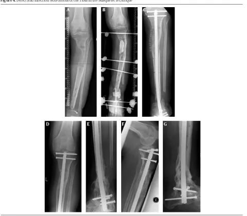
This technique is used when there is suspicion of infection or a non-union with a defect gap of > 2 cm (5 cm). In the first operation, the defect gap is filled with polymethylmethac-rylat (PMMA) cement and removal of avital tissue is con-ducted. In the presence of a known infection, an antibiotic can be mixed with the cement. The PMMA cement should be applied over the bordering vital bone (Figures 3 and 4).
Through a reaction with the cement, the induction of a highly vital membrane similar to natural periost occurs, which is responsible for the circulation of the later trans-planted graft material. This first step can be repeated as of-ten as necessary until sanitation of the infection occurs (46). The adjoining osteosynthesis material should be com-pletely removed if there is suspicion of a previous infec-tion and the extremity should be secured up until the second operation with an external fixator. Adherent bacteria on the implant surface could be observed with a sonication. In this way, the exact microbiological spec-trum can be defined and treated with antibiotics. The debrided bone tissue can be evaluated microbiological-ly and pathologicalmicrobiological-ly. If cultures are positive, additional debridement and adaption of local antibiotics is neces-sary until the infection is cleared (46).
After sanitation of the infection and a four-to-six-week interval, the defect is reconstructed with bone regenera-tion and in certain cases re-osteosynthesis happens. The constructed Masquelet membrane is opened lengthwise, PMMA cement is removed carefully, and the defect is filled with a combination of MSC-rich autologous cancel-lous bone (e.g. cancelcancel-lous bone from the iliac crest or RIA material), tricalcium phosphate (TCP) and an osteoinduc-tive growth factor (e.g. BMP-7 or BMP-2) (58).
3.8.2. Follow-Up
Table 1. Score for Measuring an Individual’s Risk of Developing a Non-Union (5)
Sub-Item Scores
Localization
Humerus Prox. 4 points Diaph. 6 points Distal 2 points
Forearm Prox. 4 points Diaph. 6 points Distal 2 points
Femur Prox. 4 points Diaph. 6 points Distal 8 points
Tibia Prox. 6 points Diaph. 8 points Distal 4 points
Soft Tissue
General 1° open 4 points 2° open 6 points 3° open 10 points
General Fasciotomy 4 points a Previous fracture 8 points a Neurological disorder 6 points b
Smoking
General Smoker 15 points Previous smoker 5 points Non- smoker 0 points
Comorbiditiy/Medication
General NSAID 4 points Bisphosphonate 6 points Diabetes 4 points
Type 1 < 10 points Low risk NA
Type 2 10 - 20 points Middle risk NA
Type 3 > 20 points High risk NA
Abbreviations: diaph, diaphyseal; prox, proximal; NA, not available. aAffected bone.
bAffected limb.
Table 2. Non-Union Scoring System (27)
Criteria Score a Max. Score
The Bone Quality of the bone
Good 0 NA
Moderate (e.g. mildly osteoporotic) 1 NA
Poor (e.g. severe porosis or bone loss) 2 NA Very poor (necrotic, appears avascular or septic) 3 3
Primary injury – open or closed fracture
Closed 0 NA
Open 1° grade 1 NA
Open 2 – 3° A grade 3 NA
Open 3° B – C grade 5 5
Number of previous interventions on this bone to procure healing
None 1 NA
< 2 2 NA
< 4 3 NA
> 4 4 4
Invasiveness of previous interventions
Minimally-invasive: Closed surgery (screws, k wires) 0 NA
Internal intra-medullary (nailing) 1 NA
Internal extra-medullary 2 NA
Any osteosynthesis which includes bone grafting 3 3
Adequacy of primary surgery
Adequate stability 0 NA
Inadequate stability 1 1
Weber and cech group
Hypertrophic 1 NA
Oligotrophic 3 NA
Atrophic 5 5
Bone alignment
Anatomic alignment 0 NA
Non-anatomic alignment 1 1
Bone defect – Gap
0.5 – 1 cm 2 NA
1 – 3cm 3 NA
> 3cm 5 5
Soft Tissues Status
Intact 0 NA
Previous uneventful surgery, minor scarring 2 NA Previous treatment of soft tissue defect (e.g. skin loss, local flap cover, multiple incisions, compartment syndrome,
old sinuses) 3 NA
Previous complex treatment of soft tissue defect (e.g. free flap) 4 NA Poor vascularity: absence of distal pulses, poor capillary refill, venous insufficiency 5 NA Presence of actual skin lesion/defect (e.g. ulcer, sinus, exposed bone or plate) 6 6
The Patient ASA Grade
1 or 2 0 NA
3 or 4 1 1
No 0 NA Yes – well controlled (HbA1c < 10) 1 NA Yes – poorly controlled (HbA1c >10) 2 2
Blood tests: FBC, ESR, CRP
FBC: WCC > 12 1 NA
ESR > 20 1 NA
CRP > 20 1 3
Clinical infection status
Clean 0 NA
Previously infected or suspicion of infection 1 NA
Septic 4 4
Drugs
Steroids 1 NA
NSAIDs 1 2
Smoking status
No 0 NA
Yes 5 5
Abbreviation: NA, not available.
aHigher score implies a more-difficult-to-procure union.
Figure 2. Reamer Irrigator Aspirator Method
3.9. Soft Tissue Covering and Improvement of
Vas-cularization
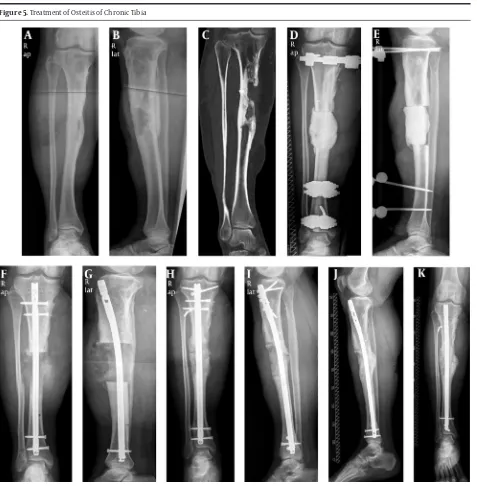
Sufficient soft tissue covering and improvement of blood circulation of the affected extremity are necessary for successful non-union therapy and use of the above mentioned techniques (Figure 5). An interdisciplinary therapy concept in cooperation with plastic and vascular surgeons is meaningful and often necessary.
Ideally, complex cases should be discussed interdisci-plinary and a therapy concept should be agreed upon, for example in an interdisciplinary extremity board. The chance of healing is higher with interventional angiog-raphy and sufficient soft tissue covering with a vascular-ized surgical flap e.g. anterolateral thigh (ALT) flap before definitive defect filling.
Atrophic Tibia Non-Union
Defect < 1 cm or No Clinical Signs of
Infection
Defect < 1 cm or Clinical Signs of
Infection
One - Step Treatment Two - Step TreatmentAccording to Masquelet
Figure 3. Diagram for Treatment Decisions (39)
Figure 4. Defect and Infection Non-Unions of the Tibia in the Masquelet Technique
Figure 5. Treatment of Osteitis of Chronic Tibia
A - C, preoperatively; D and E, postoperative step I; F and G, postoperative step II; H and I, postoperative: 1 year; J and K, recall postoperative: 2 years.
4. Conclusions
Through sufficient soft tissue management and stabile primary osteosynthesis, the development of a non-union can be avoided. A useful prophylaxis against infections and non-unions, especially in open lower leg fractures, is a combination of BMP-2 and antibiotic covered medul-lary nails (ETN PROtect) (55, 59, 60).
In delayed fracture healing, there are simple techniques for improving the chances of bone consolidation such as raising weight bearing or the dynamization of static medullary nails.
In delayed fracture healing, consolidation can often be reached with simple measures such as increasing the load or dynamization of the statically locked medullary nail. Additionally, a local ultrasound treatment in the early phase of delayed fracture healing can be used.
often complications and pain in the area. Alternatively, RIA can be taken out of the femur with lower extraction morbidity. Through correct placement of the guide wire, rare complications such as perforation or fracture of the femur can be avoided. If consolidation after cancellous bone is still not present, the further use of BMPs in com-bination with cancellous bone is indicated. In complex cases, consequential and radical removal of the infection often requires improved circulation through interven-tional angiography and use of the Masquelet technique as well as a tissue covering to avoid treatment failure. In complex cases, a close interdisciplinary team involving an extremity board comprised of an orthopedist/trauma surgeon, vascular surgeon, radiologist and plastic sur-geons can be useful.
Footnote
Authors’ Contribution:Study concept and design, analysis and interpretation of data, drafting of the man-uscript, critical revision of the manuscript for impor-tant intellectual content and statistical analysis: Arash Moghaddam and Gerhard Schmidmaier. Correction of the paper: Claudia Ermisch.
References
1. Einhorn TA. The cell and molecular biology of fracture healing. Clin Orthop Relat Res. 1998;(355 Suppl):S7–21. [PubMed: 9917622] 2. Schmidmaier G, Schwabe P, Wildemann B, Haas NP. Use of bone
morphogenetic proteins for treatment of non-unions and future perspectives. Injury. 2007;38 Suppl 4:S35–41. [PubMed: 18224735] 3. Giannoudis PV, Einhorn TA, Schmidmaier G, Marsh D. The
dia-mond concept--open questions. Injury. 2008;39 Suppl 2:S5–8. doi: 10.1016/S0020-1383(08)70010-X. [PubMed: 18804574]
4. Rothman RH, Klemek JS, Toton JJ. The effect of iron deficiency anemia on fracture healing. Clin Orthop Relat Res. 1971;77:276–83. [PubMed: 5140454]
5. Moghaddam A, Zimmermann G, Hammer K, Bruckner T, Grutzner PA, von Recum J. Cigarette smoking influences the clin-ical and occupational outcome of patients with tibial shaft frac-tures. Injury. 2011;42(12):1435–42. doi: 10.1016/j.injury.2011.05.011. [PubMed: 21665205]
6. Keating JF, O’Brien PI, Blachut PA, Meek RN, Broekhuyse HM. Reamed interlocking intramedullary nailing of open fractures of the tibia. Clin Orthop Relat Res. 1997;(338):182–91. [PubMed: 9170379]
7. Clifford RP, Lyons TJ, Webb JK. Complications of external fixation of open fractures of the tibia. Injury. 1987;18(3):174–6. [PubMed: 3508844]
8. Ruedi TP, Luscher JN. Results after internal fixation of commi-nuted fractures of the femoral shaft with DC plates. Clin Orthop Relat Res. 1979;(138):74–6. [PubMed: 445920]
9. Wolinsky PR, McCarty E, Shyr Y, Johnson K. Reamed intramedul-lary nailing of the femur: 551 cases. J Trauma. 1999;46(3):392–9. [PubMed: 10088839]
10. Ricci WM, Bellabarba C, Evanoff B, Herscovici D, DiPasquale T, Sanders R. Retrograde versus antegrade nailing of femoral shaft fractures. J Orthop Trauma. 2001;15(3):161–9. [PubMed: 11265005] 11. Kontakis GM, Papadokostakis GM, Alpantaki K, Chlouverakis G,
Hadjipavlou AG, Giannoudis PV. Intramedullary nailing for non-union of the humeral diaphysis: a review. Injury. 2006;37(10):953– 60. doi: 10.1016/j.injury.2006.02.050. [PubMed: 16777105] 12. Rommens PM, Kuechle R, Bord T, Lewens T, Engelmann R, Blum
J. Humeral nailing revisited. Injury. 2008;39(12):1319–28. doi: 10.1016/j.injury.2008.01.014. [PubMed: 18417134]
13. Wright RR, Schmeling GJ, Schwab JP. The necessity of acute bone grafting in diaphyseal forearm fractures: a retrospective review. J Orthop Trauma. 1997;11(4):288–94. [PubMed: 9258828]
14. Wei SY, Born CT, Abene A, Ong A, Hayda R, DeLong WG, Jr. Diaphy-seal forearm fractures treated with and without bone graft. J Trauma. 1999;46(6):1045–8. [PubMed: 10372622]
15. Ring D, Allende C, Jafarnia K, Allende BT, Jupiter JB. Ununited diaphyseal forearm fractures with segmental defects: plate fixa-tion and autogenous cancellous bone-grafting. J Bone Joint Surg Am. 2004;86-a(11):2440–5. [PubMed: 15523016]
16. Richard MJ, Ruch DS, Aldridge JM, 3rd. Malunions and nonunions of the forearm. Hand Clin. 2007;23(2):235–43. doi: 10.1016/j. hcl.2007.02.005. vii. [PubMed: 17548014]
17. Krzykawski R, Krol R, Kaminski A. The results of locked intramed-ullary nailing for non-union of forearm bones. Ortop Traumatol Rehabil. 2008;10(1):35–43. [PubMed: 18391904]
18. Moghaddam-Alvandi A, Zimmermann G, Buchler A, Elleser C, Biglari B, Grutzner PA, et al. [Results of nonunion treatment with bone morphogenetic protein 7 (BMP-7)]. Unfallchirurg. 2012;115(6):518–26. doi: 10.1007/s00113-011-2100-0. [PubMed: 22476375]
19. Gaston MS, Simpson AH. Inhibition of fracture healing. J Bone Joint Surg Br. 2007;89(12):1553–60. doi: 10.1302/0301-620X.89B12.19671. [PubMed: 18057352]
20. Moghaddam A, Weiss S, Wolfl CG, Schmeckenbecher K, Wentzensen A, Grutzner PA, et al. Cigarette smoking decreases TGF-b1 serum concentrations after long bone fracture. Injury. 2010;41(10):1020–5. doi: 10.1016/j.injury.2010.03.014. [PubMed: 20471641]
21. McKibbin B. The biology of fracture healing in long bones. J Bone Joint Surg Br. 1978;60-B(2):150–62. [PubMed: 350882]
22. Tonna EA. Histologic and histochemical studies on the peri-osteum of male and female rats at different ages. J Gerontol. 1958;13(1):14–9. [PubMed: 13514031]
23. Macey LR, Kana SM, Jingushi S, Terek RM, Borretos J, Bolander ME. Defects of early fracture-healing in experimental diabetes. J Bone Joint Surg Am. 1989;71(5):722–33. [PubMed: 2659600]
24. Cruess RL, Sakai T. Effect of cortisone upon synthesis rates of some components of rat bone matrix. Clin Orthop Relat Res. 1972;86:253–9. [PubMed: 4261729]
25. Schoierer O, Bloess K, Bender D, Burkholder I, Kauczor HU, Schmidmaier G, et al. Dynamic contrast-enhanced magnetic resonance imaging can assess vascularity within fracture non-unions and predicts good outcome. Eur Radiol. 2014;24(2):449– 59. doi: 10.1007/s00330-013-3043-3. [PubMed: 24145951] 26. Bode G, Strohm PC, Sudkamp NP, Hammer TO. Tibial shaft
frac-tures - management and treatment options. A review of the cur-rent literature. Acta Chir Orthop Traumatol Cech. 2012;79(6):499– 505. [PubMed: 23286681]
27. Calori GM, Phillips M, Jeetle S, Tagliabue L, Giannoudis PV. Clas-sification of non-union: need for a new scoring system? Injury. 2008;39 Suppl 2:S59–63. doi: 10.1016/s0020-1383(08)70016-0. [PubMed: 18804575]
28. Abumunaser LA, Al-Sayyad MJ. Evaluation of the calori et Al non-union scoring system in a retrospective case series. Orthopedics. 2011;34(5):359. doi: 10.3928/01477447-20110317-31. [PubMed: 21598896] 29. Pneumaticos SG, Panteli M, Triantafyllopoulos GK, Papakostidis C, Gi-annoudis PV. Management and outcome of diaphyseal aseptic non-unions of the lower limb: a systematic review. Surgeon. 2014;12(3):166– 75. doi: 10.1016/j.surge.2013.10.007. [PubMed: 24309558]
30. Calori GM, Colombo M, Mazza EL, Mazzola S, Malagoli E, Marelli N, et al. Validation of the Non-Union Scoring System in 300 long bone non-unions. Injury. 2014;45 Suppl 6:S93–7. doi: 10.1016/j.in-jury.2014.10.030. [PubMed: 25457326]
31. Roussignol X, Currey C, Duparc F, Dujardin F. Indications and results for the Exogen ultrasound system in the management of non-union: a 59-case pilot study. Orthop Traumatol Surg Res. 2012;98(2):206–13. doi: 10.1016/j.otsr.2011.10.011. [PubMed: 22424956] 32. Giannoudis PV, Einhorn TA, Marsh D. Fracture healing: the dia-mond concept. Injury. 2007;38 Suppl 4:S3–6. [PubMed: 18224731] 33. Calori GM, Colombo M, Mazza E, Ripamonti C, Mazzola S, Marelli
34. Giannoudis PV, Calori GM, Begue T, Schmidmaier G. Bone regen-eration strategies: current trends but what the future holds? Injury. 2013;44 Suppl 1:S1–2. doi: 10.1016/S0020-1383(13)70002-0. [PubMed: 23351862]
35. Giannoudis PV, Ahmad MA, Mineo GV, Tosounidis TI, Calori GM, Kanakaris NK. Subtrochanteric fracture non-unions with implant failure managed with the "Diamond" concept. In-jury. 2013;44 Suppl 1:S76–81. doi: 10.1016/S0020-1383(13)70017-2. [PubMed: 23351877]
36. Calori GM, Colombo M, Ripamonti C, Bucci M, Fadigati P, Mazza E, et al. Polytherapy in bone regeneration: clinical applications and preliminary considerations. Int J Immunopathol Pharmacol. 2011;24(1 Suppl 2):85–90. [PubMed: 21669144]
37. Pountos I, Georgouli T, Pneumaticos S, Giannoudis PV. Frac-ture non-union: Can biomarkers predict outcome? Injury. 2013;44(12):1725–32. doi: 10.1016/j.injury.2013.09.009. [PubMed: 24075219]
38. Calori GM, Giannoudis PV. Enhancement of fracture healing with the diamond concept: the role of the biological chamber. Injury. 2011;42(11):1191–3. doi: 10.1016/j.injury.2011.04.016. [PubMed: 21596376]
39. Moghaddam A, Zietzschmann S, Bruckner T, Schmidmaier G. Treatment of atrophic tibia non-unions according to 'diamond concept': Results of one- and two-step treatment. Injury. 2015;46 Suppl 4:S39–50. doi: 10.1016/S0020-1383(15)30017-6. [PubMed: 26542865]
40. Giannoudis PV, Jones E, Einhorn TA. Fracture healing and bone repair. Injury. 2011;42(6):549–50. doi: 10.1016/j.injury.2011.03.037. [PubMed: 21474131]
41. Schmidmaier G, Schwabe P, Strobel C, Wildemann B. Carrier systems and application of growth factors in orthopaedics. In-jury. 2008;39 Suppl 2:S37–43. doi: 10.1016/S0020-1383(08)70014-7. [PubMed: 18804572]
42. Dinopoulos C. Re: The article "Dinopoulos H, Dimitriou R, Gi-annoudis PV. Bone graft substitutes: what are the options? Sur-geon. 2012 Aug;10(4):230-9. Surgeon. 2013;11(5):293. doi: 10.1016/j. surge.2013.05.008. [PubMed: 23932800]
43. Kouroupis D, Baboolal TG, Jones E, Giannoudis PV. Native multi-potential stromal cell colonization and graft expander multi-potential of a bovine natural bone scaffold. J Orthop Res. 2013;31(12):1950–8. doi: 10.1002/jor.22438. [PubMed: 23868185]
44. Kanakaris NK, Morell D, Gudipati S, Britten S, Giannoudis PV. Reaming Irrigator Aspirator system: early experience of its mul-tipurpose use. Injury. 2011;42 Suppl 4:S28–34. doi: 10.1016/S0020-1383(11)70009-2. [PubMed: 21939800]
45. Cox G, McGonagle D, Boxall SA, Buckley CT, Jones E, Giannoudis PV. The use of the reamer-irrigator-aspirator to harvest mes-enchymal stem cells. J Bone Joint Surg Br. 2011;93(4):517–24. doi: 10.1302/0301-620X.93B4.25506. [PubMed: 21464493]
46. Schmidmaier G, Moghaddam-Alvandi A. [cited 2015 10.06]; Was gibt es Neues 2014 in der Pseudarthrosen-Therapie? 2014 Available from: http://www.ecme-center.org/WebObjects/ECMECenter. woa/cms/1001057/Kurssuche.html?courseDetails=1003662. 47. Cuthbert R, Boxall SA, Tan HB, Giannoudis PV, McGonagle D, Jones
E. Single-platform quality control assay to quantify multipoten-tial stromal cells in bone marrow aspirates prior to bulk manu-facture or direct therapeutic use. Cytotherapy. 2012;14(4):431–40. doi: 10.3109/14653249.2011.651533. [PubMed: 22268519]
48. Fayaz HC, Giannoudis PV, Vrahas MS, Smith RM, Moran C, Pape HC, et al. The role of stem cells in fracture healing and nonunion. Int Orthop. 2011;35(11):1587–97. doi: 10.1007/s00264-011-1338-z. [PubMed: 21863226]
49. Blokhuis TJ, Calori GM, Schmidmaier G. Autograft versus BMPs for the treatment of non-unions: what is the evidence? In-jury. 2013;44 Suppl 1:S40–2. doi: 10.1016/S0020-1383(13)70009-3. [PubMed: 23351869]
50. Schmidmaier G, Herrmann S, Green J, Weber T, Scharfenberger A, Haas NP, et al. Quantitative assessment of growth factors in reaming aspirate, iliac crest, and platelet preparation. Bone. 2006;39(5):1156– 63. doi: 10.1016/j.bone.2006.05.023. [PubMed: 16863704]
51. Calori GM, Colombo M, Mazza EL, Mazzola S, Malagoli E, Mineo GV. Incidence of donor site morbidity following harvesting from iliac crest or RIA graft. Injury. 2014;45 Suppl 6:S116–20. doi: 10.1016/j.injury.2014.10.034. [PubMed: 25457330]
52. Schmidmaier G, Wildemann B. The role of BMPs in current orthopedic practice. IBMS BoneKEy. 2009;6(7):244–53. doi: 10.1138/20090386.
53. Giannoudis PV, Kanakaris NK, Dimitriou R, Gill I, Kolimarala V, Montgomery RJ. The synergistic effect of autograft and BMP-7 in the treatment of atrophic nonunions. Clin Orthop Relat Res. 2009;467(12):3239–48. doi: 10.1007/s11999-009-0846-2. [PubMed: 19396502]
54. Aymar M, Ewerbeck V. Standardverfahren in der Operativen Or-thopädie und Unfallchirurgie. In: Uberarb V, Aufl E, editors. 4 ed. New York: Thieme, Stuttgart; 2014. p. 898.
55. Schmidmaier G, Lucke M, Wildemann B, Haas NP, Raschke M. Prophylaxis and treatment of implant-related infections by anti-biotic-coated implants: a review. Injury. 2006;37 Suppl 2:S105–12. doi: 10.1016/j.injury.2006.04.016. [PubMed: 16651063]
56. Giannoudis PV, Chris Arts JJ, Schmidmaier G, Larsson S. What should be the characteristics of the ideal bone graft substitute? Injury. 2011;42 Suppl 2:S1–2. doi: 10.1016/j.injury.2011.06.001. [PubMed: 21700284]
57. Janicki P, Schmidmaier G. What should be the characteristics of the ideal bone graft substitute? Combining scaffolds with growth factors and/or stem cells. Injury. 2011;42 Suppl 2:S77–81. doi: 10.1016/j.injury.2011.06.014. [PubMed: 21724186]
58. Schmidmaier G, Capanna R, Wildemann B, Beque T, Lowenberg D. Bone morphogenetic proteins in critical-size bone defects: what are the options? Injury. 2009;40 Suppl 3:S39–43. doi: 10.1016/s0020-1383(09)70010-5. [PubMed: 20082789]
59. Fuchs T, Stange R, Schmidmaier G, Raschke MJ. The use of gen-tamicin-coated nails in the tibia: preliminary results of a pro-spective study. Arch Orthop Trauma Surg. 2011;131(10):1419–25. doi: 10.1007/s00402-011-1321-6. [PubMed: 21617934]