Vol. 1, No.1, 55-60, Winter/Spring 2011
*Corresponding author: mahtabyarmohammadi@gmail.com © 2011 Progress in Biological Sciences
Tel.:+98-1325743724: fax: +98-132574-3722
AFLP reveals no sex-specific markers in Persian sturgeon
(Acipenser persicus) or beluga sturgeon (Huso huso) from
the southern Caspian Sea, Iran
Mahtab Yarmohammadi1*, Mohammad Pourkazemi1, Ahmad Ghasemi2, Mohammad Hassanzadeh
Saber1 and Fereidoon Chakmehdouz1
1
Genetic Department, Dr. Dadman International Sturgeon Research Institute, Rasht, Iran 2
Persian Gulf Research and Studies Center, Persian Gulf University, Boushehr, Iran
The late sexual maturity in sturgeon and the absence of morphological differences between males and females makes sex discrimination difficult. Identification of sex at an early life stage is of high interest in caviar production because it allows efficient selection of females. In this study, the ge-nome of 10 mature male and 10 mature female specimens of Persian sturgeon (Acipenser persicus)
and beluga sturgeon (Huso huso) were screened using AFLP and 100 primer combinations. Re-sults showed a total of 3771 and 3779 scoreable bands in A. persicus and H. huso, respectively. Approximately 30% of markers in A. persicus and 29.6% H. huso were polymorphic. No sex spe-cific makers were identified. The results of the present study suggest that the sex chromosomes are not extensively differentiated in sturgeon species, or possibly the methods utilized were not suffi-ciently sensitive to recognize them. © 2011 Progress in Biological Sciences, Vol 1, No.1, 55-60.
Key words: Persian sturgeon, beluga, sex determination, AFLP, Acipenser persicus, Huso huso
INTRODUCTION
Sturgeon, the source of caviar, inhabit the north-ern hemisphere exclusively (Billard and Lecoin-tre, 2001). Overfishing, deterioration of natural spawning grounds and pollution have seriously threatened sturgeon species to the verge of ex-tinction (Pourkazemi, 2006). The decline in nat-ural populations has increased interest in aqua-culture caviar production, and the number of aquaculture farms is rapidly increasing. Howev-er, fishes can be sexed only after several years
of rearing (Logan et al., 1995, Bahmani and
Ka-zemi, 1998), and the rearing of males for the first years significantly increases production
costs, up to 30% of the total cost (Wuertz et al.,
2006). Even after maturity, sexing by ultra-sound, endoscopy, or by measuring plasma lev-els of hormone is time consuming and often stressful for the animals (Hett and Ludwig, 2005). The identification of a molecular marker
and the consequent development of a PCR-based method that allows sex identification at early life stages would be a valuable innovation, significantly benefiting aquaculture and reduc-ing depletion of natural populations. Hetero-morphic sex chromosomes have not been re-vealed in any sturgeon species (Fontana and Co-lombo, 1974; Holcik, 1986; Van Eenennaam, 1997). However, studies of gynogenetic females indicated a heterogamety (WZ/ZZ) mechanism
in white sturgeon A. transmontanus (Van
Ee-nennaam et al., 1999), bester sturgeon (H. huso
female × A. ruthenus male) (Omoto et al., 2005)
and shortnose sturgeon Acipenser brevirostrum
(Flynn et al., 2006). Identification of sex-linked
(Keyvanshokooh et al., 2007) approaches with-out satisfactory results. This lack of evidence suggests that sex determination may be
envi-ronmental (in Lake Sturgeon (Acipenser
fulves-cens) McCormick et al., 2008). However, the
hypothesis of genetically based sex determina-tion in sturgeon is supported by the balanced sex ratio consistently observed with artificial repro-duction (Van Eenennaam, 1997).
Among marker systems currently available, AFLP is dominant, providing a high level of po-lymorphism detection and multilocus and
ge-nome-wide marker profiles (Vos et al., 1995).
Amplified Fragment Length Polymorphism has been used to assess sex-specific markers in the
three-spined stickleback (Gasterosteus
aculea-tus) (Griffiths et al., 2000) and rainbow trout
(Onchorhynchus mykiss) (Sato et al., 2001). It
also widely used for studying sex specific mark-ers in avian species (Griffith and Orr, 1999).
Persian sturgeon A. persicus Borodin 1897 and
the beluga sturgeon Huso. huso L. 1758 are the
most important species in the Caspian Sea and are considered candidate species for aquacul-ture. Persian sturgeon is chiefly distributed in the southern Caspian Sea (Pourkazemi, 2006). Considering the endangered status of these spe-cies, artificial reproduction and stocking is ne-cessary for their survival.
Male and female genomes of A. persicus and H.
huso have been randomly screened by AFLP
(Vos et al., 1995). The present study presents the
first comparative genome analysis of male and female Persian and beluga sturgeons from the Caspian Sea using AFLP technique. The search for possible sex-linked markers was performed despite previous negative results with other sturgeon species, as sex determination mechan-isms may be different even among closely re-lated fish species.
MATERIAL AND METHODS Fish sampling and DNA extraction
Fin tissue samples were prepared from 20 adult beluga and 20 adult Persian sturgeons of both sexes (10 of each sex) and preserved in ethanol. Spawners were caught in March 2007 from the Caspian Sea coastline and transferred to the Shahid Beheshti Sturgeon Propagation
Com-plex, located in Rasht, Iran. Sex of spawners was identified by examination of testes and ova-ries.
Total genomic DNA was extracted from pre-served fin tissue using the phenol–chloroform
procedure (Pourkazemi et al., 1999). The
quanti-ty and qualiquanti-ty of extracted DNA was assayed using Nanodrop (ND1000, USA) and 1% aga-rose electrophoresis.
Polymerase chain reaction
The AFLP procedure was conducted as
de-scribed by Vos et al. (1995). An aliquot of total
DNA (250 ng) was digested using 10U MseI and
EcoRI in Tango buffer (Fermentas, France) in a
20 µl reaction at 37oC for 90 min. Ligation was
immediately initiated with a 5 µl mix of MseI
(50 mM) and EcoRI (5 mM) adapters, T4 DNA
Ligase, and 1X ligation buffer and carried out at
37oC for 3 h. Preamplification PCR was
per-formed on 15 µl volumes, with 5 µl diluted re-stricted/ligated DNA, 1X PCR buffer, 1.5 mM
MgCl2, 0.2 mM of each dNTPs, 10 pmol MseI+
C and EcoRI+A primers (Table 1).
All PCR amplifications were carried out with an Eppendorf thermocycler (Mastercycler ep gra-dient, 96 plus, Eppendorf, Germany) with 30 sec
denaturation at 94oC, 30 sec annealing at 56oC,
and 1 min extension at 72oC, for 22 cycles. For
selective amplifications, preliminary trials were conducted using 100 primer combinations. Se-lective PCR amplifications were performed on 15 µl volumes, with 3 µl diluted (1:10) pream-plification products as template, 1X PCR buffer
with 1.5 mM MgCl2, 0.2 mM of each dNTPs
(CinnaGene, Tehran, Iran), and 10 pmol M+4
and E+3 primers (Table 1). PCR consisted of 1
min denaturation at 94oC, 10 cycles of 30 sec at
94oC, 30 sec at 63oC, and 2 min at 72oC with the
annealing temperature decreasing from 63oC in
1oC increments in cycles 2-10. This was
fol-lowed by 26 cycles of 30 sec at 94oC, 30 sec at
54oC, and 2 min at 72oC.
Electrophoresis
After PCR, an equal volume of loading buffer (98% formamide, 10 mM EDTA, 0.1% bromo-phenol blue, 0.1% xylene cyanol) was added. The reaction mixture was then heated for 10 min
57
was pre-electrophoresed at 55oC, 95 W for 20
min, then 5 µl of the amplified DNA was loaded and run on a 6% polyacrylamide gel (19:1 acry-lamidae/bisacrylamidae; 7.5 M Urea; 1X TBE buffer) with 1X TBE buffer on a vertical gel electrophoresis system (BIO-RAD sequi Gen, GT, 38 ×30 cm/ PowerPAC 5000, USA) at 85
W and 50oC for 90 min. After electrophoresis,
the bands were visualized using silver staining. Clear and unambiguous bands of lengths rang-ing from 40 to 1000 bp were considered for fur-ther analysis.
Data analysis
One hundred combinations of primer sets were tested and only the recognizable AFLP markers were scored. The scored AFLP bands were dark, consistent, and repeatable in individuals across polymerase chain reactions and gels. The ob-tained results from male and female individuals were scored as presence (1) and absence (0) of band in each polymorphic locus. The number of polymorphic, monomorphic, and total alleles per each primer combination for each species was manually scored.
RESULTS
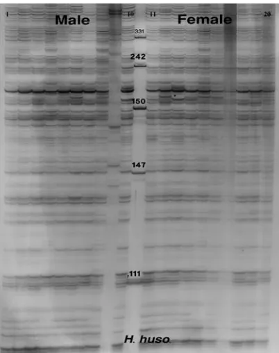
Figures 1 and 2 show the denatured acrylamide gel of male and female Persian and beluga
sturgeons using E+3/M+4 primer combination
of AFLP technique. Amplified Fragment Length
Polymorphism band patterns in A. persicus and
H. huso showed high levels of variation among
individuals, unrelated to sex.
A set of 100 (E+3 and M+4) primer
combina-tions in A. persicus and H. huso yielded a total
of 3771 and 3779 scoreable bands, respectively,
of which 30% in A. persicus and 29.6% in H.
huso were polymorphic. The fragments ranged
from 50 to 600 bp without revealing any sex-specific markers (Table 2).
DISCUSSION
The 100 primer combinations used and the markers analyzed, ranging from 50 to 600 bp, revealed no sex markers in the sturgeon species studied. Previously, Wuertz et al. (2006) used AFLP to search for sex-specific DNA markers
in four sturgeon species, A. naccarii, A. baerii,
A. gueldenstaedtii, and A. ruthenus. Although
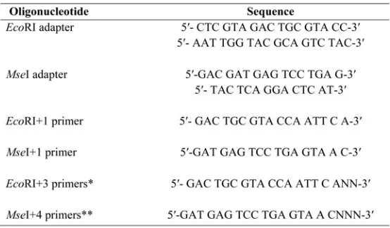
Table 1. Sequences of primers and adaptors for DNA-AFLP analysis.
*EcoRI+3 primers are: E-AAT, ACA, ATT, ACC, AAC, AAA, ACG, ATA, ATC, and AAG
**MseI+4 primers are: M-CACA, CGAA, CGAT, CCTT, CATA, CATT, CGTC, CTGC, CCGT, and CATT
Fig. 1. AFLP generated from DNA samples of 20 Huso huso using a set of Eco+3 / Mse+4 primers. 1-10 male and 11-20 female, (M) ladder, pUC Mix Marker, 8 (Ferments, France).
1100-9230 bands were screened per species, sex-specific markers were not detected.
Several molecular techniques have been used for the determination of sex in fish, including sex-specific markers in rainbow trout using RAPD
and FISH (Fluorescence In Situ Hybridization)
techniques (Ittura et al., 1998) and the
three-spined stickleback Gasterosteus aculeatus using
AFLP (Griffith et al., 2000). DNA markers from a gynogenetic triploid ginbuna genome were studied using representational differences
analy-sis (RDA), and sex-specific markers in Takifugo
rubripes were characterized by a cDNA-AFLP
technique (Cui et al., 2006).
However, all techniques tested have failed to find sex-specific markers in sturgeon, including
RAPD (Wuertz et al., 2006; Keyvanshokooh et
al., 2007, 2009), single-strand conformation
po-lymorphisms (SSCP), AFLP, inter-simple
se-quence repeats (ISSR) (Wuertz et al., 2006),
RDA, subtractive hybridization (McCormick et
al., 2008), and a candidate gene approach (Hett
and Ludwig, 2005; Hett et al., 2005 ). In addi-tion to screening at the genomic level, studies of
Fig. 2. AFLP generated from DNA samples of 20
Aci-penserpersicus using a set of Eco+3/Mse+4 primers. 1-10
male and 11-20 female, (M) ladder, pUC Mix Marker, 8 (Ferments, France).
expression patterns such as proteomics have also failed to find sex-specific markers (Keyvansho-kooh et al., 2009).
McCormick et al. (2008) suggested that an
envi-ronmental sex determination system may exist in sturgeon. However, environmental sex deter-mination leads to variations in sex ratios (Bull, 1983), and the sex ratio in adult populations of sturgeon is 1:1, both in natural and aquaculture populations (Chapman et al., 1996).
59
of markers in the studied species (Keyvansho-kooh and Gharaei, 2010).
Alternatively, it may be that homologous chro-mosomes carrying the sex-determining factors are not extensively differentiated in sturgeon species, or that the methods utilized did not have
the required sensitivity (Wuertz et al., 2006).
Due to these findings, we believe that alternative approaches based on gene expression at differ-ent stages of gonadal maturation should be used.
Acknowledgments
This research was funded by the Iranian Fisheries Re-search Organization (project number 2-025-200000-03-0000-84029). We thank the personnel of the Genetic and Physiology Departments of the International Sturgeon Research Institute for technical support and Mr. Hossein Mohammadi Parashkooh for providing us with the sam-ples required for the study. We are highly indebted to Mrs. Arshad for providing language help and Dr. Congiu for proofreading the article.
REFERENCES
Billard R, Lecointre G (2001) Biology and conservation of sturgeon and paddlefish. Rev Fish Biol 10, 355-392
Bahmani M, Kazemi R (1998) Histological study of go-nads in young farmed sturgeons. Iran Sci Fish J 1, 1-16
Bull JJ (1983) Evolution of sex determining mechanisms. (Menlo Park, CA, USA: Benjamin Cumming)
Chapman FA, Van Eenennaam JP, Doroshov SI (1996) The reproductive condition of white sturgeon, Acipenser
transmontanus, in San Francisco Bay, California. Fishery
Bulletin 94, 628-634
Cui JZ, Shen XY, Gang QL, Yang GP, Gu QQ (2006) Identification of sex markers by cDNA-AFLP in
Takifu-gu rubripes. Aquacult 257, 30-36
Flynn SR, Matsuoka M, Martin-Robichaud DJ, Benfey TJ (2006) Gynogenesis and sex determination in short-nose, Acipenser brevirostrum LeSuere. Aquacult 253, 721-727
Fontana F, Colombo G (1974) The chromosomes of Ital-ian sturgeons. Experientia 30, 739-742
Griffiths R, Orr KJ (1999) The use of amplified fragment length polymorphism (AFLP) in the isolation of sex-specific markers. Mol Ecol 8, 671-674
Griffith R, Orr KJ, Adam A, Barber I (2000) DNA sex identification in the three–spined stickleback. J Fish Biol 57, 1331-1334
Hett A, Pitra C, Jenneckens I, Ludwig A (2005) Charac-terization of sox9 in European Atlantic sturgeon (
Aci-penser sturio). J Hered 96, 150-154
Hett AK, Ludwig A (2005) SRY-related (Sox) genes in the genome of European Atlantic sturgeon (Acipenser
sturio). Genome 48, 181-186
Holcik J (1986) The Freshwater fishes of Europe – Gen-eral introduction to fishes, Acipenseriformes. vol. I, part II (Aula Verlag: Wiesbaden, Germany)
Iturra P, Medrano JF, Bagley M, Lam N, Vergara N, Martin JC (1998) Identification of sex chromosome mo-lecular markers using RAPDs and fluorescent in situ hy-bridization in rainbow trout. Genetica 101, 209-213
Keyvanshokooh S, Pourkazemi M, Kalbasi MR (2007) The RAPD technique failed to identify sex- specific se-quences in beluga (Huso huso). J Appl Ichthyol23, 1-2
Keyvanshokooh S, Kalbassi MR, Hosseinkhani S, Vaziri B (2009) Comparative proteomics analysis of male and female Persian sturgeon (Acipenser persicus) gonads. Anim Reprod Sci 11, 361-368
Keyvanshokooh S, Gharaei A (2010) A review of sex determination and searches for sex-specific markers in sturgeon. Aquacult Res 41, 1-7
Logan SH, Johnston WE, Doroshov SI (1995) Econom-ics of joint production of sturgeon (Acipenser
transmon-tanus Richardson) and roe for caviar. Aquacult 130,
299-316
McCormick CR, Bos DH, DeWoody JA (2008) Multiple molecular approaches yield no evidence for sex-determining genes in Lake Sturgeon (Acipenser fulves-cens). J Appl Ichthyol 24, 643-645
Omoto N, Maebayashi M, Adachi S, Arai K, Yamauchi K (2005) Sex ratios of triploids and gynogenetic diploids induced in the hybrid sturgeon, the bester (Huso huso
female× Acipenser ruthenus male). Aquacult 245, 39- 47
Sato T, Yokomizo S, Matsuda M, Hamaguchi S, Sakai-zumi M (2001) Gene-centromere mapping of medaka sex chromosomes using triploid hybrids between Oryzias
latipes and O. luzonensis. Genetica 111, 71–75
Van Eenennaam AL (1997) Genetic analysis of sex de-termination mechanism of white sturgeon (Acipenser
transmontanus Richardson). PhD dissertation,
Universi-ty of California, Davis
Van Eenennaam AL, Van Eenennaam JP, Medrano JF, Doroshov SL (1999) Evidence of female heterogametic genetic sex determination in White sturgeon. J Hered 90, 231-233
Vos P, Hogers R, Bleeker M, Reijans M, Van de Lee T, Hornes M, Frijters A, Pot J, Peleman J, Kuiper M, et al. (1995) AFLP: a new technique for DNA fingerprinting. Nucleic Acids Res 23, 4407-4414