MUSEI NATIONALIS PRAGAE
www.aemnp.eu
ISSN 1804-6487 (online) – 0374-1036 (print)
R E S E A R C H P A P E R
New Oriental and Australasian taxa of Colobathristidae
(Hemiptera: Heteroptera) in the collection
of the State Natural History Museum Stuttgart
Viktor HARTUNG1,2)
1) State Museum of Natural History Stuttgart (SMNS), Rosenstein 1, D-70191 Stuttgart, Germany.
2) Museum für Naturkunde – Leibniz-Institute for Evolution- and Biodiversity Science, Invalidenstrasse 43, 10115 Berlin, Germany;
e-mail: viktor.hartung@mfn.berlin
Abstract. TheColobathristidae are a poorly studied family of Lygaeoidea distributed in the tropics and subtropics of Australasian, Neotropical, and Oriental biogeographic regions.
Phaenacantha Horváth, 1904 is the largest genus of the family, with 33 described species.
Here, two new species of Phaenacantha are described from Borneo (P. grimmae sp. nov. and
P. nigrispina sp. nov.) and one from New Caledonia (P. paveli sp. nov.), all of which represent
the fi rst record of the genus from these islands. Also, a specimen of genus Symphylax
Hor-váth, 1904 from Borneo is described that has strong similarities to S. handschini Kormilev,
1953 from the same island, but differs from it in some minor features and is considered a new
subspecies, S. handschini kinabaluensis subsp. nov.
Key words. Hemiptera, Heteroptera, Colobathristidae, new subspecies, Phaenacantha, Sym-phylax, new species, true bugs, Borneo, Indonesia, New Caledonia
Zoobank: http://zoobank.org/urn:lsid:zoobank.org:pub:E04E98C4-9A0D-4285-9924-2A441CA756F1
© 2019 The Authors. This work is licensed under the Creative Commons Attribution-NonCommercial-NoDerivs 3.0 Licence. Accepted:
18th June 2019
Published online: 15th August 2019
doi: 10.2478/aemnp-2019-0031
Introduction
Colobathristidae is a relatively small family of
pentato-momorphan Heteroptera, with only 96 species (BERGROTH
1910, 1914; BREDDIN 1904; CARVALHO & HENRY 1986;
COSCARÓN & DELLAPÉ 2003; GHAURI 1968; HORVÁTH
1904, 1908, 1912, 1914, 1916, 1922; HSIAO et al. 1977;
KIRKALDY 1907; KORMILEV 1949a,b, 1951, 1953; ŠTYS
1966a, 1974, 1977a, ; ŠTYS & EXNEROVÁ 2013; ŠTYS &
HENRY 2015; DELLAPÉ & HENRY 2019). These herbivore
insects are slender middle-sized bugs with thin extremi-ties, somewhat resembling Hymenoptera. They inhabit tropics and subtropics of the Australasian, Neotropical, Oriental and southeastern Palaearctic Regions, where some are considered pests of sugarcane, maize, abacá (Musa textilis Née) (GHAURI 1968) and other
monocot-yledons (SWEET 2000). Most colobathristid genera are
monotypic or include only a few species, but the
Orien-tal-Australasian Phaenacantha Horváth, 1904, with 33
species, is the largest genus in the family. Despite their
economic relevance and existence of excellent keys for
the genera in the Western (ŠTYS & HENRY 2015, HENRY
et al. 2015) and Eastern Hemispheres (ŠTYS & EXNEROVÁ
2013), the biology of the species is poorly known. The
last comprehensive review by HORVÁTH (1904) was in
Latin, and without any genital or habitus illustrations. Even the latest species was described based on the single
specimen collected a hundred years ago (ŠTYS & HENRY
2015); ŠTYS & HENRY (2015) also mention that fresh
ma-terial of Colobathristidae is rare in European collections. Thus the finding of recent (collected in 1990s to
2000s) specimens of Phaenacantha and one Symphylax
specimen and the collector is a coleopterist. Species of
Phaenacantha were not known from either Borneo or New Caledonia (although the genus is present on most of the neighbouring islands). Careful study of the available literature and type specimens revealed that this latest material represents three new species, which are described in the present article.
Material and methods
Measurements were taken using a Leica microscope with an eyepiece micrometer. Some specimens were remounted to make certain morphological features better visible; the specimens were relaxed and removed from the glue using a commercially available window cleaning agent. The genital segments were relaxed and softened in
10% solution of KOH overnight. The specimen of P. paveli
with excessive amounts of glue, was soaked for several hours in acetone and then in 100% ethanol; this treat-ment was unsuccessful, but a subsequent bath in window cleaning agent removed the specimen from its card. It was then soaked for 3 hours in warm 0.5% solution of sodium triphosphate to restore the abdomen that was seriously defl ated (after the method described in HANSON PRITCHARD
& KRUSE 1983).
Digital photographs of whole specimens or their larger body parts were prepared on Leica Microscope DMC 5400 using the focus-stacking software LAS X and assembled with Helicon Focus 6.7.1. Photos of genitalia were taken with Keyence microphotography system VHX-5000; to hold the fi ne parts in place, the approach of SU (2016) using
alcohol-based hand sanitizer was applied. The parameres were photographed as close to the natural position in lateral or median view in situ as possible. All photos were enhanced using Adobe Photoshop CS5.1.
The pronotum of Colobathristidae is considered tri-partite, following ŠTYS & EXNEROVÁ (2013) and ŠTYS &
HENRY (2015); consisting of anterior collar, middle lobe
and posterior lobe. The declination angle of the scutellar spine is measured in relation to the meso- and metanotum, not to the body axis, since this can vary depending on factors as e.g. mounting condition. Homologization of veins is made for Phaenacantha after ŠTYS (1966b) and for
Symphylax after ŠTYS & EXNEROVÁ (2013). Terminology of
metathoracic glands and accompanying structures follows KMENT & VILÍMOVÁ (2010).
The following acronyms of the collections mentioned in the text are:
MMBC Moravian Museum, Brno, Czech Republic;
SDEI Senckenberg German Entomological Institute Müncheberg, Germany;
SMNS Staatliches Museum für Naturkunde Stuttgart, Germany; USNM National Museum of Natural History, Smithsonian Institution,
Washington, USA;
ZMHB Museum of Natural History Berlin, Germany.
Where label texts are reproduced, a single slash (/) means a new line and a double slash (//) a new card; notes by the present author are given in square brackets.
Results
Phaenacantha grimmae sp. nov.
(Figs 1–11, 35–36, 41–42, 47)
Type material. HOLOTYPE: (SMNS), collection number SMNS_
HET_00005: “BORNEO: SABAH Crocker / Range N.P., NW Keningau, / 900-1200 m., 18.XI.1996, leg. / D. Grimm [printed in black ink on a yellow paper label]“; specimen mounted on a card, pygophore with gen-italia (left paramere separated) dissected and stored in a plastic vial with glycerol attached to the same pin. PARATYPES: 2 (SMNS) (Figs 1–5),
collection numbers SMNS_HET_00006 and SMNS_HET_00007: same collection information (except collection date, 17.XI.1996) and mounting as the holotype, pygophore and genitalia intact; 3 and 3 (Figs 6–11, 47) (1 2 MMBC, 2 1 USNM): “MALAYSIA, Sabah; Borneo / bog nr. km 56, Keningau / Hwy., montane forest / CL 2037 VIII-6-85 / J.T. & D.A.Polhemus [printed in black ink on a white label, s. insert in Fig. 6–11] // loan x gift / J. Polh. 90 [Pavel Štys’ handwriting in black ink on a white label]“; intact specimens mounted on triangles, found among the material left by late Professor Pavel Štys.
Differential diagnosis. Absence of the triangular cell in the wing (a character that occurs within Colobathristidae
only in Phaenacatha and Discocentrus Horváth, 1922) and
absence of a spine on the posterior lobe of the pronotum
(that only Discocentrus possess) characterize the species
as belonging to the genus Phaenacantha. A singular medial
sulcus on the vertex in front of the ocelli places it in the
nominotypical subgenus. Within the genus, P. grimmae
(together with P. nigrispina described below) belongs to
a small group of species (most of them from New Guin-ea) with a completely black pronotum (the middle lobe + collar – or anterior lobe, as it was seen by HORVÁTH (1904)
– are black in almost all other Phaenacantha, whereas the
posterior lobe is normally pale brown-yellowish). These are P. ambigua Horváth, 1904; P. biroi Horváth, 1904; P. consobrina Horváth, 1904; P. distincta (Distant, 1901); and
P. suturalis Horváth, 1904. Only for P. biroi (the type spe-cies of the genus) is there data on the structure of genitalia,
provided by GHAURI (1968). Phaenacantha biroi clearly
differs from P. grimmae in the structure of hypandrium
and parameres, but also in some somatic characters such
as colour of femora (black in P. biroi, light brown in P.
grimmae). Differences in other species named above are in the somatic characters: the head is almost completely
black in P. grimmae (with only two brown stripes on the
vertex each of which start at ocellus and end between the
respective antennal tubercle and eye), whereas P. ambigua,
P. consobrina, P. distincta and P. suturalis have only two dark stripes and in some cases also the bases of the clypeus and the gula are black; the pleura and sterna of the thorax
are all dark in P. grimmae, whereas in the other species
the supracoxal lobes, and peritremes of the metathoracic
glands or metepimera are pale. Phaenacantha grimmae
also differs from P. distincta, P. consobrina and P. ambigua
by the dark brown abdomen with a yellowish connexivum (abdomen reddish-testaceous in the other species), the smaller body size (up to 7.5 mm) from P. distincta, P. su-turalis and P. ambigua (8–9 mm), the posterior pronotal lobe being only twice as long as the collar + middle lobe
(2.5 to 3.0 times as long in P. ambigua, P. consobrina and
P. suturalis), the fourth antennomere being longer than the
P. consobrina and P. suturalis. Phaenacantha grimmae also differs from P. distincta and P. ambigua by its subvertical scutellar spine (60° oblique in the two species).
Description. Male. Coloration. Head (Fig. 3) overall black, except for two brown or yellowish-brown stripes starting on each side at ocellus and ending between the
eye and antennal socket (adjacent parts sometimes dark brown instead of black), and very thin brown semicircular region adjacent to the frontomedial margin of the eye; ocelli red, eyes brown; labium dark brown, with tips of segments I to III paler and tip of the terminal segment black; antennae light brown, with the tip of antennomere
Figs 6–11. Phaenacantha grimmae sp. nov., paratypes, abdomen with terminalia in situ. 6–8 – paratype male, 9–11 – paratype female. 6, 9 – dorsal view. 7, 10 – lateral view. 8, 11 – caudal view. Abdominal segments are indicated with Roman numbers. Insert – labels of the female paratype. Male paratype illustrated is the same as in Figs 1–5. Abbreviations: ag = dorsoabdominal gland, hyp = hypandrium, pyg = pygophore, t = tergite, val = valvula, vf = valvifer.
III and antennomere IV slightly darker; coxae (Fig. 4) with dark brown basis and yellowish brown tip, trochan-ter to tarsus yellowish brown, except for darker trochan-terminal tarsomere; posterior pronotal lobe dark brown, rest of the thorax greyish black, except yellowish-brown hind mar-gin of scutellum (Fig. 3) and basal two thirds of scutellar spine (tip brown to black) and brown hind dorsal angle of metepimeron; fore supracoxal lobes (Fig. 4) purplish brown, middle supracoxal lobe purplish brown anteriorly
II black, tergites III and IV lighter brown and abdominal
glands orifi ces (between segments III/IV, IV/V and V/VI)
yellowish; connexiva yellow (Figs 6–7, 9–10) except in segments II and VII concolorous with rest of the segment; pygophore dark brown.
Vestiture and texture. Head (except the two symmetrical depressions posterolaterally of ocelli), pronotal collar and middle lobe as well as the propleura, prosternum and abdo-men covered with short adherring silvery setae (Figs 3–4), relatively sparse on head and dense on the forementioned parts of prothorax (sometimes at least partly obliterated on the middle lobe of the pronotum). Pronotum punctate; punctures on posterior lobe of pronotum larger than on middle lobe and each with a short seta, but regions between punctures glabrous and shiny. Punctation and vestiture of scutellum similar to that of pronotum. Meso- and meta-pleura and -sterna as densely punctate as posterior pronotal lobe with little vestiture, but not shiny due to mycoid microsculpture typical for evaporatoria covering thoracic region. Legs (Figs 4–5) with short adhering light-coloured setae easily visible on dark background but merging with it on yellowish tibiae; tibiae also with some slender short spines. Setae on antennae slightly darker and mostly very short. Wing veins with short, sparse, light setae. Pilosity overall on pygophore short (Figs 35–36, 41).
Structure. Head (Figs 3–4) with a median sulcus starting anteriad ocelli and reaching middle of vertex. Sulcus with a slim row of setae anteromedially not reaching posterior end of sulcus. Two other depressions on the head starting in front of ocelli and extending posterolaterad, symmetri-cal on each side, with hind margin sinuate and building a sharp edge and anterolateral slope being very smooth (Fig. 3). Ridges on the head not pronounced, only a tripartite elevation just behind antennal socket recognizable, median area of vertex convex. Eyes substylate. Angle between gula and clypeus sharp, gula oblique (Fig. 4). Labium almost reaching middle coxae. Antennae thin, longer than body (Figs 1–2), most segments (especially I and IV) slightly to moderately curved. Foremost part of pronotum (Fig. 3) forming a collar, the lowest and the thinnest part of prono-tum. Middle lobe with convex lateral outline, broader than collar; posterior lobe with a weakly convex lateral outline, broader than the middle lobe. Middle and posterior lobe in the same plane, separated only by a shallow transverse impression; this plane’s degree of ascension not steep. Hind third of posterior pronotal lobe sloped posteriad, building an obtuse angle with the rest of the pronotum. Hind margin of pronotum weakly bisinuate, concolorous with the rest of the pronotum. Scutellar spine subequal in length to posterior pronotal lobe (Fig. 4), slightly inclined anteriad (the angle to the plane of meso- and metanotum 90–100°), slightly curved posteriad. Legs as long as or longer than body, hind legs the longest; femora slightly bent. Fore femora with slightly curved spine subtermi-nally (Fig. 5). Fore tibia thickened on tip, semicircularly incised medially and with a comb of setae. Fore and hind wings well developed (all specimens regardless of sex macropterous), but much shorter than abdomen (Fig. 6), often not reaching hind margin of tergite V (last abdominal
gland opening thus visible even with wings in repose), but sometimes almost reaching hind margin of tergite VI. Ab-domen slimmest around segment II and thickest at segment V. Most abdominal segments clearly separate, only ventral border between segments IV and V indistinct, but dorsal border more distinctive; all segment borders smooth and not elevated. Lateral segment outlines straight or slightly and broadly convex at most (Fig. 1). Hind border of seg-ment VII in lateral outline straight (Figs 7, 10). Segseg-ments VIII and IX (pygophore) sunken into segment VII, whereas small part of segment VIII still visible in lateral view (Fig. 7). Pygophore with hypandrium (Figs 35–36, 41) broadly elevated and expanded terminally, not building parandria; posterior margin of hypandrium broadly rounded, not an-gulate. Paramere (Fig. 42) oblong, of complicated relief, terminating in a process directed posteriad.
Female. Similar to male in coloration, vestiture, sculp-ture and strucsculp-ture. Differences in abdominal strucsculp-ture, with abdomen broadest at segment IV and not V as in male, maximal abdominal width also being slightly higher in female. Abdominal segment borders all clear, even between sternites IV and V; smooth except those between segments IV–V, V–VI and VI–VII slightly bulging (Fig. 9). Outlines of abdominal segments similar to male except segment IV clearly convex, segment V concave and segment VI bisinuate (all straight in male). Sternite VII broadly in-cised medially (Fig. 47). Valvula I long and broad, dark brown, but with paler median margins (Fig. 10). Valvula I indistinct in unprepared specimen. Valvifer II long and slim, uniformly brown as tergite IX. Other parts of genital apparatus sunken deep into segment VII, dark coloured and indistinct in intact specimen (Fig. 11).
Measurements.Body length
6.60–7.10 mm,
7.40–7.70 mm; head width (= max. body width)
1.23–1.37 mm,
1.30–1.34 mm. Total antennae length
9.01–10.37 mm (average segment ratios, I to IV:1.00-1.45-1.72-2.15),
9.42–9.69 mm (average segmentratios, I to IV: 1.00-1.49-1.77-2.02). Ratio antenna : body
1.36–1.47 in
, 1.26–1.27 in
. Labium length
1.40–1.60 mm,
1.55–1.63 mm. Distance between ocelli
0.07–0.08 mm,
0.06–0.10 mm, between ocellusand eye
0.26–0.30 mm,
0.29–0.32 mm, ratio in
3.50–4.21, in
2.95–4.92. Length of collar+middlelobe of pronotum
0.50–0.52 mm,
0.48–0.54 mm;length of posterior pronotal lobe
0.77–0.97 mm,
0.90–1.05 mm, ratio of the two measurements 1.54–1.94
in
and 1.67–2.10 in
. Scutellar spine length
0.84–1.08 mm,
0.86–0.96 mm; ratio to length ofposterior pronotal lobe 0.92–1.20 in
and 0.90–1.07 in
. Measurements of the hind tarsus (segments I / II / III,respectively):
0.90–1.16 / 0.19–0.24 / 0.27–0.32 mm,
1.04–1.07 / 0.20–0.22 / 0.30–0.33 mm, total lengthof the hind tarsus
1.39–1.70 mm,
1.57–1.62 mm.Maximum width of abdomen
0.87–0.95 mm,
1.20–1.23 mm, minimum width
0.60–0.65 mm,
0.65–0.71 mm, ratio 1.38–1.57 in
and 1.72–1.85 in
.Etymology. The species is named after Dorothee Grimm (then SMNS), who fi rst collected it in Borneo.
Phaenacantha nigrispina sp. nov.
(Figs 12–22, 37–38, 43–44, 48)
Type material. HOLOTYPE: (SMNS), collection number SMNS_
HET_T00002: “BORNEO: SABAH / Kinabalu N. P.: Poring / 500m, 29.XI.-2.XII.1996 / leg. W. SCHAWALLER [printed in black ink on yellow label]”. Specimen glued to a card, pygophore with genitalia (left paramere separated) dissected and stored in a plastic vial with glycerol attached to the same pin. Terminal fl agellomere on right antenna broken, the detached part glued to the same card with the specimen. PARATYPES:
1 (SMNS) (Figs 20–22, 48), collection number SMNS_HET_T00003: same collection data as the holotype. Intact specimen, glued to a triangle. 1 (SMNS), collection number SMNS_HET_T00004: same collection data as the holotype. Glued to a card; terminal fl agellomere absent, left front terminal tarsomere detached and glued to the same card as the specimen; VII abdominal segment slightly damaged (genitalia intact).
Differential diagnosis. Characters demonstrating the
affi liation to the nominotypical subgenus of the genus
Phaenacantha are the same as in the case of P. grimmae.
The completely black scutellar spine is a unique character within the genus and discerns this species from all others.
Also as in P. grimmae, all three parts of pronotum are
black, a rare character which immediately separates the two species from many others in the genus, including all
described after HORVÁTH (1904) published his key to the
genus. According to this key, the only species that seems to be close to P. nigrispina is P. biroi (they share the dark colouring of all parts of the pronotum and of the femora
that, according to HORVÁTH (1904), are black in P. biroi
but are dark brown in P. nigrispina). Phaenacantha biroi
is easy to discern from P. nigrispina by the structure of
the genitalia (GHAURI 1968), for example, the paramere
with a posteriordorsal process in P. nigrispina and without
such in P. biroi, the much more slender overall design of
the paramere in P. biroi; parandria much more slender in
P. nigrispina and directed dorsoanteromediad whereas in
P. biroi each parandrium is directed laterad. Additional characters include the pronotum less elevated above the head in P. nigrispina than in P. biroi (compare the Fig. 15
in the present paper with the Fig. 1 from GHAURI 1968);
distance between an ocellus and an eye is at least 4 times the distance between ocelli in P. biroi and 5–6 in P. nigrispina
(although, as ŠTYS 1974 indicated in his note on P. pallida
(Stål, 1870), this character is not very reliable in species with ocelli set very close to each other); third antennal segment twice as long as the fi rst in P. biroi and longer than the fi rst, but not so long in P. nigrispina; fourth segment is longer than the third in P. nigrispina and shorter than the third in P. biroi (characters assessed after GHAURI 1968).
The species differs from P. grimmae in many genitalic
features: male terminalia little more protruding in P.
grim-mae (segment VIII clearly visible in lateral view, Fig. 8)
than in P. nigrispina (segment VIII hardly visible in lateral
view, Fig. 18); pilosity of pygophore long in P. nigrispina
(Fig. 43) and short in P. grimmae (Fig. 41); hypandrium
with well-developed parandria directed dorsoanteriad in
P. nigrispina (Figs 37–38, 43) and without pronounced
parandria in P. grimmae (Figs 35–36, 41); paramere much
more slender in P. grimmae (Fig. 42) than in P. nigrispina
(Fig. 44); posterior process of paramere directed posteriad in P. grimmae (Figs 36, 41) and dorsomedioanteriad in P. nigrispina (Figs 38, 43); female sternite VII with broad
median incision in P. grimmae (Fig. 47) and without such
an incision in P. nigrispina (Fig. 48); valvula I longer and
with lighter coloured median margin in P. grimmae (Fig.
11), shorter and of uniform dark brown colour in P.
ni-grispina (Fig. 22); valvula II inconspicuous in unprepared specimens in P. grimmae (Fig. 11) and clearly visible in P. nigrispina (Fig. 22); valvifer II thinner, longer and
uniform-ly brown in P. grimmae (Fig. 11) and shorter, broader and
with lighter brown-coloured region bordering tergite IX in
P. nigrispina (Fig. 22). Somatic characters separating the two species include the femora (dark brown in P. nigrispina
and testaceous in P. grimmae, Figs 1 and 12), the posterior
lobe of the pronotum (more highly elevated in P. nigrispina
than in P. grimmae, Figs 4 and 15), the hind pronotal margin
(completely black or dark brown in P. grimmae, and much
paler in P. nigrispina,Figs 3 and 14); the hind margin of scutellum and the base of the scutellar spine (yellowish in
P. grimmae, black in P. nigrispina,Figs 3 and 14).
Description. Male. Coloration. Head (Fig. 14) largely dark brown to black, except for two broad yellow-ish-brown stripes starting thin on each side at ocellus and becoming broader anteriorly, reaching anteromedial margin of eye and antennal socket and fusing with the yellowish-brown region at the base and sides of clypeus anteromedially; also parts of genae immediately ventrad and anteriad from eyes yellowish-brown. Ocelli red, eyes
brown; labium brown (Fig. 15), with tips of segments I to III paler and tip of terminal segment black; antennae and
legs brown (Figs 12–13), fi rst antennomeres and femora
darker than more distal parts; terminal tarsomeres con-colorous with more proximal ones; thorax almost com-pletely black to purplish black (including scutellar spine), except hind margin of pronotum (brown to transparently whitish) (Fig. 14), posterior halves of fore and middle supracoxal lobes as well as hind supracoxal lobes and metepimera yellowish brown, peritreme of metathoracic gland dark brown (Fig. 15); fore wings brown at the very base (Fig. 14) with yellowish-brown veins on corium and clavus (somewhat darker on clavus and posterior margin of corium), regions between the veins trans-parent on corium and clavus and with brownish hue in membrane, and the veins (developed as folds) brown and with the immediate borders whitish; hind wings almost completely transparent except the divergence point of R and M brown and the membrane brownish as in the fore wing; abdominal sternites (Fig. 13) dark brown, tergites dark brown in the middle and reddish-brown on lateral margins (tergites I and II almost black); segments III and IV slightly paler (sternite IV in the female paratype with large yellow spot in the middle) and abdominal gland
orifi ces (between segments III/IV, IV/V and V/VI) paler
brown (Fig. 17); connexivum yellow except segments II (concolorous with the rest of the segment) and VII yel-lowish only anteriorly, but for the most part concolorous with rest of the segment (Figs 17–18, 20–21); pygophore and segment VIII dark brown (Fig. 19).
Vestiture and texture. Head (except two symmetrical depressions posterolaterally of ocelli), pronotal collar and middle lobe as well as propleura and prosternum covered with short silvery setae (Fig. 14), relatively sparse on head and dense on forementioned parts of pronotum. Prono-tum punctate; punctures on posterior lobe of pronoProno-tum larger than on middle lobe and each with short seta on the bottom, with regions between punctures glabrous and shiny. Punctation and vestiture of scutellum similar to that. Meso- and metapleura and sterna (Fig. 15) as densely punctate as posterior pronotal lobe, with as little vestiture, but not shiny due to mycoid microsculpture typical of evaporatoria. Other body regions except wings with short adhering light-coloured setae easily visible on dark background but merging with it, e.g., on yellowish connexivum (Figs 17–22). Tibiae also with short slender spines, more numerous distally and almost absent proxi-mally (Fig. 16).
Figs 17–22. Phaenacantha nigrispina sp. nov., holo- and paratype, abdomen with terminalia in situ. 17–19 – holotype male, 20–22 – paratype female. 17, 20 – dorsal view. 18, 21 – lateral view. 19, 22 – caudal view. Abbreviations: ag = dorsoabdominal gland, hyp = hypandrium, pyg = pygophore, t = tergite, val = valvula, vf = valvifer. Abdominal segments are indicated with Roman numbers.
clypeus. Area of vertex bordered by the ridges fl at or
even slightly concave. Eyes substylate. Angle between gula and clypeus sharp, gula oblique (Fig. 15). Labium reaching middle coxae. Antennae thin, longer than body, most segments (especially I and IV) slightly to mod-erately curved (Figs 12–13). Collar the lowest and the thinnest part of pronotum (Fig. 14). Middle lobe with convex lateral outline, broader than collar; posterior lobe with a weakly sinuate lateral outline, broader than middle lobe. Middle and posterior lobe not quite in the same plane (posterior lobe convex in dorsal outline,
with a terminal comb of setae. Fore and hind wings well developed (all specimens regardless of sex macropter-ous), somewhat shorter than abdomen, reaching the middle of tergite VI (last abdominal gland opening not visible with wings in repose) (Figs 17, 20). Abdomen thinnest around segment II and broadest at segment V. No abdominal segments fused, segment borders smooth and not elevated. Lateral segment outlines straight or slightly and broadly convex at most. Hind border of segment VII in lateral outline slightly concave (Figs 18, 21). Segments VIII and IX (pygophore) sunken into segment VII, segment VIII indistinct in lateral view (Fig. 18). Ventral pilosity on pygophore long (Figs 19, 43). Hypandrium broadly elevated and expanded terminally, building short parandria directed dorsoanteriad (Figs 37–38, 43); posterior margin of hypandrium in lateral view broadly rounded, not angulate. Paramere with an expanded middle part, terminating in a process directed dorsoposteriomediad (Figs 38, 43–44).
Female. Very similar to male in coloration, vestiture, sculpture and structure. Differences in abdominal struc-ture, with abdomen broadest at segment IV and not V as in male, maximal abdominal width also being slightly higher in female. Abdominal segment borders (Fig. 20) smooth except for slight bulging between segments VI–VII. Outlines of abdominal segments similar to male except segment IV and V convex and segment VI concave (all straight in male). Sternite VII without medial incision, ventral side of the segment being ca. as long as the dorsal one (Fig. 48). Valvula I short, dark brown, valvula II well visible, dark brown to black (Fig. 22). Valvifer II short and broad, brown, but the median margins testaceous as neighbouring parts of tergite IX.
Measurements. Body length
7.80–7.90 mm,
8.50mm; head width (= max. body width)
1.34–1.38 mm,
1.50 mm. Total antenna length
9.06–10.20 mm,
10.54–10.62 mm (average segment ratios for both sexes together, I to IV: 1.00-1.46-1.72-1.98). Ratio antenna : body
1.16–1.29 in
, 1.24 in
. labium length
1.75–1.86mm,
2.13 mm. Distance between ocelli
0.05–0.06mm,
0.06 mm, between ocellus and eye
0.30–0.31mm,
0.35 mm, ratio in
5.08–6.10, in
5.75. Lengthof collar+middle lobe of pronotum
0.46–0.47 mm,
0.50 mm; length of posterior pronotal lobe
0.94–1.00mm,
1.10 mm, ratio of the two measurements in
2.00–2.17 and in
2.20. Scutellar spine length
0.96–1.04 mm,
1.12 mm; ratio to length of posterior pronotallobe in
0.96–1.11 and in
1.02. Measurements ofthe hind tarsus (segments I / II / III, respectively):
1.17–1.27 / 0.20 / 0.29–0.30 mm,
1.22 / 0.21 / 0.30mm, total length of the hind tarsus
1.66–1.77 mm,
1.73 mm. Maximum width of abdomen
1.13–1.17mm,
1.37 mm, minimum width
0.52 mm,
0.60mm, ratio in
2.17–2.25 and in
2.28.Etymology. The specifi c epithet is derived from the Latin
niger (black) and spina (spine), referring to the coloration of the scutellar spine in this species, which is unique among
the known Phaenacantha species; noun in apposition.
Distribution. Malaysia (Sabah).
Phaenacantha paveli sp. nov.
(Figs 23–34, 39–40, 45–46)
Type material. HOLOTYPE: (SMNS), collection number SMNS_HET_
T00001: “Nouvelle Calédonie / La Foa, Pocquereux / 24.II.2006 – m 50 / Mauro Daccordi leg. [printed in black ink on white card]”. Three terminal segments of the right antenna missing, abdomen strongly defl ated and distorted, the wall of the abdomen damaged because of distortion. Initially mounted on a rectangular card, now on a triangle; abdominal segment VIII and pygophore with genitalia (left paramere separated) and the 8th abdominal segment dissected and stored in a plastic vial with glycerol attached to the same pin.
Differential diagnosis. Same characters as for the two
aforementioned species demonstrate the affi liation of P.
paveli to the nominotypical subgenus of the genus Phae-nacantha. The differences to the known Phaenacantha
species were estimated using the key from HORVÁTH (1904)
and species descriptions therein, as well as subsequent
descriptions of Phaenacantha species by HORVÁTH (1908,
1914), KIRKALDY (1908) and BERGROTH (1914). The key
by HORVÁTH (1904) groups the new species, due to the
bicolorous pronotum, subvertical scutellar spine, 4th antennomere longer than the third and body size under 8 mm together with P. conviva, P. mehelyi and P. sedula;
Phaenacantha species described after HORVÁTH’s (1904)
key was published are P. androgyna Bergroth, 1914,
P. australiae Kirkaldy, 1907, P. dignota Horváth, 1914,
P. marcida Horváth, 1914, P. soror Horváth, 1914 and
P. trilineata Horváth, 1908. Phaenacantha australiae is the only species for which genital structure is described
(ŠTYS 1966b), along with many characters relevant for
genus or family level but not for comparison with other
Phaenacantha, such as proportions of the pronotal lobes, relative length of antennal segments etc. Parameres in
P. paveli and P. australiae are not dissimilar, although
the anterior lobe is more rectangular in P. paveli and the
posterior lobe forms a more obtuse angle in it compared to P. australiae (cf. the present Fig. 46 with Figs 13 and
14 from ŠTYS 1966b). Especially striking is the difference
in the structure of hypandrium which bears two small
pointed parandria in P. australiae and two paddle-like
large parandria in P. paveli (Figs 39–40, 45). Diagnostic
characters that discern P. paveli from other species listed
above include: body length just 6.70 mm in P. paveli (at
least 7.40 mm in P. mehelyi, 7.60 mm in P. sedula, 10.00
mm in P. dignota, 8.25 mm in P. marcida and 7.75 in
P. soror and P. trilineata); distance between ocellus and eye ca. 7 times larger than that between ocelli in P. paveli
(4 to 5 times larger in P. mehelyi, P. sedula, P. dignota,
P. marcida, P. soror and P. trilineata); posterior lobe of the pronotum being only 1.35 the length of the middle lobe +
collar (twice as long in P. conviva, P. mehelyi, P. sedula,
P. androgyna, P. marcida and P. soror); antenna being 1.35 the length of the body in P. paveli (1.50 in P. conviva
and P. mehelyi, 2.00 in P. dignota, 1.20 in P. marcida and shorter than the body in P. trilineata); scutellar spine being subvertical in P. paveli (oblique at 60° in P. androgyna and
Figs 23–28. Phaenacantha paveli sp. nov., holotype. 23 – habitus, dorsal view, untreated. 24 – habitus, dorsal view, after treatment with sodium phosphate. 25 – habitus, lateral view, untreated. 26 – thorax, dorsal view, untreated. 27 – head and thorax, lateral view, untreated. 28 – head, dorsal view, untreated. Insert – specimen label. Abbreviations: col = pronotal collar, mp = middle lobe of the pronotum, pp = posterior lobe of the pronotum.
fuscous longitudinal stripes on the posterior pronotal lobe absent in P. paveli, but present in P. androgyna, P. soror and
P. trilineata; posterior part of the posterior pronotal lobe
building an angle to the rest of the segment in P. paveli,
but lying in the same plane in P. conviva, P. mehelyi and
P. soror; fourth antennomere being clearly longer than the
third in P. paveli (subequal to it in P. conviva and P. mehelyi
or shorter than that in P. dignota); the head with black longi-tudinal stripes on the vertex and black gula in P. paveli and without any black pattern in P. conviva; scutellum is mostly black in P. paveli and rufous-testaceous in P. marcida.
feature of P. paveli before treatment with sodium triphos-phate, was not used as a diagnostic character, since the colour was not stable and thus could have been present
also in Phaenacantha individuals from other species and
subsequently become lost in the dry specimens. However, this character should be tested on new material, since no
other Phaenacantha specimens that were surveyed in
dif-ferent collections demonstrated such coloration.
Genital structure separates P. paveli well from the Bor-nean species described above – for instance, the medial concavity of the 7th tergite (Figs 29–30), the male segment
VIII protruding even more than in P. grimmae (cf. Figs
7 and 31) the large paddle-like parandria (Figs 30, 33, 39–40, 45) or the paramere with a broad dorsal surface (Fig. 46). Somatic characters as e.g. smaller body size, generally lighter coloured body, posterior pronotal lobe
yellowish-brown and not dark, scutellar spine not curved and clearly shorter than the prosterior pronotal lobe help discerning this species from P. grimmae and P. nigrispina.
Description. Male. Coloration. Initially, as seen in fi gures 23, 25–28 and 29–34, a strong reddish hue was present in many lighter coloured body parts of the specimen such as extremities, abdomen or head. The treatment in solution
of sodium triphosphate, while reinfl ating the abdomen
and making many morphological traits accessible, led to disappearance of the reddish hue, so that the specimen is now looking as in the Fig. 24. The coloration before the sodium triphosphate treatment is described, with notes on changes after the treatment in brackets.
Head (Fig. 28) yellowish brown, with only several re-gions dark brown: between and behind ocelli; two median stripes reaching from ocelli to antennae; gula, bucculae and neighbouring part of genae. Ocelli and eyes red (Fig. 28);
labium yellowish brown with tip black; fi rst antennomere
(Fig. 25) reddish (yellow after sodium triphosphate treat-ment), antennomeres II–III yellowish-brown with dark tips, antennomere IV infuscate; femora reddish (yellow after the treatment), coxae, trochanters, tibiae and tarsi (except the dark brown terminal tarsomeres) yellowish-brown; collar and middle lobe of prothorax (Fig. 26) black, posterior lobe whitish medially and reddish-testaceous laterally (brown after treatment) with punctuation partly dark, partly con-colorous; hind margin whitish; scutellum black, except for apex and posterior margin yellowish (transitional regions dark brown), scutellar spine yellowish with black tip; tho-racal pleura and sternites (Fig. 27) black, with supracoxal lobes, dorsoposterior margin of mesopleuron, metepimera and peritreme of metathoracic gland yellowish. Fore wings (accessed after treatment) with most veins yellowish, ex-cept Cu, Pcu and the branch connecting Cu and R brown, claval organ orange, veins in membrane (formed as folds) brownish and set off against the very slightly yellow-ish-transparent membranous parts as well as on the clavus. Hind wings weakly yellowish-transparent except the light brown divergence point of R and M. Abdomen (except connexivum; Figs 25, 29–31) with segments I–II black, segment III partly brownish partly reddish and segments IV to VII almost completely reddish (all reddish parts be-coming yellowish-brown after the treatment); connexivum yellowish in segments II and III and concolorous in others; pygophore yellowish (Fig. 33).
Vestiture and texture. Head (except two symmetrical depressions posterolaterally of ocelli) (Fig. 28), pronotal collar and middle lobe as well as propleura and prosternum (Fig. 26) covered with short adhering yellowish setae, relatively sparse on head and somewhat denser on the forementioned parts of pronotum. Pronotum punctate (Fig. 26); punctures on posterior lobe larger and with a short seta (vestiture of punctation on middle lobe more variable), regions between punctures glabrous but dull. Punctation and vestiture of scutellum similar to that of middle pro-notal lobe, with setae not limited to punctures. Meso- and metapleura and -sterna (Fig. 27) as densely punctate as posterior pronotal lobe and with as little vestiture, but also mycoid microsculpture typical for evaporatoria covering
them. Antennae and legs with dense seta cover, darker and shorter on basal antennomere and femora and lighter and longer on more distal parts. Abdomen with adhering light vestiture except pygophore (incl. hypandrium) with much longer setae posteromedially (Figs 33, 45).
Structure. Head (Fig. 28) with a median sulcus starting anteriad of ocelli and reaching middle of vertex. Sulcus with slim row of setae anteromedially, not quite reaching posterior end of sulcus. Two other depressions on the head, symmetrical on each side, with the hind margin bisinuate and building a sharp edge, anterolateral slope smooth. Weak ridges reaching from each side of sulcus to respective antennal socket, more recognizable on a row of setae than elevation; median part of vertex convex. Eyes substylate. Angle between gula and clypeus sharp, gula subparallel to horizontal (Fig. 27). Labium reaching base of middle coxae. Antennae thin, longer than body, most segments (especially I and IV) slightly to moderately curved. Middle lobe of pronotum (Fig. 26) with convex lateral outline, broader than collar; posterior lobe with almost straight lateral outline, broader than middle lobe. Middle and posterior lobe not quite in the same plane (Fig. 27), both lobes slightly convex in dorsal outline, sepa-rated by a shallow transverse impression; posterior lobe not much elevated above head. Hind third of posterior pronotal lobe sloped posteriad, building almost right angle with rest of the lobe. Hind margin of pronotum weakly bisinuate (Fig. 26). Scutellar spine clearly shorter than posterior pronotal lobe (Fig. 27), vertical (the angle to the plane of meso- and metanotum ca. 90°), almost straight. Legs as long as or longer than body, hind legs the longest (Figs 23–25); femora slightly bent. Fore femora subtermi-nally with curved spine (Fig. 32). Fore tibia thickened on tip, semicircularly incised and carrying a comb of setae (Fig. 34). Fore and hind wings well developed (specimen macropterous), fore wings almost reaching tip of the
ab-domen (Figs 29–31). Abab-domen strongly defl ated in dry
condition (Figs 23–24), with connexiva almost touching
each other dorsally at segment V; while infl ated under
Measurements. Body length 6.70 mm; head width (= max. body width) 1.16 mm. Total antenna length 9.10 mm (segment ratios, I to IV: 1.00-1.41-1.61-1.75). Ratio antenna : body 1.36; labium length 1.34 mm. Distance be-tween ocelli 0.04 mm, bebe-tween ocellus and eye 0.28–0.29 mm, ratio of the two measurements 7.16. Length of collar + middle lobe of pronotum 0.55 mm; length of posterior pronotal lobe 0.75 mm, ratio of the two measurements 1.36. Scutellar spine length 0.55 mm; ratio to length of posterior pronotal lobe 0.73. Measurements of the hind tarsus (segments I / II / III, respectively): 1.17 / 0.18 / 0.30 mm, total length of the hind tarsus 1.65 mm. Width
of abdomen not measured because of its strong defl ation
and distortion.
Etymology. I dedicate this fi rst Phaenacantha known from New Caledonia to the late Professor Pavel Štys, who greatly promoted the study of Colobathristidae, among
many other fi elds. We initially planned to describe this
species together.
Distribution. New Caledonia.
Symphylax handschini kinabaluensis subsp. nov.
(Figs. 49–62)
Type material. HOLOTYPE: (SMNS), collection number SMNS_
HET_00008: “BORNEO: SABAH, Kinabalu / N. P., Poring, 450-700m; / 30.XI.1996, leg. D. Grimm [printed in black ink on yellow label]”. Mount-ed on a triangle; pygophore with genitalia (left paramere separatMount-ed) are dissected and stored in a plastic vial with glycerol attached to the same pin.
Differential diagnosis. With the key from ŠTYS & EX -NEROVÁ (2013) the specimen is clearly identifi ed as a
Symphylax Horváth, 1904 and with the key from ŠTYS
(1977b) as Symphylax handschini Kormilev, 1953, based
on the structure of genitalia. However, although the hypandrium of the SMNS specimen is very much alike that of the holotype (and the only described specimen) of
S. handschini, the structure of paramere shows distinct differences. The concavity on the dorsal surface seems to be closer to the posterior margin than in S. handschini,
and the posterior lobe is less well developed than in S.
handschini. Although the type specimen of S. handschini
was not available, KORMILEV’s (1953) description of S.
handschini shows also differences in somatic characters
from the SMNS specimen: “the hind border” of the head is
black in the holotype, according to KORMILEV (1953), but
in the SMNS specimen the head is black as far anterior as the front margin of the eye (Fig. 51); supracoxal lobes are whitish-yellow in the holotype but brown in the SMNS specimen (Fig. 52); the color of abdomen is orange anteri-orly and yellow-brown or reddish-brown posterianteri-orly in the holotype but brown to blackish anteriorly and yellowish posteriorly with some reddish hue medially in the SMNS specimen; labium “almost reaches the middle coxae” (KORMILEV 1953) in the holotype (ŠTYS 1977b corrects:
“reaches apices of mesocoxae”) and reaches beyond middle coxae in SMNS specimen.
Description. Male. Coloration. Head (Fig. 51) black dorsally from hind margin to anterior margin of eyes, gula black, rest of head yellow with some irregular light brown pattern. Ocelli and eyes red; labium (Fig. 52) yellow with
black tip; antennae brown, with fi rst antennomere lighter
than the others, its base being almost yellow (Figs 49–50). collar and middle lobe of pronotum black (Fig. 51), posterior lobe yellow except small brownish black macula medially on front margin indicating a start of longitudinal stripe present in some other species; two dark brown maculae on humeral angles; posterior margin of pronotum whitish-transparent. Punctation concolorous on collar, middle pronotal lobe and scutellum and dark on posterior pronotal lobe. Scutellum black at base, getting brown closer to top and yellow on top, as well as base of scutellar spine, its apical two thirds light to dark brown (Fig. 52). Meso- and metapleura and sternites greyish black (initially covered by dried rests of metathoracic gland secretion), supracoxal lobes dark brown anteriorly and lighter brown posteriorly, peritreme of metathoracic gland brownish black. Bases of fore wings (Figs 49, 51) dark brown, rest of fore wings mostly light-coloured: veins yellowish brown, except dark brown distal parts of Cu and Pcu, claval organ, proximal part of RP and distal part of dividing vein; membranous parts transparent. Coxae dark brown at base anteriorly and lighter brown posteriorly, yellow at apex; other leg segments yellowish brown with distal parts of middle and hind tibiae and all tarsi darker brown (Figs 49–50, 52). Abdomen yellowish for most part, except segments I and II greyish black to brown, sternite
Figs 47–48. Female abdomen tip in new Phaenacantha spe-cies, ventrolateral (oblique) view; note the deep broad semi-circular median incision of the 7th sternite in 47. 47 – P. grim-mae sp. nov. 48 – P. nigrispina
Figs 49–54. Symphylax handschini kinabaluensis subsp. nov., holotype. 49 – habitus, dorsal view. 50 – habitus, lateral view. 51 – head and thorax, dorsal view. 52 – head and thorax, lateral view; meso- and metapleura covered by rests of dried metathoracal gland secretion. 53 – right fore femur with the spine. 54 – right fore tibia with a comb of setae. Insert – specimen label. Abbreviations: col = pronotal collar, mp = middle lobe of the pronotum, pp = posterior lobe of the pronotum.
and tergite III with large dark brown medial area and sternite IV and tergite IV with dark brown area medioanteriorly; connexivum yellow (Figs 55–56) except segment II dark brown. Pygophore yellow (Fig. 57).
Vestiture and texture. Head (except the two symmetrical depressions posterolaterally of ocelli and medial sulcus anteriad of them), pronotal collar and middle lobe as well as propleura and prosternum covered by short, relatively sparse, adhering, yellowish setae (Fig. 51). Pronotum with
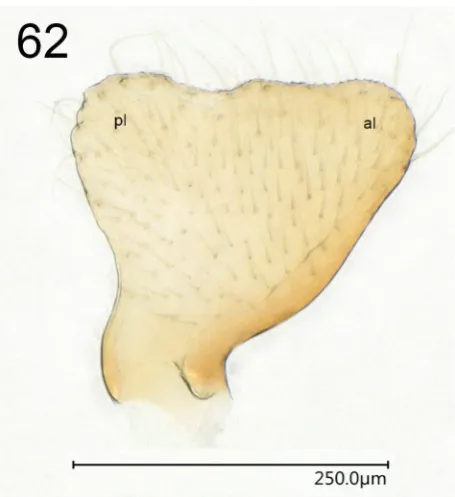
Figs 55–61. Symphylax handschini kinabaluensis subsp. nov., holotype, genitalia. 55 – abdomen tip, dorsal view. 56 – abdomen tip, lateral view. 57 – abdomen tip, caudal view. 58 – pygophore with genitalia, dorsal view. 59 – pygophore with genitalia, lateral view. 60 – pygophore with genitalia, caudal view. 61 – hypandrium, lateral view. Abbreviations: hyp = hypandrium, p = paramere, par = parandrium, pp = posterior process of sternite VIII, pyg = pygophore. Abdominal segments are indicated with Roman numbers.
cover of short light setae getting slightly longer and darker on tarsi; sparse short setae on veins. Abdomen with adher-ing light longer setae on distal segments and especially on hypandrium (Figs 56–57, 61).
Structure. Head (Fig. 51) of a length comparable to
Phaenacantha species, with a median sulcus starting
52). Labium reaching beyond middle coxae. Antennae thin, much longer than body, most segments (especially I and IV) slightly to moderately curved, left antennomere IV vestigial (Fig. 49). Pronotum with collar similar to that in
Phaenacantha species (Fig. 51). Middle lobe of pronotum with convex lateral outline, broader than collar; posterior lobe with almost straight lateral outline, same width as middle lobe. Middle and posterior lobe ca. in the same plane (Fig. 52), both lobes clearly convex in dorsal out-line, separated by a deep transverse impression; posterior lobe only slightly elevated above the head. Hind third of posterior pronotal lobe gradually roundedly sloped poste-riad; hind margin with weakly concave median part and two small rounded lateral lobes (Fig. 51). Scutellar spine clearly longer than posterior pronotal lobe, slightly inclined anteriad (the angle to the plane of meso- and metanotum 95–100°), somewhat curved posteriad. Legs generally at least as long as or longer than body, hind legs the longest (Figs 49–50); femora slightly bent and widened distally, fore femora distally with a long straight spine and two small accessory spinules (Fig. 53). Fore tibiae widened distally, with a median groove carrying terminally a comb of setae (Fig. 54). Fore and hind wings well developed (specimen macropterous), fore wings ending very close to tip of abdomen. Abdomen slimmest at segments II and III and broadest at V, difference between broadest and thinnest part larger than in studied Phaenacantha species (Fig. 49). Bor-ders between abdominal segments clear, segment borBor-ders smooth and not elevated. Lateral segment outlines straight or almost straight to slightly convex. Posterior border of segment VII in lateral outline almost straight (Fig. 56). Segments VIII and IX (pygophore) sunken into segment VII, sternite VIII with posterior median process sticking out (Figs 56–57). Pilosity on pygophore short, except setae on dorsal region of hypandrium (Figs 58–61). Hypandrium
building sharply ending parandria directed dorsoanteriad (Figs 58, 60–61); pygophore rounded posteriorly (Fig. 59); posterior margin of hypandrium in lateral view almost building an angle (Fig. 61). Paramere (Figs 59, 62) spade-like, with broad dorsal margin with a concavity slightly posteriad from the middle, not forming processes, with anterior and posterior lobes well developed.
Measurements. Body length 6.80 mm; head width (= max. body width) 1.31 mm. Total antenna length 10.47 mm (average segment ratios, I to IV: 1.00-1.49-1.52-1.98). Ratio antenna : body 1.54. Labium length 2.01 mm. Dis-tance between ocelli 0.12 mm, between ocellus and eye 0.22 mm, ratio 1.83. Length of collar + middle lobe of pronotum 0.47 mm; length of posterior pronotal lobe 0.82 mm, ratio of the two measurements 1.74. Scutellar spine tip broken, therefore the length not measurable. Measurements of the hind tarsus (segments I / II / III, respectively): 0.93 / 0.17 / 0.25 mm, total length of the hind tarsus 1.35 mm. Maximum width of abdomen 1.23 mm, minimum width 0.43 mm, ratio 2.86.
Etymology. The name of this subspecies refers to Kinabalu National Park where this specimen was collected; adjective.
Distribution. Malaysia (Sabah).
Discussion
Genus Phaenacantha, whose 33 representatives
de-scribed to date were recorded from Southern India and Shandong Province of China to New Guinea, Australia and Fiji, was not previously known from Borneo or New Caledonia. It is no surprise to fi nd representatives on these islands, but still this fi lls some gaps in yet very incomplete knowledge of this taxon. It is interesting that
morphologi-cally both P. grimmae and P. nigrispina from Borneo and
P. paveli from New Caledonia have affi nities mostly to species from New Guinea: P.ambigua, P. biroi, P. conso-brina, P. distincta and P. suturalis, in case of the species from Borneo; and P. conviva, P. mehelyi and P. sedula, in case of P. paveli. Of all these species, only P. ambigua is
known from Ceylon and P. sedula from Burma, Singapore
and Sumatra; the rest were described from New Guinea. This information might be useful for future workers trying to hypothesize phylogenetic relationships of the genus.
Patterns of speciation that led to the diversity of Phae-nacantha are poorly studied (as are many other aspects of the family Colobathristidae). This genus includes species
with large geographic ranges, e.g., Phaenacantha (
Anoryg-ma) lobulifera Horváth, 1916, that was initially described from Shandong Province in China and was later found in
Fujian, Guangxi and Yunnan (HSIAO 1977) or even Thailand
(material viewed at MMBC) – along with species occurring almost sympatrically, as e.g. many species known from
New Guinea or P. nigrispina and P. grimmae, whose type
localities are within the same mountain range, separated by only about 100 kilometers. It would be interesting to learn what features of biology allow some species main-tain their unity across thousands of kilometers (if they are indeed single species and not complexes of cryptic species), whereas others obviously experienced speciation events on much smaller spatial scale. The third dimension
– elevation above the sea level – might play a role. New
Guinea, the island with the highest known Phaenacantha
diversity, is famous for its mountains. The type localities of P. nigrispina (500 m above sea level) and P. grimmae
(900–1200 m) in Borneo clearly differ in elevation. Host specifi ty might also infl uence speciation.
Phaenacantha may include many more undescribed species, as the survey of museum collections at SDEI, MMBC, SMNS or ZMHB has demonstrated. In contrast to the statement of ŠTYS & HENRY (2015), it was found
that at least the collections visited by the author do contain
dozens of quite fresh Phaenacantha specimens (as does the
collection in National Museum, Prague – P. Kment, pers. comm.) – although in most cases Colobathristidae were collected only incidentally, as, e.g., the specimens found in Borneo by Wolfgang Schawaller and Dorothee Grimm. The collection at SMNS harbours two more specimens of
Phaenacantha, a female from Borneo and a male from New Guinea. Both most likely belong to new species, but it was decided not to describe them until Horváth’s type specimens and more fresh material from Borneo (possibly including males) can be examined.
Symphylax is the second most diverse Oriental genus of Colobathristidae, with nine described species. In contrast to Phaenacantha where no representatives were known
from Borneo, several species of Symphylax are recorded
for the island. The specimen from SMNS looks similar to
one of them, S. handschini (see the differential diagnosis
above), but at the same time there is a number of differences in the structure of parameres, coloration or the length of
labium. Bearing in mind some cases of intraspecifi c
char-acter variation reported by ŠTYS (1977a) that especially
concern coloration and length of labium and allopatric occurrence of the SMNS specimen (Kinabalu NP, northern
part of Sabah, Malaysia) and the holotype of S. handschini
(Pelawan Besar in Kalimantan Timur, Indonesia, some 300 kilometers to the south across a mountain range), it was concluded that the SMNS specimen most likely belongs
to a new subspecies of S. handschini. Careful study of the
holotype along with any fresh material from Borneo would be helpful to clarify the status of this taxon further.
Trichocentrus Horváth, 1904 is the third best
represent-ed colobathristid genus (after Phaenacantha and
Symphy-lax) in the collections visited in preparation for this study.
SMNS material also includes a specimen of Trichocentrus
similar to T. gibbosus Horváth, 1904.
It is worth noting for later studies on Phaenacantha
(and most likely other Colobathristidae as well) that the
restoration technique of defl ated specimens using sodium
triphosphate (e.g., HANSON PRITCHARD & KRUSE 1983) might
lead to the loss of original coloration of the animals, as
happened to the holotype of P. paveli in the present study
– although the foregoing incubation in acetone and alcohol might have had an impact, too.
Acknowledgements
Lars Krogmann (State Museum of Natural History, Stuttgart) kindly provided access to SMNS collection. Many of the staff of the same institution helped with
advice, equipment, materials, instructions on using the software etc., among them Susanne Grube, Michael Haas, Milan Pallmann, Hossein Rajaei and Dominic Wanke; I am especially grateful to Tanja Schweizer for lending an experienced hand in some complicated cases of specimen preparation. Wolfgang Schawaller and Dorothee Grimm shared valuable information on their collecting trip to
Borneo and Susanne Leidenroth (SMNS) helped fi nding
contact information of D. Grimm. Jürgen Deckert (Museum of Natural History, Berlin) granted access to the collection of ZMHB and kindly provided advice, material and equip-ment, as did Lukas Kirschey from the same institution. Stephan Blank and Angelika-Katrin Wanda Weirauch (both German Entomological Institute, Müncheberg) kindly provided access to the collection of SDEI and loaned
specimens of Phaenacantha, as did Petr Baňař (Moravian
Museum, Brno, Czech Republic) from the MMBC collec-tion; he also provided access to much of the material left by the late Professor Štys. I also thank Petr Baňař, Jürgen Deckert, Petr Kment (National Museum, Prague, Czech Republic), Péter Kóbor (National Museum, Budapest, Hungary) and Maria Tavano (Museum of Natural History Giacomo Doria, Genoa, Italy) for informative discussions that helped in writing this article. I am especially grateful to Petr Kment for inviting me to submit an article for the volume honouring Professor Pavel Štys and for his help and patience as its editor. Notes and suggestions of Thomas J. Henry (National Museum of Natural History, Smithsonian Institution, Washington D.C., USA) and Elöd Kondorosy (University of Pannonia, Keszthely, Hungary), who reviewed the fi rst version of this article, helped very much to improve its quality.
References
BERGROTH E. 1910: Remarks on Colobathristidae with descriptions of three new genera. Annales de la Société Entomologique de Belgique
54: 297–305.
BERGROTH E. 1914: Three new Heteroptera from Ceylon. Annales de
la Société Entomologique de Belgique58: 183–188.
BREDDIN G. 1904: Noch einiges über Colobasiastes Bredd. (Rhynchota heteroptera). Wiener Entomologische Zeitung 23: 245–250. CARVALHO J. C. M. & HENRY T. J. 1986: Sobre um gênero novo
peculiar da família Colobathristidae (Hemiptera) da região de Carajás (Pará, Brasil). Boletim do Museu Paraense Emílio Goeldi. Zoologia
2: 85–91.
COSCARÓN M. D. C. & DELLAPÉ P. M. 2003: A new species of Trichocentrusfrom Ecuador (Heteroptera: Colobathristidae), with a key to the species of the genus. Zootaxa 213: 1–8.
DELLAPÉ P. M. & HENRY T. J. 2019: Lygaeoidea species fi le. Retrieved from http://lygaeoidea.speciesfi le.org/HomePage/Lygaeoidea/Home-Page.aspx on 07.05.2019
GHAURI M. S. K. 1968: Notes on Colobathristidae (Heteroptera), including descriptions of new species and a suspected virus vector of Musa in Sabah. Proceedings of the Royal Entomological Society
of London B37: 80–88.
HANSON PRITCHARD M. & KRUSE G. O. 1984: Making the best of things: reclaiming specimens. Systematic Parasitology 6: 253–255. HENRY T. J., DELLAPÉ P. M. & DE PAULA A. S. 2015: The big-eyed
bugs, chinch bugs, and seed bugs (Lygaeoidea). Pp. 459–514. In: PANIZZI A. R. & GRAZIA J. (eds.): True bugs (Heteroptera) of the
Neotropics. Springer, Dordrecht, Heidelberg, New York, London, 904 pp.
HORVÁTH G. 1904: Monographia Colobathristinarum. Annales Musei
HORVÁTH G. 1908: Colobathristinae et Heterogastrinae novae in Museo Nationali Hungarico. Annales Musei Nationalis Hungarici
6: 591–595.
HORVÁTH G. 1912: Hemipteren aus Java. Tijdschrift voor Entomologie
55: 338–346.
HORVÁTH G . 1914: Miscellanea hemipterologica, XIII–XVII. XIII. Colobathristidarum species novae. Annales Musei Nationalis
Hun-garici12: 623–660.
HORVÁTH G. 1916: Colobathristidarum species nova. Annales Musei
Nationalis Hungarici14: 422.
HORVÁTH G. 1922: Colobathristidae novae in Musei nationali hungar-ico. Annales Musei Nationalis Hungarici19: 154–160.
HSIAO T. Y. 1977: Colobathristidae. Pp. 288–290, 305. In: HSIAO T. Y., REN SH.-ZH., ZHENG L.-Y., JING H.-L. & LIU S. L. (eds.): A
Hand-book for the Determination of the Chinese Hemiptera-Heteroptera.
Vol. I. Science Press, Beijing, 382 pp. (in Chinese, English summary),
KIRKALDY G. W. 1908: Memoir on a few heteropterous Hemiptera from eastern Australia. Proceedings of the Linnean Society of New
South Wales32: 768–788.
KMENT P. & VILÍMOVÁ J. 2010: Thoracic scent efferent system of Pentatomoidea (Hemiptera: Heteroptera): a review of terminology.
Zootaxa2706: 1–77
KORMILEV N. A. 1949a: Notas sobre los Colobathristidae de Bolivia con la descripción de un género y una especie nuevos (Hemiptera).
Notas del Museo de La Plata14, Zoologia124: 167–176.
KORMILEV N. A. 1949b: La familia Colobathristidae Stål en la Ar-gentina con la descripción de tres especies nuevas neotropicales (Hemiptera). Acta Zoológica Lilloana7: 359–383.
KORMILEV N. A. 1951: Notas sobre ‘‘Colobathristidae’’ neotropicales (Hemiptera), con la descripción de tres géneros y siete especies nuevas. Revista Brasileira de Biologia11: 63–84.
KORMILEV N. A. 1953: Notes on Colobathristidae from Borneo and Java in the collections of the Museum of Natural History in Basle (Hemiptera). Mitteilungen der Schweizerischen Entomologischen
Gesellschaft 26: 287–292.
ŠTYS P. 1966a: Revision of the genus Dayakiella Horv. and notes on its systematic position (Heteroptera: Colobathristidae). Acta
Entomo-logica Bohemoslovaca63: 27–39.
ŠTYS P. 1966b: Morphology of the wings, abdomen and genitalia of Phaenacantha australiae Kirk. (Heteroptera, Colobathristidae) and notes on the phylogeny of the family. ActaEntomologica Bohemoslo-vaca63: 266–280.
ŠTYS P. 1974: A synopsis of the genus Phaenacantha Horv., I. Subgenus Anorygma Horv. (= Tagalisca Horv.) (Heteroptera, Colobathristi-dae). Annales Historico-Naturales Musei Nationalis Hungarici66: 225–234.
ŠTYS P. 1977a: Revision of Symphylax (Heteroptera: Colobathristidae).
Acta Zoologica Academiae Scientiarum Hungaricae23: 427–451.
ŠTYS P. 1977b: Symphylax horvathi sp. n. from Borneo (Heteroptera, Colobathristidae). Reichenbachia16: 229–232.
ŠTYS P. & EXNEROVÁ A. 2013: A new genus and species of Oriental Colobathristidae (Hemiptera: Heteroptera) with a key to Eastern Hemisphere genera and morphological and functional considerations.
EntomologicaAmericana118: 53–65.
ŠTYS P. & HENRY T. J. 2015: A new genus and species of Colobath-ristidae (Hemiptera: Heteroptera) from Peru, a replacement name for the preoccupied genus Labradoria Kormilev, and a key to the Neotropical genera. Proceedings of the Entomological Society of
Washington 117: 27–36.
SU Y. N. 2016: A simple and quick method of displaying liquid-preserved morphological structures for microphotography. Zootaxa 4208(6): 592–593.
SWEET M. H. 2000: Seed and chinch bugs. Pp. 143–264. In: SCHAEF-FER C. W. & PANIZZI A. R. (eds.): Heteroptera of economic
im-portance. CRC Press, Boca Raton, London, New York, Washington,