1
Dr. Venkata Naga Prahalada Karnati
2
Sreekar kumar reddy .R
CORRESPONDING AUTHOR
1
Dr.Venkata Naga Prahalada Karnati
Lecturer,
Department of physiotherapy, College of Applied Medical Sciences, Shaqra University, Shaqra,
Saudi Arabia.
Int J Physiother. Vol 2(1), 352-360, February (2015) ISSN: 2348 - 8336
ABSTRACT
Background: Conventional back care exercises are advocated to treat the pain and to strengthen the involved muscles. There will always be the possibility of the pain getting recurred due to disproportionate balance and stability in the muscles. The core stabilization is major trend in rehabilitation. It aims at improving stability during functional activities, balance, flexibility, strength training and effectively manage the pain as well.
Methods: Forty patients with chronic Mechanical Low back pain were randomly assigned into control group that received conventional back exercises and SWD (n=20), experimental group received core stabilization and SWD (n=20). Both the groups received SWD, along with conventional back exercises for one-group and core stabilization for the other group 3 days a week for 6 weeks .The treatment outcome was assessed using visual analogue scale, Rolland Morris Disability Questionnaire and Lumbar range of motion by using goniometer.
Results:After a 6 week training period the core stabilization group scored significantly higher than the conventional group for VAS (p=0.05) and RMDQ (p=0.05) where as ROM improved higher in conventional group (p=0.05)
Conclusion: After the treatment sessions Core stabilization group registered a significant improvement when compared to conventional back care exercises in improving function and in relieving pain.
Key words: core stabilization, conventional exercises, Mechanical low back pain, Physio ball, VAS, RMDQ, and ROM.
2Professor,
Narayana College of Physiotherapy, Nellore, Andhra Pradesh, India.
Received 14th October 2014, revised 24th November 2014, accepted 02th December 2014
DOI: 10.15621/ijphy/2015/v2i1/60041
INTRODUCTION
Low back pain is defined as the pain that occurs in an area with boundaries between the lowest rib and the crease of the buttocks.1 Low backache is a discomfort in the area of the lower part of the back and spinal column.2 Low back pain is associated with deconditioning of spine and trunk due to lack of core strength and stability in which 60- 80% of
general population suffer with high recurrence
rates of 60 – 85 % within following three years.3 Chronic Low back pain is the pain that persists longer than the expected time period for healing,
with a duration of more than three months.4
Most low back injuries are not the result of a single exposure to a high magnitude load, but instead due to cumulative trauma from sub-failure-magnitude loads like repeated small loads (e.g. bending) or a sustained load (e.g. sitting). Low back injury results from repetitive motion at end range as a result of a history of excessive loading which gradually, but progressively, reduces the tissue failure tolerance.
6Mechanical low back pain is a cumulative process
resulting from chronic poor posture coupled with sedentary habits that put the back under severe
mechanical stress.7A wide range of conservative
interventions has been advocated for the treatment of low back pain when it is chronically symptomatic. These interventions include orthotic bracing, flexion exercises, abdominal trunk curls, hamstring stretching, pelvic tilt exercises, and general aerobic exercise such as swimming and walking.8
These conventional back care exercises decrease the pain and increase the strength of involved muscles, but results in frequent recurrence rates because of their effectiveness only up to one year and patients are left out with some residual pain and disability.
The conventional back exercises strengthen the involved muscles like abdominals, which are ineffective after 45 degrees of trunk curls.9 The human spine buckles invitro during a compressive load of 90 N but the spine is loaded of about 4000 – 6000 N, while administering various back extension exercises like prone lying and lifting one leg, alternate leg and arm lifts, lifting upper trunk and both legs off the floor.10 The efficacy of general back exercises however, appears limited in achieving these goals.11
Lumbar instability is considered to be a significant factor in patients with chronic low back pain.12 Spinal instability is described as a significant decrease in the capacity of the stabilizing systems of the spine to maintain the intervertebral neutral zones within physiological limits so that there is no
neurological dysfunction, no major deformity, and no incapacitating pain.
A conceptual model of the spinal stabilization system was introduced by Punjabi, which describes the interaction between components providing stability in the spine. This model redefined the notion of spinal instability in terms of a region of laxity around the neutral resting position of a
spinal segment, that he terms the ‘neutral zone.’13
The large load-carrying capacity of the spine is achieved by the participation of well-coordinated muscles surrounding the spinal column. The role of multifidus, transverses abdominus, diaphragm and pelvic floor, as well as those muscles working across the pelvic region, play an integral role in the dynamic stability of the lumbar and lumbopelvic regions.14
A link has been established between dysfunction in the local muscle system and back pain, which has lead to a concept of therapeutic exercise to enhance lumbar and lumbopelvic stabilisation, based on the specific rehabilitation of both the
global, and the local muscle system.15
A recent focus in the physiotherapy management of patients with CLBP has been the specific training of muscles surrounding the lumbar spine whose primary role is considered to be the provision of dynamic stability and segmental control to the
spine40. These are the deep abdominal muscles
(internal oblique) and transversus abdominis and the lumbar multifidus. The importance of LM muscle regarding its potential to provide dynamic control to the motion segment in its neutral zone is
now well acknowledged.16
The deep abdominals, in particular the TA, are primarily involved in the maintenance of intraabdominal pressure, while imparting tension to the lumbar vertebrae through the thoracolumbar fascia.17 It is considered that the role of the deep abdominal muscles acting in co-contraction with the LM is to provide a stiffening effect on the lumbar spine through its attachment to the thoracolumbar fascia, in conjunction with an
increase in intraabdominal pressure. In addition,
there is increasing evidence that these muscles are preferentially affected in the presence of low back
pain and lumbar instability.18
The aims of core stability training is to effectively recruit the trunk musculature and then learn to control the position of the lumbar spine during
dynamic movements.19
Core stabilization exercises facilitate
transfer the loads equally and to make the patient functionally active. Swiss ball exercise can improve nervous system function that results in functional
strength gain.20The abdominal hollowing exercises
decrease the compressive loads on the spine by 40%.
Many recent studies have proved that spinal stabilization exercises are more effective than
conventional back exercises in improving
functional status and lessen the behavioral, cognitive and disability aspects of low back pain syndrome. But there are some conflicting reports that core strengthening is not significant to
decrease the low back pain.21
Core stabilization is most effective on dynamic surfaces in order to recruit proprioceptive,
kinesthetic and balance system.22
Though conventional back care exercises and core stabilization exercises are proved to be effective in chronic mechanical low back pain patients, no literature comparing the effectiveness on each other were found which necessitated the present study to compare the outcome of conventional and core stabilization exercises in chronic mechanical low back pain.
METHODOLOGY
40 subjects with age group between 30-50 years, Both male and female patients, Postural
predisposition (both mechanical and
occupational), were taken selected from the outpatient department of physiotherapy, patients with cardio –pulmonary diseases, tumor, infection and fracture, rheumatic and inflammatory condition, disc disease, Lumbar strain or spra, Lumbar canal stenosis, Bowel and bladder dysfunction were excluded, divided in two groups of 20 each selected randomly both male and female of age group 30-50 with the diagnosis of chronic mechanical low back pain. in two groups of 20 each selected randomly both male and female of age group 30-50 with the diagnosis of chronic mechanical low back pain, divided into two groups, Group A: Control group 20 patients, Group B: Experimental group 20 patients
Ethical Clearance was obtained from the concerned authorities of the institution.
Informed consent was taken from the patients prior to the evaluation and treatment sessions. 40 patients were randomly selected and equally divided into control and experimental groups of 20 each. An Orthopaedic evaluation was done prior to the study to rule out other causes of backache. Pain was measured on visual analog Scale and each patient was asked to fill the Rolland Morris low back pain and disability questionnaire.
Group A
Short wave diathermy was given for 15 minutes prior to starting the exercises to relieve pain.51 The patients in the control group were treated with conventional back exercise program for 3 days a week for 6 weeks.
Exercise 1: supine lying – Leg lifts
The patient in supine lying was asked to lift one leg first and hold it for five seconds and return to neutral position and repeat the same for other leg. Later both the legs were made to lift simultaneously, holding them for five seconds and bringing them back to neutral position.
Exercise 2: Abdominal crunches in crook lying position
The patient in crook lying was asked to place the hands behind the head and lift the trunk upwards, rotate to either side to reach the knees and hold the position for five seconds then bring them back to neutral position.
Exercise 3: Prone lying – Leg lifts
The patient in prone lying was asked to lift one leg first and hold it for five seconds then bring it to neutral position and repeat the same for other leg. Later made to lift both the legs simultaneously, hold them for five seconds, and then bring them back to neutral position.
Exercise 4: Prone lying – Trunk lifts
The patient in prone lying was asked to keep the hands along the side of the body, lift the trunk off the floor and hold the position for five seconds, then bringing it back to neutral position.
*Each of these exercises was given for ten repetitions per session.
Group B:
Short wave Diathermy was given for 15 min before the exercise session to relieve pain. Patients in experimental group were treated with core stabilization exercises for 30 min of 10 repetitions each with 10 sec hold and adequate rest was given between each repetition. The training session was scheduled for 3 days a week for 6 weeks.
The Exercises given were as follows: Exercise 1:
Exercise 2:
Patient lying on his back with calves resting on the ball was asked to rock very slowly side-to-side with normal breathing.
Exercise 3:
The patient in supine lying on the floor with feet on the ball and ankles together, arms behind the buttocks, using the thigh and abdominals asked to straighten the legs and hold it for 10 seconds then bring them back to neutral position.
Exercise 4:
The patient in prone lying on physio ball was asked to lift one leg and contralateral arm, d hold it for 10 seconds, bring them back to neutral position. *Each of these exercises was given for ten repetitions per session.
After 6 weeks of training program, the patients were reassessed on the basis of pain rating on VAS and disability rating on the Rolland Morris Disability Questionnaire and ROM by using Goniometer.
STASTISTICAL ANALYSIS
A group of 40 patients were randomly assigned into two groups of 20 in each (n=20) into Control group
(n=20), Experimental group (n=20), which were analyzed for their normality and homogeneity by using one-way ANOVA. This analysis has shown that all the groups were homogeneous and hence were analyzed for their significance by using independent t- test. This analysis has shown significance in relation to decrease in pain, improving the functional outcome and disability at p=0.05 in core stabilization group when compared to control group.
RESULTS
The following is the statistical analysis done in this study:
A total of 40 patients (n=40) were randomly assigned to Group-A (n=20) and Group-B (n=20). The data collected were analyzed for the following outcome measures as variables.
1. Rolland Morris Disability Questionnaire
2. Visual Analogue Scale
3. Lumbar Range of motion
All these variables were tested for normality of data using graphical analysis.
It was further evaluated for consistency of data
Rolland Morris Disability Questionnaire
Table-1: Test for Homogeneity of Pre-test variables for RMDQ scale Sum of
Squares Df
Mean
Square F Sig. Std. deviation t-value P value
Between Groups 27.950 6 4.658 1.433 .275 1.394538 2.308 0.05
Within Groups 42.250 13 3.250 6.734827
Total 70.200 19
The homogeneity of the data in the two groups was analyzed by using one-way ANOVA, which showed that the significance was greater than p=0.05 and hence both the groups were homogenous, for pre-test RMDQ scores.
VAS GROUP
Table-2: Test for Homogeneity of Pre-test variables for VAS scale Sum of
Squares df
Mean
Square F Sig.
Std.
Deviation t-value P value
Between Groups 9.583 4 2.396 2.331 .103 0.933 4.098 0.05
Within Groups 15.417 15 1.028 0.994
Total 25.000 19
The data collected was analyzed for homogeneity between the groups and within the groups using ANOVA table holding control group as the defining variable.
RANGE OF MOTION FLEXION GROUP
Table -3: Test for Homogeneity of Pre-test variables for Flexion Group Sum of
Squares df
Mean
Square F Sig.
Std.
Deviation t-value P value
Between Groups 41.783 6 6.964 1.541 .241 3.704 0.293 0.05
Within Groups 58.767 13 4.521 2.673
Total 100.550 19
The data collected was analyzed for homogeneity between the groups and within the groups using ANOVA table holding control group as the defining variable.
It was shown that all the values calculated had a significance greater than p=0.05 and hence the data are considered homogenous for Flexion Group.
EXTENSION GROUP
Table-4: Test for Homogeneity of Pre-test variables for Extension Group Sum of
Squares df
Mean
Square F Sig.
Std.
Deviation t-value P value
Between Groups 45.217 8 5.652 .539 .805 3.281 0.855 0.05
Within Groups 115.333 11 10.485 3.726
Total 160.550 19
The data collected was analyzed for homogeneity between the groups and within the groups using ANOVA table holding control group as the defining variable.
It was shown that all the values calculated had a significance greater than p=0.05 and hence the data are considered homogenous for Extension Group.
RIGHT SIDE FLEXION GROUP
Table-5: Test for Homogeneity of Pre-test variables for Right Side Flexion Group Sum of
Squares df
Mean
Square F Sig.
Std.
Deviation t-value P value
Between Groups 33.786 6 5.631 .725 .638 2.384 1.186 0.05
Within Groups 101.014 13 7.770 2.723
Total 134.800 19
The data collected was analyzed for homogeneity between the groups and within the groups using ANOVA table holding control group as the defining variable.
It was shown that all the values calculated had a significance greater than p=0.05 and hence the data are considered homogenous for Right Side flexion Group.
LEFT SIDE FLEXION GROUP
Table-6: Test for Homogeneity of Pre-test variables for Left Side Flexion Group Sum of
Squares df
Mean
Square F Sig.
Std.
Deviation t-value P value
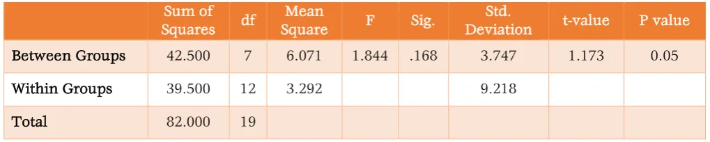
Between Groups 42.500 7 6.071 1.844 .168 3.747 1.173 0.05
Within Groups 39.500 12 3.292 9.218
The data collected was analyzed for homogeneity between the groups and within the groups using ANOVA table holding control group as the defining variable.
It was shown that all the values calculated had a significance greater than p=0.05 and hence the data are considered homogenous for Left Side Flexion Group.
RIGHT ROTATION GROUP
Table-7: Test for Homogeneity of Pre-test variables for Right Rotation Group Sum of
Squares df
Mean
Square F Sig.
Std.
Deviation t-value P value
Between Groups 53.000 7 7.571 1.284 .335 3.883 1.173 0.05
Within Groups 70.750 12 5.896 4.773
Total 123.750 19
The data collected was analyzed for homogeneity between the groups and within the groups using ANOVA table holding control group as the defining variable.
It was shown that all the values calculated had a significance greater than p=0.05 and hence the data are considered homogenous for Right Rotation Group.
LEFT ROTATION GROUP
Table-8: Test for Homogeneity of Pre-test variables for Left Rotation Group Sum of
Squares df
Mean
Square F Sig.
Std.
Deviation t-value P value
Between Groups 35.943 9 3.994 .415 .901 2.874 2.067 0.05
Within Groups 105.867 11 9.624 4.193
Total 141.810 20
The data collected was analyzed for homogeneity between the groups and within the groups using ANOVA table holding control group as the defining variable.
It was shown that all the values calculated had a significance greater than p=0.05 and hence the data are considered homogenous for Left Rotation Group.
Note: The ANOVA table gives the F-values for significance of variance and as all the values have significance greater than 0.05 hence the groups are considered homogenous.
Data Analysis for significance of improvements between the groups
Group- A data analysis
The data showed that the mean improvements in
conventional training group is 5.35 0.933 for VAS
scale, 10.55 1.395 for RMDQ, 15.43.704 for
flexion, 16.853.281 for extension, 222.384 for Right side flexion, 24.43.747 for Lt side flexion, 25.73.883 for Right rotation, 26.052.875 for Lt rotation. This clearly indicates that all the patients in this group have showed improvements in all the three categories of outcome measures.
Group- B data analysis
The data in this group of patients showed mean improvements in all categories with VAS
improvements being 6.6 0.995, for RMDQ 14.1
6.735, 15.1 2.673 for flexion, 15.9 3.726 for extension, 21.05 2.723 for Right side flexion, 22.85 9.218 for Lt Side flexion, 24.45 4.773 for Right rotation, 23.7 4.193 for Lt rotation.
The calculated p value showed a significance of difference in improvement at p=0.05, which indicates that conventional group has higher gains
in improvement in left rotation than the Core strengthening group.
Table-9: Mean Improvements between the Groups
Parameter Group A Group B t-Values
Mean S.D Mean S.D
VAS 5.35 0.933 6.6 0.995 4.0983
Rolland Morris 10.55 1.395 14.1 6.735 2.308
Flexion 15.4 3.704 15.1 2.673 0.2936
Extension 16.85 3.281 15.9 3.726 0.855
Rt. Side Flex 22 2.384 21.05 2.723 1.173
Lt. Side Flex 24.4 3.747 22.85 9.218 0.696
Rt. Rotation 25.7 3.883 24.45 4.773 0.872
Lt. Rotation 26.05 2.875 23.7 4.193 2.067
Figure-1: Mean Improvements
DISCUSSION
This study is done on 20 patients in each group, with 11 males and 9 females in conventional group and 11 males and 9 females in core stabilization group.
The patients in group A showed improvements in VAS score with a mean of 5.35 and in Rolland Morris Disability Questionnaire with a mean of 10.55. These patients also shown improvements in flexion, extension, side flexion and rotation at p=0.05.
The patients in Group B also showed
improvements in VAS scores with a mean of 6.6 and Rolland Morris Disability Questionnaire with a mean of 14.1. These patients also shown improvements in flexion, extension, side flexion and rotation at p=0.05.
In case of Group A improvements in ROM is slightly higher than that of Group B, this could be attributed to the reason that in Group A, the concentration is on strengthening the isolated
muscles, where as in group B the concentration is on strengthening the group muscles.
Though conventional back care exercises and core stabilization exercises are proved to be effective in chronic mechanical low back pain patients, the group that received core stabilization exercises shown more improvements in VAS with significance at p=0.05.
This is in accordance to the Mc Gill’s study that performing exercises on labile surfaces increased abdominal muscle activity, which changes both the level of muscle activity and the way that the muscles co- activate to stabilize the spine and the whole body. This suggests a much higher demand on motor control system, which may be desirable for rehabilitation program.
Group B patients showed improvements in their disability levels measured by Rolland Morris Disability Questionnaire as core stabilization creates a “girdle” of protection for the low back that
Mean Improvements
0 5 10 15 20 25 30
VAS
Rol land
Mor ris
Flexi on
Ext ensi
on
Rt. Side
Fle x
Lt. S ide
Flex
Rt. Rot
atio n
Lt. R otat
ion
challenge balance, postural trunk muscles, flexibility and coordination.
The results of this study support the initial hypothesis that specific exercise training of the "stability" muscles of the trunk is effective in reducing pain and functional disability in patients with chronically symptomatic low back pain. Analysis of the pain and functional disability revealed that there is a difference in improvements between both the groups. This treatment approach was more effective than other conservative treatment approaches which mainly involved conventional exercise programs.
This is in support of Punjabi’s hypothesis that the stability of the lumbar spine is dependent not solely on the basic morphology of the spine, but also the correct functioning of the neuromuscular system. Therefore, if the basic morphology of the lumbar spine is compromised, as in the case with symptomatic CLBP, the neuromuscular system may be trained to compensate, to provide dynamic stability to the spine during the demands of daily living.
Consistent with these findings, McGill reported
that lumbar stability is maintained in vivo by
increasing the activity (stiffness) of the lumbar
segmental muscles, and highlighted the
importance of motor control to coordinate muscle recruitment between large trunk muscles and small intrinsic muscles during functional activities, to ensure stability is maintained.
The other advantages of core stability
strengthening program is that, they apart from improving core strength and stability also improved flexibility, posture, ease of movement,
heightened body awareness, balance and
coordination
CONCLUSION
Supporting evidence from the literature though seems to be controversial in certain areas, the out come of this study with highly significant statistical changes will lead us to the conclusion of accepting the research hypothesis which could be stated as “Core stabilization program is more effective in the management of chronic mechanical low back pain than conventional exercises”.
REFERENCES
1. Shaughnessy, M. 1: Caulfield, B. A pilot study to
investigate the effect of lumbar stabilization exercise training on functional ability and quality of life in patients with chronic low back pain. International Journal of Rehabilitation Research.2004; 27(4): 297-301.
2. Hides JA, Jull GA, and Richardson CA:
Long-term effects of specific stabilizing exercises for first-episode low back pain. Spine. 2001; 26(11):243-8.
3. Carpenter DM, Nelson BW: Low back
strengthening for the prevention and treatment of low back pain, Med Sci Sports Exerc.1999; 31(1): 18-24.
4. Panjabi MM: Clinical spinal instability and low
back pain, J Electromyogr Kinesiol.2003;13(4): 371-9.
5. McGill SM: Low back exercises: evidence for
improving exercise regimens, Phys Ther. 1998;78(7):754-65.
6. Nelson BW, O'Reilly E: The clinical effects of intensive, specific exercise on chronic low back pain: a controlled study of 895 consecutive patients with 1-year follow up, Orthopedics. 1995; 18(10): 971-81.
7. Hodges, Paul W. Richardson: etal, Inefficient
Muscular Stabilization of the Lumbar Spine Associated with Low Back Pain: A Motor Control Evaluation of Transversus Abdominis Spine.1996; 21(22): 2640-2650.
8. Granata KP, Wilson SE: Trunk posture and
spinal stability. Clin Biomech (Bristol, Avon). 200;16(8): 650-9.
9. O'Sullivan PB: Lumbar segmental 'instability':
clinical presentation and specific stabilizing exercise management. Man Ther. 2000; 5(1):2-12.
10.Van Dieen JH, Cholewicki J, Radebold: A.
Trunk muscle recruitment patterns in patients with low back pain enhance the stability of the lumbar spine. Spine.2003; 28(8): 834-41.
11.Akuthota V, Nadler SF: Core strengthening,
Arch Phys Med Rehabil. 2004;85(3 Suppl 1): S86-92.
12.Yilmaz F, Yilmaz A: Efficacy of dynamic lumbar
stabilization exercise in lumbar
microdiscectomy, J Rehabil Med.2003; 35(4): 163-7.
13.Johannsen F, Remvig L: Exercises for chronic
low back pain: a clinical trial, J Orthop Sports Phys Ther.1995; 22(2): 52-9.
14.Gardner-Morse MG, Stokes IA: The effects of
abdominal muscle co activation on lumbar spine stability, Spine.1998;23(1): 86-91.
15.Francisco J Vera-Garcia, Stuart M McGill etal: Abdominal Muscle Response During Curl-ups on Both Stable and Labile Surfaces. Phys Ther. 2000 Jun;80(6):564-9.
16.R S Jemmett: Rehabilitation of lumbar
17.Carpenter DM, Nelson BW: Low back strengthening for the prevention and treatment
of low back pain, Med Sci Sports
Exerc.1999;31(1): 18-24.
18.Mior S: Exercise in the treatment of chronic pain, Clin J pain.2001;17(4): S77-85.
19.Richardson, Carolyn, et al, Therapeutic
Exercise for Spinal Segmental Stabilization in Low Back Pain. Churchill Livingstone, Edinburgh, 1999.
20.Marshall PW, Murphy BA. Core stability
exercises on and off a Swiss ball. Arch Phys Med Rehabil.2005; 86(2): 242-9.
21.Cosio-Lima LM etal: Effects of physioball and conventional floor exercises on early phase adaptations in back and abdominal core stability and balance in women. J Strength Cond Res. 2003;17(4): 721-5.
22.Koes BW, Bouter LM, Beckerman H, van der
Heijden GJ, Knipschild PG. Physiotherapy exercises and back pain: a blinded review. BMJ, 1991;302: 1572-6.
Citation
Dr. Venkata Naga Prahalada Karnati, Sreekar kumar reddy .R. (2015). CORE STABILIZATION PROGRAM AND CONVENTIONAL EXERCISES IN THE PATIENTS WITH LOW BACK PAIN”-A