NECROTISING FASCIITIS; IMPORTANCE IN MAXILLOFACIAL SURGERY
A REVIEW WITH CASE REPORT
Dr. Dilip Kumar, R., *Dr. Madhukar Natekar
Dr. Prashanth
Department of Oral and Maxillofacial Surgery
Kumaraswami Layout,
ARTICLE INFO ABSTRACT
Necrotising
necrotizing process that has high rate of fatality. It can occur in an occult fashion wherein the necrosis progresses within the fascia and subcutaneous tissue
intact. Unless a high degree of suspicion is expressed by the surgeon this condition can go unnoticed and lead to death. In maxillofacial region, infections of odontogenic origin and peri
are the main causative factors, responsible for this condition .The possibility of acquiring this disease is accentuated in patients with on low immunity status like diabetes. In this review we analyse a detailed review of this morbid disease and present two
Copyright©2017, Dr. Dilip Kumar et al. This is an open access article distributed under the Creative Commons Att use, distribution, and reproduction in any medium, provided the original work is properly cited.
INTRODUCTION
Necrotizing fasciitis is a rare life-threatening, multi
soft tissue infection characterized by progressive, usually rapid, necrotizing process of the sub-cutaneous tissues and fascial planes, with resulting skin gangrene and systemic toxicity (Reed and Anand, 1992; Scott et al., 1994
1988; Valko et al., 1990; Freischlag et al
terminology, Necrotizing refers to "causing the death of tissues" and fasciitis signifies “inflammation of the fascia of a muscle or organ”. Necrotizing fasciitis is rarely seen in head and neck region and when it so occurs, it tends to have a high morbidity owing to its proximity to vital structures in this region (Shaikh et al., 2012). It occurs as a spread of infection from teeth or pharynx. Occlusion of vascular
compromise, spread of infection to plural and mediastinal spaces are known to occur with the progression of the disease (Carter and Banwell, 2004; Vaid et al., 2002
1997). Patients with low immunity, especially those
with Diabetes Mellitus, have been found to play a major role in initiation and progression of this disease (Shindo
Hence, early recognition and surgical intervention are the most
*Corresponding author:Dr. Madhukar Natekar,
Department of Oral and Maxillofacial Surgery, Dayananda Sagar College of Dental Sciences, Shavige Mallige Hills, Kumaraswami Layout, Bangalore 560078
ISSN: 0975-833X
Article History:
Received 17th December, 2016
Received in revised form 14th January, 2017
Accepted 20th February, 2017
Published online 31st March,2017
Citation: Dr. Dilip Kumar, R., Dr. Madhukar Natekar, Dr. H. P. Raghuveer, Dr. Shobha, E. S., Dr. Prashanth, N. T. and Dr. Vinod Rangan “Necrotising fasciitis; Importance in maxillofacial surgery a review with case report
Key words:
Review, Necrotising fascitis, Maxillofacial surgery, Odontogenic infection.
REVIEW OF LITERATURE
NECROTISING FASCIITIS; IMPORTANCE IN MAXILLOFACIAL SURGERY
A REVIEW WITH CASE REPORT
Madhukar Natekar, Dr. H. P. Raghuveer,
Prashanth, N. T. and Dr. Vinod Rangan
Department of Oral and Maxillofacial Surgery, Dayananda Sagar College of Dental Sciences,
Kumaraswami Layout, Bangalore-560078
ABSTRACT
Necrotising fasciitis is a rare soft tissue infection of soft tissues characterized by rapidly progressive, necrotizing process that has high rate of fatality. It can occur in an occult fashion wherein the necrosis progresses within the fascia and subcutaneous tissues leaving the upper muscular and dermal layer intact. Unless a high degree of suspicion is expressed by the surgeon this condition can go unnoticed and lead to death. In maxillofacial region, infections of odontogenic origin and peri
the main causative factors, responsible for this condition .The possibility of acquiring this disease is accentuated in patients with on low immunity status like diabetes. In this review we analyse a detailed review of this morbid disease and present two case reports with their effective management.
is an open access article distributed under the Creative Commons Attribution License, which use, distribution, and reproduction in any medium, provided the original work is properly cited.
threatening, multi-microbial soft tissue infection characterized by progressive, usually rapid, cutaneous tissues and fascial planes, with resulting skin gangrene and systemic toxicity 1994; Balcerak et al.,
et al., 1985). By
Necrotizing refers to "causing the death of fasciitis signifies “inflammation of the fascia of a s rarely seen in head and neck region and when it so occurs, it tends to have a high morbidity owing to its proximity to vital structures in this . It occurs as a spread of infection Occlusion of vascular structures, airway compromise, spread of infection to plural and mediastinal spaces are known to occur with the progression of the disease 2002; Ali and Zayed, with low immunity, especially those patients with Diabetes Mellitus, have been found to play a major role in Shindo et al., 1997). Hence, early recognition and surgical intervention are the most
Department of Oral and Maxillofacial Surgery, Dayananda Sagar College of Dental Sciences, Shavige Mallige Hills, Kumaraswami Layout,
Bangalore-important steps in determining the prognosis of this condition (Freischlag et al., 1985; Carter
Synonyms
It has been also called by other names including hospital gangrene (Brooks, 1966), Meleney’s gangrene (Meleney, 1924), hemolytic streptococcal gangrene, gangrenous erysipelas, flesh eating disease and synergetic necrotizing cellulitis (Shaikh et al., 2012; Carter
Historical background
Suppuration in the tissue beneath the skin
of pus spreading to surrounding healthy tissues has been mentioned in SusruthaSamhita
disease was narrated as early a Hippocrates. An army surgeon, Joseph Jones,
gangrene’ extensively and noticed 50% death ratein 2642 cases (Shaikh et al., 2012). The first reported case of this infection affecting the perineum was given by Baurienne in 1764 and Banwell, 2004). In 1883, a study conducted by Fournier who described and reported this infection in 5 young adults (Fournier's gangrene) (Shaikh et al
2004; Vaid et al., 2002). In 1924 Meleney reported on 20 cases of ‘necrotising erysipelas’ in China and was the first to
International Journal of Current Research
Vol. 9, Issue, 03, pp.48440-48444, March, 2017
INTERNATIONAL
OF CURRENT RESEARCH
Dr. Dilip Kumar, R., Dr. Madhukar Natekar, Dr. H. P. Raghuveer, Dr. Shobha, E. S., Dr. Prashanth, N. T. and Dr. Vinod Rangan
fasciitis; Importance in maxillofacial surgery a review with case report”, International Journal of Current Research
NECROTISING FASCIITIS; IMPORTANCE IN MAXILLOFACIAL SURGERY
, Dr. Shobha, E. S.,
agar College of Dental Sciences, Shavige Mallige Hills,
fasciitis is a rare soft tissue infection of soft tissues characterized by rapidly progressive, necrotizing process that has high rate of fatality. It can occur in an occult fashion wherein the necrosis s leaving the upper muscular and dermal layer intact. Unless a high degree of suspicion is expressed by the surgeon this condition can go unnoticed and lead to death. In maxillofacial region, infections of odontogenic origin and peri-tonsillar regions the main causative factors, responsible for this condition .The possibility of acquiring this disease is accentuated in patients with on low immunity status like diabetes. In this review we analyse a case reports with their effective management.
ribution License, which permits unrestricted
important steps in determining the prognosis of this condition and Banwell, 2004).
It has been also called by other names including hospital gangrene (Brooks, 1966), Meleney’s gangrene (Meleney, 1924), hemolytic streptococcal gangrene, gangrenous erysipelas, flesh eating disease and synergetic necrotizing
Carter and Banwell, 2004)
Suppuration in the tissue beneath the skin (sopha), with danger of pus spreading to surrounding healthy tissues has been mentioned in SusruthaSamhita (Shaikh et al., 2012). This disease was narrated as early as 5th century BC by . An army surgeon, Joseph Jones, studied ‘hospital gangrene’ extensively and noticed 50% death ratein 2642 cases The first reported case of this infection he perineum was given by Baurienne in 1764 (Carter . In 1883, a study conducted by Fournier who described and reported this infection in 5 young adults
et al., 2012; Carter and Banwell,
In 1924 Meleney reported on 20 cases of ‘necrotising erysipelas’ in China and was the first to INTERNATIONAL JOURNAL
OF CURRENT RESEARCH
establish the haemolytic streptococcus as the aetiological agent (Shaikh et al., 2012)The term necrotizing fasciitis was given by Wilson in 1952 when he described the principle chareteristics of the disease which included inflammation and necrosis of fat and deep fascia which did not effect the muscles (Carter and Banwell, 2004; Vaid et al., 2002).
Epidemiology and risk factors
The Incidence of Necrotizing fasciitis is 0.40 cases per 100,000 people in US population (Shaikh et al., 2012). It has been found that patients aged 50 years and older are at higher risk with incidence levels reaching as high as 12 per 100,000 over the age of 80 (Moss et al., 1990). No significant difference in incidence is observed between men or women, but children and younger individuals are also at risk of developing necrotizing fasciitis, varicella infection being a common predisposing factor (Moss et al., 1990; Lancerotto et al., 2012). Although it can occur anywhere in the body, the condition is more commonly seen in the upper (48%) & lower extremities (28%), abdominal wall & perineum (21%) (Lancerotto et al., 2012). Head and neck region has occurrence rate of 5% with the commonest cause of cervical necrotising fasciitis being dental infections (Deganello et al., 2009). The other causes include peritonsillar and pharyngeal abscesses, osteoradionecrosis, neck surgery &steroid neck injections (Lancerotto et al., 2012; Deganello et al., 2009). Necrotizing fasciitis has always been associated with at least one disease that increases their susceptibility to infection (Vaid et al., 2002; Moss et al., 1990). Apart from Diabetes, the other diseases thatare implicated with increased incidence of causing necrotizing fasciitis include chronic renal failure, peripheral vascular disease, alcohol abuse, malignancy, immunosuppressive therapy, HIV infection, and chronic cardiac and pulmonary diseases (Carter and Banwell, 2004).
Microbiology
Necrotizing Fasciitis in most cases is a polymicrobial infection.A, β-hemolytic streptococci and staphylococci were previously thought to be the main agents in this condition. However research shows that anaerobes too includingthose such as Provetelladentiola, Bacteroidesfragilis, also playa very important role. Other organisms such as Streptococcus, Staphylococcus, Enterococcus species, Lactobacillus, and Corinebacterium seem to be the most common aerobic organisms (Lancerotto et al., 2012; Wang and Lim, 2014). Gram negative aerobic pathogens such as E. coli,A.
hydrophila,V. vulnificus are encountered more frequently as
well. More recently, an increase incidence of
methicillin-resistant Staphylococcus aureus (MRSA) and methicillin-sensitive Staphylococcus aureus (MSSA) species have been frequently isolated from necrotizing fasciitis infections (Wang and Lim, 2014).
Clinical features
Necrotizing fasciitis is can be diagnosed by a plethora of findings such as Local erythema and swelling accompanied by
pain and raise in local temperature, with an ill-defined border
is seen initially. Infective process can progress within a few hours and local pain could be replaced by numbness or analgesia as a result of cutaneous nerve destruction. Demarcation between the involved and uninvolved tissue also develops in most cases. As the disease progresses, it assumes a
pale purple hue, which later tends to become hemorrhagic and gangrenous in nature. Late signs of necrotising fasciitis include large hemorrhagic bullae, skin necrosis, fluctuancy, crepitus and sensory and motor deficits. Surgical emphysema may be elicited during palpation which may indicate presence of gas-forming organisms. Based on the clinical signs NF can be staged as Stage I (early) includes clinical signs of tenderness, erythema, and swelling. Stage II (intermediate) involves blistering or bullae & Stage III involves crepitus, skin anesthesia, and skin necrosis (Vaid et al., 2002; Lancerotto et al., 2012; Wang and Lim, 2014; Weiss et al., 2011).
Investigation and diagnosis
Laboratory findings in necrotizing fasciitis are non-specific but with increased white blood cell count (WBC), Creatine phosphokinase, Albumin, Sodium, prothrombin time, or activated partial thromboplastin time have been suggested as useful parameters to identify this condition but are not exclusive. Wall et al. suggested White Blood Cells> 15.4 X 10⁹ and Sodium levels of<135mmol/L, the former more important than the latter, as laboratory parameters to distinguish necrotizing fasciitis from non-necrotizing soft tissue infections (Weiss et al., 2011; Harrison and Kapoor, 2016). In 2004, Wong et al published the results of their Laboratory Risk Indicator for Necrotizing Fasciitis (LRINEC) (Table-1) which is a useful diagnostic adjunct, but less useful in achieving an early diagnosis (Harrison and Kapoor, 2016; Wong et al., 2004).
Imaging
Currently along with lab-investigations Ultrasound (US), Computed tomography (CT), and Magnetic Resonance Imaging (MRI) are recommended diagnostic methods used (Kehrl, 2014) diffuse thickening of fascial planes, abnormal fluid collections along the fascial plane and irregularity of the fascia can be detected using ultrasonography. In cervical necrotizing fasciitis, Computerized Tomographic (CT) scanning is useful in demarcating the spread of infection, along the fascial planes (Tsai et al., 1996; Maisel and Karlen, 1994). Magnetic Resonance Imaging (MRI) with sensitivity 90% -100% and specificity of about 50% - 85% is considered by many as the diagnostic test of choice radiologically, in differentiating necrotizing and non-necrotizing soft tissue infections Hyper-intense signal on T2-weighted images at the deep fascia and within muscles with Soft tissue thickening is characteristic feature of the MRI finding that would help to establish the diagnosis. MRI provides a better tissue contrast, is more sensitive to soft-tissue fluid accumulation, with increasing visual access of pathologic process (Edlich et al., 2010; Green et al., 1996).
Histopathology
muscles and the overlying skin. The degree of infection is generally larger than what is exhibited on the skin surface (Stamenkovic and Lew, 1984; Barker et al., 1987).
Pathophysiology
Necrotizing fasciitis is characterized by a minor injury fast progressing to necrotizing infection of the superficial fascia. It spreads through the fascial planes without involvement of skin. The bacteria accumulates at the site of initial trivial injury & into the superficial fascia, proliferating and producing an enzyme called hyaluronidase (Andreasen et al., 2001; Majeski and Majeski, 1997). This enzyme catalyzes, degradation and necrosis of the fascial planes; promoting growth of bacteria and enhancing spread of infection. The onset of vertical spread of infection marks the involvement of the deeper fascia, muscles, subcutaneous tissues and skin signifies the further progress of disease (Majeski and Majeski, 1997). Infiltration of leukocytes into the deeper fascial planes and thrombosis of arteries and veins results in blocking of nutrient vessels of the dermis leading to ischemia and necrosis of the skin above (Cheung et al., 2009).
Management
Administration of broad-spectrum antibiotics, followed bysurgical debridement after early diagnosis is the main line of treatment for necrotizing fasciitis. The first step of management may involve tracheotomy in order to secure the airway, combination of electrolyte fluids, vasoactive drugs & insulin before any surgical intervention is carried out (Durrani and Mansfield, 2003). In the early stages (Stage I) it may be difficult to diagnose necrotizing fasciitis but when it involves deeper fascial planes, surgical intervention must be carried out to establish diagnosis that is to be followed by early debridement that provides a better prognosis (Durrani and Mansfield, 2003; Ord and Coletti, 2009).
Surgical Debridement
Timing and adequacy of surgical debridement is a crucial factor influencing the outcome and must be done at the earliest. Surgical debridement is a mandatory life-saving step and should be performed as soon as possible. There is a nine-fold increase in mortality rate when debridement is delayed for more than a day after hospital admission (Wong et al., 2004) Debridement must be carried out until brisk bleeding occurs
from adjacent tissues and underlying muscles, if they are involved, further removal of necrotic tissues decreases the bacterial load, thus reducing the inflammation that leads to recovery. Anaerobic bacteria may also be lysed as surgical exploration exposes tissues to oxygen. In areas where infections have extended to pocket areas in subcutaneous & / or
sub-muscular regions, blunt probing of the wound multi-directionally is generally advocated. Wound debridement
should be repeated subsequently in the next 24 - 48 hours, or longer, depending on the viability of the tissues and severity (Durrani and Mansfield, 2003; Ord and Coletti, 2009; Sarani et al., 2009).
Antibiotic Therapy
Broad-spectrum antibiotics must be initiated when necrotizing fasciitis is suspected. Multidrug combinations have also been prescribed including high-dose Clindamycin/ Penicillin/ Aminoglycoside / Metronidazole for coverage of Gram-negative bacteria including penicillin-resistant staphylococci. Antibiotics to combat Methicillin resistant streptococcus aureus such as Linezolid or Daptomycin are often considered when deemed necessary (Miller et al., 2005). Hyperbaric oxygen (HBO) therapy, Intravenous immunoglobulin therapy are gaining importance in the treatment of necrotizing fasciitis as adjuvant modes of managing this condition.
Case Report 1
A 45 years old female patient reported with swelling on the right side of face with existing extra-oral sub-mandibular and sub-mental incision, previously treated elsewhere with an incision and drainage surgical procedure. The swelling had increased since then with purulent discharge seen from site of incision. The patient’s right side of the face, neck and upper part of the chest were swollen with tense, warm skin and crepitus could be felt. Per oral examination was not possible owing to severe trismus and pain. There was a bilateral, tender, diffuse, edematous swelling on lower third of the face and neck region with local raise in temperature. Patient had fever of 100 °F. The patient was admitted in the ICU and intravenous line was secured and carefully monitored. Her random blood sugar was 518mg/dl. Intravenous antibiotics such as Ceftriaxone, Cloxacillin and Metronidazole were advised along with Intravenous hydration. Medical consultation was sought regarding her uncontrolled diabetes and soluble insulin was started with frequentmonitoring of
Classification
Types of NF incidence Aetiology Organism Clinical progress Outcome/mortality
Type I (70-80% of cases)
Immunocompromised
patients/complicated abdominal surgery/perianal abscess Polymicrobial synergistic, often bowel flora derived
Mixed aerobes and anaerobes (E. coli,
Psuedomonasspp, Bacteroides
More indolent better
prognosis, easier to
recognize clinically
variable: depends on
underlying comorbidities
Type II (20-30% of cases)
skin or throat derived
direct inoculation from
trauma/intramuscular injection
pharyngitis/vaginitis/proctitis often monomicrobial
Usually group A – β hemolytic streptococcu
Occasionally S .aureus
Aggressive protean
presentation easily
missed. Rapidly
progresses to
myonecrosis
Depends if associated with myositis or toxic shock syndrome(STSS) STSS negative- <32% STSS positive >67% Type III (commoner in
Asia)
Gram negative, often marine related organisms
Vibrio sppmainly Aeromonashydrophilia Enterobacteriaceae
seafood ingestion or water contamination of wounds
30-40% despite prompt diagnosis and aggressive therapy
Type IV (fungal) Usually trauma/burns
Associated
immunocompetent patients
Candidia spp. Immuno-compromised
patients. Zygomycetes in
immunocompetent patients
Aggressive with rapid extension especially if immune-compromised
>47%(higher if
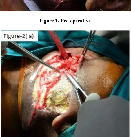
blood and urine sugar and serum electrolytes. An informed consent was obtained and the patient was taken up for debridement under general anaesthesia. With a patent airway being maintained with an elective Tracheostomy. There were plenty of dirty grayvery foul smelling pus (Figure 1) pockets in almost all the cavities of the neck extending above up to the zygomatic space. Wound debridement and drainage were performed (Figure 2). Post-operatively; the wound was cleaned twice daily with hydrogen peroxide and povidone-iodine (Figure 3). The wound healed by secondary intention after 2 weeks.
Figure 1. Pre-operative
Figure 2 a) Intra operative debridement in progress
[image:4.595.51.278.191.352.2]Figure 2. b) Debridement completed
Figure 3. Postoperative wound dressing
Case Report 2
A 70 year old male patient reported with bilateral swelling in the submandibular and submental region with history of 1week duration. There was draining sinus in the left submandibular region with pus discharge. Skin over the swelling appeared edematous and with bluish black discoloration in the midline. The other medical parameters were investigated and found to be normal. Patient was febrile with elevated WBC count, had difficulty in swallowing. Patient was admitted and the pus discharge was sent for culture and sensitivity testing. On the 3rd day of hospital admission, ulceration of the skin with copious pus discharge were noticed in the midline. The skin around the ulceration appeared necrotic. Patient was taken up for debridement of necrotic tissue and 7cm and 5cm of skin and underlying fascia was debrided. Culture reported streptococcus sensitive to amoxicillin with clauvinic acid. On the 6h post-operative day, the wound was grafted with split thickness skin graft which had taken up. The patient recovered uneventfully within 3 weeks.
Conclusion
An early diagnosis and vigorous surgical intervention are the key factors in the management of necrotizing fasciitis. Initially the patient may not present with any distinguishing features of necrotizing fasciitis. Therefore it is essential to have a highly suspicious view especially when diabetic or immuno-compromised patients present with symptoms of pain, inflammation, and infection with no apparent predisposition. Necessary resuscitation, Radical surgical debridement and secondary exploration/debridement when needed, coupled with appropriate antibiotic therapyplay a very important role in management of Necrotizing Fasciitis.
REFERENCES
Ali M H. and Zayed M E; 1997. Necrotizing fasciitis of the head and neck: Report of three cases. Ann Saudi Med., 17(6):641-45.
Andreasen, T. J. et al. 2001. “Massive Soft Tissue Injury: Diagnosis and Management of Necrotizing Fasciitis and PurpuraFulminans,” Plastic and Reconstructive Surgery, 107(4) 1025-35.
[image:4.595.50.276.325.561.2] [image:4.595.59.272.595.774.2]Barker FG, Leppard BJ, Seal DV. 1987. Streptococcal necrotising fasciitis: comparison between histological and clinical features. J Clin Pathol., 40(3):335–41.
Carter PS. and Banwell PE. 2004. Necrotising fasciitis: a new management algorithm based on clinical classification.
International Wound Journal, 1(3):189-98.
Cheung, J. P. Y., B. Fung, W. M. Tang, W. Y. Ip. 2009. A Review of Necrotizing Fasciitis in the Extremities.
HongKong Medical Journal, 15 (1): 44-52.
Deganello A, Gallo O, Gitti G, De Campora E. 2009. Necrotizing fasciitis of the neck associated with Lemierre syndrome. ActaOtorhinolaryngol Ital., 29(3):160-3. Durrani MA, Mansfield JF. 2003. Anesthetic implications of
cervicofacial necrotizing fasciitis. J Clin Anesth., 15(5): 378-81.
Edlich RF, Cross CL, Dahlstrom JJ, Long WB III. 2010. Modern concepts of the diagnosis and treatment of necrotizing fasciitis. J Emerg Med., 39(2):261–65.
Freischlag JA, Ajalat G, Busuttil RW. 1985. Treatment of necrotizing softtissue infections: the need for a new approach. Am J Surg., 149(6):751-5.
Green RJ, Dafoe DC, Raffin TA. 1996. Necrotizing fasciitis.
CHEST Journal, 110(1):219-29.
Harrison WD. and Kapoor B. 2016. Necrotizing soft tissue infection: principles of diagnosis and management.
Orthopaedics and Trauma, 30(3):223-31.
Kehrl T. 2014. Point-of-care ultrasound diagnosis of necrotizing fasciitis missed by computed tomography and magnetic resonance imaging. The Journal of Emergency
Medicine, 47(2):172-5.
Lancerotto L, Tocco I, Salmaso R, Vindigni V, Bassetto F. 2012. Necrotizing fasciitis: classification, diagnosis, and management. Journal of Trauma and Acute Care Surgery, 72(3):560-6.
Maisel RH. and Karlen R. 1994. Cervical necrotizing fasciitis.
The Laryngoscope,104(7):795-8.
Majeski J. and E. Majeski, 1997. Necrotizing Fasciitis: Improved Survival with Early Recognition by Tissue Biopsy and Aggressive Debridement. Southern Medical
Journal, 90(11):1065-68.
Miller LG, Perdreau-Remington F, Rieg G, Mehdi S, Perlroth J, Bayer AS, Tang AW, Phung TO, Spellberg B. 2005. Necrotizing fasciitis caused by community-associated methicillin-resistant Staphylococcusaureus in Los Angeles.
N Engl J Med., 352(14):1445–53.
Moss RM, Kunpittaya S, Sorasuchart A. 1990. Cervical necrotizing fasciitis: an uncommon sequela to dental infection. Annals of Otology, Rhinology & Laryngology, 99(8):643-6.
Ord R. and Coletti D. 2009. Cervico-facial necrotizing fasciitis. Oral Dis., 15(2):133–41.
Reed, JM. and Anand, VK. 1992. Odontogenic cervical necrotizing fasciitis with intrathoracic extension.
Otolaryngol Head Neck Surg., 107(4):596-600.
Sarani B, Strong M, Pascual J, Schwab CW. 2009. Necrotizing fasciitis: current concepts and review of the literature. J Am
Coll Surg., 208(2):279–88.
Scott PMJ, Dhillon, RS, McDonald, PJ. 1994. Cervical necrotizing fasciitis and tonsillitis. J Laryngol Otol., 108(05):435-437.
Shaikh N, Khawaiter J, Al-Thani H. 2012. Necrotizing fasciitis: a surgical and medical emergency. Surgical
Science, 03(11):518-25.
Shindo, Maisie L., Vincent P. Nalbone, and William R. Dougherty. 1997. Necrotizing fasciitis of the face. The
Laryngoscope, 107(8) :1071-79.
Stamenkovic I. and Lew PD. 1984. Early recognition of potentially fatal necrotizing fasciitis. The use of frozen-section biopsy. NEngl J Med., 310(26):1689–93.
Tsai CC, Lai CS, Yu ML, Chou CK, Lin SD. 1996. Early diagnosis of necrotizing fasciitis by utilization of ultrasonography. The Kaohsiung Journal of Medical
Sciences, 12(4):235-40.
Vaid N, Kothadiya A, Patki S, Kanhere H. 2002. Necrotising fasciitis of the neck. Indian Journal of Otolaryngology and
Head & Neck Surgery, 54(2):143-145 .
Valko PC, Barrett SM, Campbell JP. 1990. Odontogenic cervical necrotizingfasciitis. Ann Emerg Med., 19(5):568-71.
Wang JM. and Lim HK. 2014. Necrotizing fasciitis: eight-year experience and literature review. Brazilian Journal of
Infectious Diseases, 18(2):137-43.
Weiss A, Nelson P, Movahed R, Clarkson E, Dym H. 2011. Necrotizing fasciitis: review of the literature and case report. Journal of Oral and Maxillofacial Surgery, 69(11):2786-94.
Wong CH, Khin LW, Heng KS, Tan KC, Low CO. 2004. The LRINEC (Laboratory Risk Indicator for Necrotizing Fasciitis) score: a tool for distinguishing necrotizing fasciitis from other soft tissue infections. Critical Care
Medicine, 32(7):1535-41.