pH controlled assembly of a self-complementary
halogen-bonded dimer
†
Leonardo Maugeri, Ellen M. G. Jamieson, David B. Cordes, Alexandra M. Z. Slawin and Douglas Philp*
Phenols and their corresponding phenoxide anions can form halogen bonds with neutral iodotriazoles. The strength of these interactions depends critically on the protonation state of the oxygen atom – the interaction of the phenoxide anion is more than an order of magnitude stronger than the corresponding phenol. The assembly of a molecule bearing both an iodotriazole and a phenoxide anion into a self-complementary dimer, stabilised by two halogen bonds between the phenoxide anions and the neutral iodotriazoles has been demonstrated. The corresponding phenol shows no halogen bond mediated assembly either in the solid or in the solution state. This assembly process can be actuated simply by a change in protonation state–treatment of the phenol with one equivalent of base results in deprotonation and assembly of the dimer. The structure of the homodimer formed by the phenoxide-bearing iodotriazole has been determined in the solid state and19F NMR spectroscopy demonstrates that the assembled dimer persists in solution and that it has significant stability.19F NMR spectroscopy has also been used to demonstrate that the assembly process is completely reversible.
Introduction
Althoughrst described more than a century ago, halogen bonds1 (XBs) – the noncovalent interaction between a Lewis acidic halogen atom and a Lewis base–have only recently been inves-tigated as reliable tools2for the assembly of complex molecular and supramolecular architectures. Led by the contributions of Metrangolo and Resnati from the late 1990s, to date, XBs have found applications in crystal engineering,3medicinal chemistry,4 materials chemistry5 and nanoscience.6 The assembly of struc-tures in solution using XB interactions in organic solvents constitutes a rapidly expanding7area of research. These studies of XBs in solution are of pivotal importance for the understanding of the factors that contribute to the stability8 of an XB, whose understanding will ultimately lead to design rules for exploiting XBs that are on a similar footing to those available9for hydrogen bonds. In addition, expanding the lexicon of XB-based interac-tions that are stable in solution is of signicant importance for the realisation of XB-based supramolecular architectures10 that are persistent in solution. Previous reports concerning the stability of XB complexes formed between neutral organic XB donors, such as peruorohalocarbons8b,c(PFHCs) and aryl iodoacetylenes,8dand
a variety of neutral organic XB acceptors in organic solvents have demonstrated that the stability constants for such complexes are generally11within the range of 0.1 to 10 M1. The stability of these single point interactions is much too low to permit the formation of organised complex arrays. One strategy to enhance the halogen bonding ability of a XB donor is by rendering the interaction
“charge assisted”, i.e. using a cationic XB donor. Examples of halopyridinium, haloimidazolium and halotriazolium XB donors have been implemented successfully in mechanically interlocked molecules by the group of Beer10b–efor sensing applications and applied12in catalysis by Huber and coworkers.
Using neutral organic XB donors, it is necessary to appeal to the concepts of interactional cooperativity13 to increase the stability of assemblies. A few research groups have managed to embed10f–haromatic PFHC XB donors within multivalent plat-forms that are able to interact with multivalent XB acceptors, forming neutral complexes, stabilised by multiple XBs, with stability constants in the range 103 to 104 M1 in organic solutions.
We have become interested in applying our experience14with self-complementary replicating templates to the creation of stable halogen-bonded assemblies in solution. Although halogen bond-based dimeric designs have been reported15 previously in the solid state, the formation of such constructs in solution remains elusive. Recently, we have demonstrated16that a molecular scaffold incorporating a 5-iodo-1,4-diaryl-1H -1,2,3-triazole XB donor and a 3-oxypyridine XB acceptor and a 5-iodo-1,4-diaryl-1H-1,2,3-triazole XB donor can drive the formation of a halogen-bonded dimer in solution. The stability of this School of Chemistry and EaStCHEM, University of St Andrews, North Haugh, St
Andrews, Fife KY16 9ST, UK. E-mail: [email protected]; Fax: +44 1334 463808; Tel: +44 1334 467264
†Electronic supplementary information (ESI) available: Details of computational methods, synthesis and characterisation data for all compounds are provided. CCDC 1488032–1488035. For ESI and crystallographic data in CIF or other electronic format see DOI: 10.1039/c6sc03696a
Cite this:DOI: 10.1039/c6sc03696a
Received 17th August 2016 Accepted 17th September 2016
DOI: 10.1039/c6sc03696a
www.rsc.org/chemicalscience
Science
EDGE ARTICLE
Open Access Article. Published on 19 September 2016. Downloaded on 29/09/2016 16:42:48.
This article is licensed under a
Creative Commons Attribution 3.0 Unported Licence.
assembly was, however, disappointing and we reasoned that this molecular design could not exploit chelate cooperativity fully. Further, we reasoned that the XB interactions of pyridine acceptors were simply too weak to permit successful assembly of discrete dimeric structures in solution by aggregating only two XB interactions. In searching for an alternative XB acceptor, we were also intrigued by the possibility of exploiting an XB interaction that could be enabled and disabled by an external stimulus, such as a pH change.
Results and discussion
The ability of phenols to function as XB acceptors as well as hydrogen bond (HB) donors has been widely investigated17–19in crystal engineering. In fact, this dual character has been exploited in the realisation of crystal lattices featuring intricate networks of orthogonal HB and XB interactions. Phenoxide anions, on the other hand, despite the loss of their HB donating character, have been shown20to be stronger XB acceptors than their neutral analogues as a result of their anionic character. Nevertheless, reports of phenoxide-based XB interactions in solution are scarce.
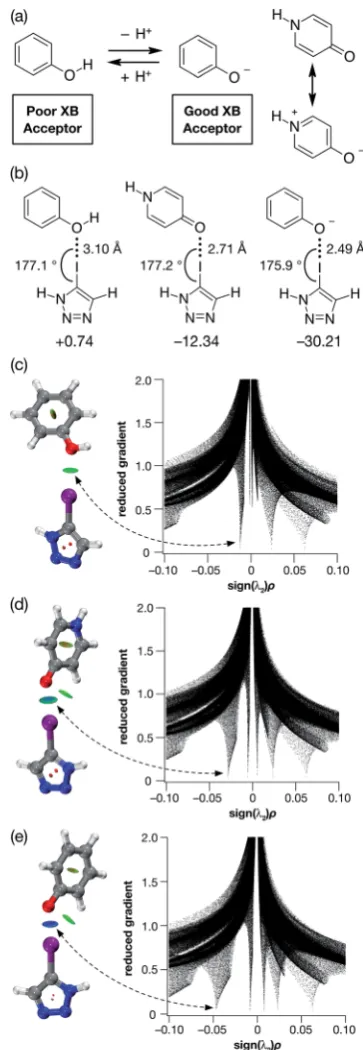
Given their different XB accepting properties, we reasoned that the phenol-phenoxide acid–base pair offered21(Fig. 1a) the ideal opportunity to create an XB interaction that could be switched on and offusing a change in pH. We also identied the possibility that 4-pyridone could provide an alternative, neutral XB acceptor with similar properties to phenoxide by virtue of its charged resonance form (Fig. 1a).
In order to probe the potential of these three acceptors to form XBs, we performed a series of calculations on the complexes formed in acetonitrile solution between phenol, 4-pyridone and the phenoxide anion and 5-iodo-1H -1,2,3-tri-azole at the TPSSh/def2-TZVP level of theory. The geometries and stabilities of these complexes (Fig. 1b) demonstrate the gradation in behaviour from phenol, through 4-pyridone, to the phenoxide anion. In the case of phenol, the O/I contact is very long (3.10 ˚A) and the enthalpy of complexation at 298 K is essentially zero at 298 K, indicating that phenol would be incapable of functioning as an XB donor in solution.
By contrast, the phenoxide anion forms an extremely short O/I contact with the iodotriazole and the complex is predicted to be very stable. As expected, 4-pyridone lies between these two extremes. Analysis of the reduced gradient22 of the electron density (Fig. 1c–e) reveals the changing nature of these inter-actions. The low density, low gradient region associated with the phenol$iodotriazole complex is van der Waals-like in nature and is barely attractive. By contrast, the low density, low gradient regions associated with the other two complexes are both signicantly attractive – particularly in the case of the phenoxide anion (sign(l2)r 0.05).
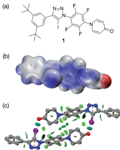
In order to validate these calculations, we focused initially on the XB capabilities of a 4-pyridone-based system. Accordingly, we designed (Fig. 2a) iodotriazole 1. The potential stability of the [1$1] dimer was assessed computationally at the TPSSh/ def2-TZVP level of theory. In order to reduce the computational cost, the tert-butyl groups present in 1 for solubility reasons
Fig. 1 (a) The phenol-phenoxide acid–base pair as pH responsive halogen bond donors. (b) Calculated O/I geometries and interaction energies (TPSSh/def2-TZVP in acetonitrile (PCM model), enthalpies of complexation at 298 K in kJ mol1) for the complexes between phenol, 4-pyridone and the phenoxide anion and 5-iodo-1H -1,2,3-triazole. Visualisation of the non-covalent interactions between 5-iodo-1H-1,2,3-triazole and (c) phenol, (d) 4-pyridone and (e) phen-oxide anion. Left: Intermolecular interaction isosurfaces generated by NCIPLOT22for s¼ 0.5 and0.05 < sign(
l2)r< 0.05 (colour scale:
attractive (blue)/repulsive (red)). Right: Plots of sign(l2)rvs.reduced
gradient highlighting the favourable interaction corresponding to the halogen bond at sign(l2)r 0.035. Atom colouring: H atoms¼white,
C atoms¼grey, N atoms¼blue, O atoms¼red, I atoms¼purple.
Open Access Article. Published on 19 September 2016. Downloaded on 29/09/2016 16:42:48.
This article is licensed under a
[image:2.595.334.516.55.581.2]were omitted from the calculated structures. These calculations reveal that the iodotriazole should possess (Fig. 2b) an electro-static potential surface that is self-complementary– a signi -cant area of positive charge is associated with theshole on the iodine atom and a signicant area of negative charge is associated with the carbonyl oxygen atom of the pyridone ring. The calculations also predict (Fig. 2c) that the iodotriazole is capable of forming an approximately centrosymmetric dimer characterised by two short I/O]C XB contacts–r(O/I)¼2.77˚A and:(O/I–C)¼173.3. The calculated enthalpy for the forma-tion of the dimer at 298 K (see ESI for details†) is43.9 kJ mol1. Analysis of the reduced gradient of the electron density reveals two low density, low gradient regions in the O/I internuclear gap associated with the halogen bonds (pale blue, Fig. 2c) and a region of weakly attractive, van der Waals-like interactions along the spine of the dimer (green, Fig. 2c). Further insight into the interactions between the two molecules1present in the dimer comes from an examination of a Natural Bond Orbital (NBO) analysis23of the system. The sum of the second order perturbation energies for the interactions between the lone pairs located on the carbonyl oxygen atoms of the pyridine ring and thes*orbital associated with the C–I bond in the iodotriazole in the dimer are 36.4 kJ mol1, suggesting that this interaction is relatively important in stabilising the homodimer.
Iodotriazole1was prepared in high yield, starting from the appropriate peruoroaryl iodotriazole and 4-hydroxypyridine in
presence of anhydrous potassium carbonate in acetonitrile, using SNAr methodology we have described16previously (see ESI for details†).
Single crystals, suitable for analysis by X-ray diffraction, were obtained by slow evaporation of a saturated toluene solution of
1. The solid state structure of1 (Fig. 3) reveals chains of the self-complementary iodotriazole units connected by XB inter-actions (r(O/I)¼2.790A;˚ :(O/I–C)¼172.1). Interestingly, the same crystal growth method afforded a second batch of crystals, which diffracted signicantly more weakly than the rst set. Although single crystal X-ray diffraction data were collected on these crystals, the data was not of sufficient quality to permit renement of the structure solution to an acceptably lowR% value. However, even with low quality data, it was clear that, within the solid state structure of these crystals, the self-complementary iodotriazole1formed a homodimeric assembly (see ESI for details†).
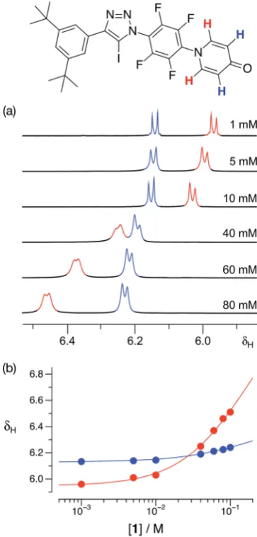
With some evidence in hand that1could form homodimers in the solid state, we wished to establish the ability of1to form homodimers in solution. Accordingly, we performed a dilution experiment using a solution of1ind8-toluene and a 500.1 MHz 1H{19F} NMR spectrum was recorded at a series of
concentra-tion steps–starting from an initial concentration of 80 mM, the solution of1was diluted progressively down to 1 mM. In this range of concentrations, the resonances arising from the pyr-idone ring protons of1experienced (Fig. 4a) signicant chem-ical shichanges. The data from this dilution experiment for both probe protons can betted24simultaneously (Fig. 4b) to a dimerisation binding model to afford a stability constant for the [1$1] dimer of 2.30.3 M1ind8-toluene at 293 K corre-sponding to a free energy of dimerisation of2.00.3 kJ mol1 at this temperature.
[image:3.595.65.260.46.289.2]The results obtained for pyridone1provided encouragement that employing the phenol-phenoxide acid–base pair should provide a viable route to achieving our objective of a pH-switchable XB assembly. Accordingly, we investigated the system shown in Fig. 5 computationally. This system incorporates a diaryl iodo-triazole XB donor and a phenol in an appropriate structural rela-tionship in order to facilitate dimerisation. The calculated
Fig. 2 (a) Design of a self-complementary iodotriazole. (b) Electro-static potential surface of1(TPSSh/def2-TZVP). Colour scale: blue¼ positive / red ¼ negative (c) Calculated structure of the [1$1] including a visualisation of the non-covalent interactions present in the dimer. Intermolecular interaction isosurfaces generated by NCIPLOT22
fors¼0.5 and0.05 < sign(l2)r< 0.05 (colour scale: attractive (blue)
/repulsive (red)). XB geometry: I/O]Cr(O/I)¼2.77A and˚ :(O/ I–C)¼173.3. Atom colouring: H atoms¼white, C atoms¼grey, N atoms¼blue, O atoms¼red, I atoms¼purple.
Fig. 3 Solid state structure of1determined from single crystal X-ray diffraction data. Halogen bonds are marked as a red dashed lines (r(O/I)¼2.790A;˚ :(O/I–C)¼172.1). Atom colouring: C atoms¼ grey, N atoms¼blue, O atoms¼red, I atoms¼purple. Hydrogen atoms and included solvent of crystallisation (toluene) are omitted for clarity.
Open Access Article. Published on 19 September 2016. Downloaded on 29/09/2016 16:42:48.
This article is licensed under a
[image:3.595.305.550.537.661.2]structure (TPSSh/def2-TZVP) of the putative phenol dimer (Fig. 5a) reveals an almost centrosymmetric homodimer in which there are two very long O/I contacts (r(O/I) ¼ 3.15 A,˚ :(C–I/O ¼ 174.9)).
The calculated enthalpy of interaction at 298 K for this dimer is4.26 kJ mol1 at this level of theory, suggesting that any halogen bonding present in this dimer is not signicantly sta-bilising. Analysis of the reduced gradient of the electron density (Fig. 5a) shows the characteristic low density, low gradient regions associated with weakly attractive interactions (green areas, Fig. 5a) both in the O/I internuclear gap and along the spine of the dimer. Further support for the relatively weak interactions between the two phenol molecules in the dimer came from an examination of a Natural Bond Orbital (NBO) analysis23of the system. The sum of the second order pertur-bation energies for the interactions between the lone pairs located on the phenol oxygen atoms and the s* orbital
associated with the C–I bond in the iodotriazole in the dimer are only 10.3 kJ mol1, well below that calculated for the pyridone-based [1$1] dimer.
Removal of a proton from the phenol affords the corre-sponding phenoxide anion (Fig. 5b) and, once again, we assessed the potential stability of the dimer formed by this anion at the TPSSh/def2-TZVP level of theory (Fig. 5b). In common with the phenol (Fig. 5a), the calculation reveals an almost centrosymmetric homodimer. In this case, however, there are two very short O/I contacts (r(O/I)¼2.51˚A,:(C– I/O¼176.7)) present and the enthalpy of dimerisation at 298 K is predicted to be50.4 kJ mol1at this level of theory–this value is only around 15% higher than the predicted enthalpy of dimerisation at 298 K for the [1$1] dimer. This observation is apparently at odds with the computational results obtained for the simple iodotriazole complexes shown in Fig. 1. In this series of simple 1 : 1 complexes, the enthalpy of complexation rises steeply as the XB acceptor changes from phenol through 4-pyridone to the phenoxide anion. However, dimer [2$2]2
suffers from strong electrostatic destabilisation as a result of the anionic nature of the two partners in the dimer. Since no counterions are included in the calculation, this effect is not mitigated in any way and the value obtained for the enthalpy of dimerisation should be viewed as a worst case value.
Analysis of the reduced gradient of the electron density (Fig. 5b) shows the characteristic low density, low gradient regions
Fig. 4 (a) Partial 500.1 MHz1H{19F} NMR spectra of iodotriazole1at various concentrations ind8-toluene at 293 K showing the
1
H reso-nances arising from the pyridone ring protons. (b) Bestfits (solid lines) of the changes in the chemical shifts of the1H resonances arising from the pyridone ring protons as a function of concentration to a1+1%
[1$1] dimerisation model.
Fig. 5 (a) Calculated structure of a putative dimer assembled from phenol bearing a diaryl iodotriazole. The structure includes a visual-isation of the non-covalent interactions present. XB geometry: I/O] Cr(O/I)¼3.15A and˚ :(O/I–C)¼173.3. (b) Calculated structure of a dimer assembled from the corresponding phenoxide anion, including a visualisation of the non-covalent interactions present. XB geometry: I/O]Cr(O/I)¼2.51A and˚ :(O/I–C)¼176.7. In both cases, the non-covalent interactions present are visualised using intermolecular interaction isosurfaces generated by NCIPLOT22for
s¼ 0.5 and0.05 < sign(l2)r< 0.05 (colour scale: attractive (blue) /
repulsive (red)). Atom colouring: H atoms¼white, C atoms¼grey, N atoms¼blue, O atoms¼red, I atoms¼purple.
Open Access Article. Published on 19 September 2016. Downloaded on 29/09/2016 16:42:48.
This article is licensed under a
[image:4.595.75.257.50.430.2] [image:4.595.311.543.50.261.2]in the O/I internuclear gap associated with signicantly attrac-tive halogen bonds (blue areas, Fig. 5b, sign(l2)r 0.05). In common with the pyridone dimer [1$1], there are other, weaker, attractive interactions along the length of the dimeric structure. NBO analysis of this system reveals a second order perturbation energy for the interaction between the lone pairs located on the phenoxide oxygen atom and thes*orbital associated with the C–I bond in the iodotriazole of 91.9 kJ mol1, which contrasts starkly with that for the corresponding phenol dimer and is indicative of the much stronger association between the two anionic species.
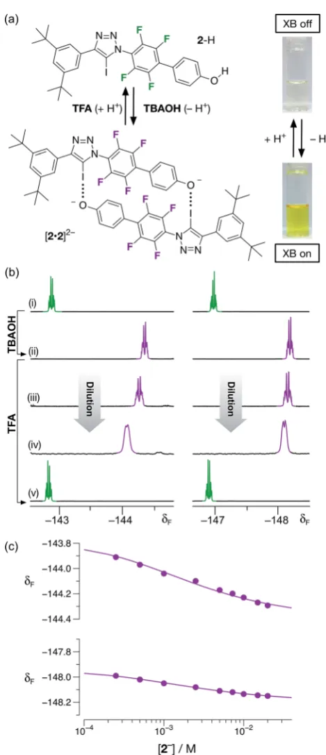
In order to test these theoretical predictions, we synthesised phenol 2–H using standard methods (see ESI for details†). Crystals of2–H, suitable for analysis by X-ray diffraction, were grown by slow evaporation of a solution of the phenol in THF. The solid state structure of2–H reveals (see ESI†) that, as ex-pected, there is no evidence of halogen bonds formed between the phenolic oxygen atom and the iodotriazole. The structure contains one solvent molecule (THF) of crystallisation and the oxygen atom of the THF molecule is hydrogen bonded to the phenolic proton. Progressive dilution of a CD3CN solution of 2–H from 20 mM to 1 mM did not result in any measurable chemical shichanges in the19F resonances arising from the peruorinated ring of2–H (see ESI†). These data suggest that
2–H is unable to undergo XB-mediated dimerisation in solution in this concentration range.
Having demonstrated that phenol 2–H was incapable of participating in halogen bonds under the conditions employed, we next turned to the potential of the corresponding phenoxide anion 2 to form a halogen-bonded dimer. Treatment of a colorless, 20 mM solution of2–H in CD3CN at room temper-ature with one equivalent of a 1 M solution of tetrabuty-lammonium hydroxide (TBAOH) in methanol resulted in the development of an intense yellow color (Fig. 6a) in the solution, consistent with the formation of the corresponding phenoxide anion through deprotonation of2–H.
The formation of the anion2was conrmed by comparison of the 470.6 MHz19F{1H} NMR spectrum of the solution with TBAOH. Aer the addition of the base, the two resonances corresponding to aryluorine atoms atd142.7 andd146.9 are replaced two new resonances signicantly upeld from those associated with2–H, atd144.3 and148.2, respectively. This observation is consistent with the formation of the phen-oxide anion. A freshly-prepared solution of2, as its tetrabutyl ammonium (TBA) salt, in CD3CN was diluted progressively (Fig. 6b(ii)/(iv)) from 20 mM to 1 mM. Downeld chemical shichanges (Dd¼+0.38 and +0.16 between 20 mM and 1 mM) of the two resonances arising from the aryluorine atoms are observed, commensurate with the presence of the [2$2]2 dimer. Simultaneous tting24 (Fig. 6c) of the chemical shi changes for the two 19F resonances at around
d 144 and
d 148 to a dimerisation binding model affords a stability constant for the [2$2]2dimer of 51030 M1, corresponding to a free energy of complexation of15.2 0.2 kJ mol1 at 293 K. In order to assess the stability of the [2$2]2homodimer in the context of both chelate cooperativity and any electrostatic repulsion experienced within [2$2]2, we sought to measure experimentally the single point association of a phenoxide
Fig. 6 (a) The monomer/dimer state of 2 can be controlled by changing the protonation state of the phenolic oxygen atom by addition ofTBAOHorTFA. (b) Partial 470.6 MHz19F{1H} NMR spectra
(298 K, CD3CN) of2: (i) 20 mM before addition ofTBAOH; (ii) 20 mM
after addition ofTBAOH; (iii) 7.5 mM after addition ofTBAOH; (iv) 1 mM after addition ofTBAOH; (v) after treatment of 20 mM solution (ii) with TFA. (c) Bestfits (solid lines) of the changes in the chemical shifts of the
19
F resonances atd144 and148 as a function of concentration to a2+2%[2$2]2dimerisation model.
Open Access Article. Published on 19 September 2016. Downloaded on 29/09/2016 16:42:48.
This article is licensed under a
[image:5.595.307.542.64.608.2]anion with a suitable diaryl iodotriazole. Unfortunately, this measurement proved to be remarkably difficult to perform. Attempts to titrate a solution of tetrabutylammonium phenolate into a model iodotriazole in CD3CN at 293 K (see ESI for details†) resulted in rapid conversion of the 5-iodotriazole to the corresponding 5-Htriazole. This reactivity is not observed with the dimeric species [2$2]2under the same conditions. Other laboratories have described25 situations in which 5-iodo-triazoles undergo formal reductive deiodination to afford their 5-Hanalogues. However, we were able to perform a titration successfully when the phenolate solution was prepared using 1,8-diaza[5.4.0]undec-7-ene (DBU) as the base. This titration (see ESI for details†) afforded an association constant (Ka) for the single point association between the phenolate anion and the model diaryliodotriazole of 6.60.2 M1. This interaction seems surprisingly weak and it is necessary to interpret the measured stability constant with caution. The phenolate counterion–DBUH+– is capable of hydrogen bonding to the phenoxide oxygen atom reducing its ability to function as a halogen bond acceptor. However, this value can be used to place an upper limit on the level of cooperativity present within the [2$2]2homodimer –the connection free energy26has an upper limit of around 6 kJ mol1and the effective molarity13for the formation of the [2$2]2homodimer has an upper limit of around 10 M.
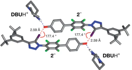
All attempts to isolate and crystallise the tetrabutyl ammo-nium salt of the [2$2]2 dimer from CD3CN solutions were unsuccessful, principally as a result of the hygroscopic char-acter of this salt. However, treatment of a concentrated solution of2–H in CD3CN with 15 equivalents of 1,8-diaza[5.4.0]undec-7-ene (DBU) (see ESI for details†) led to the spontaneous forma-tion of crystals of [2$2]$(DBUH)2suitable for analysis by single crystal X-ray diffraction.
The solid state structure of these crystals, determined from diffraction data, reveals (Fig. 7) a halogen-bonded homodimeric structure for [2$2]2in which the individual phenoxide anions are connected by remarkably short (r(O/I) ¼2.592A,˚ SvdW radii¼3.55˚A) and linear (:(C–I/O¼177.2)) I/O contacts.
There is excellent agreement between structure of [2$2]2 determined in the solid state and the calculated structure (Fig. 5). Additionally, there is a hydrogen bond from each of the
DBUH+cations to an oxygen atom in the corresponding phen-oxide anion.
Conclusions
In order for halogen bonds to achieve the status of a robust structure-directing element in the design of solution phase assemblies, it is necessary for halogen bonds to demonstrate the same level ofexibility and programmability as the ubiq-uitous hydrogen bond. Single hydrogen bonds are rarely used as a structure-directing element in the design of non-covalent assemblies – more commonly, several are used in concert to create stable structures. The aggregation of multiple hydrogen bonds in arrays are subject to clear design rules. The assembly and disassembly of the [2$2]2dimer in solution, actuated by proton transfer, represents an important step forward in the use of halogen bonds in this context. Not only does the [2$2]2 dimer have signicant stability in solution, its design was reached in a rational manner obeying clear design rules. Addi-tionally, although Sch¨ollhorn and coworkers have recently demonstrated27that both the XB accepting and donating char-acter of electrochemically active molecules can be modulated by the applied potential, the work presented here is, to the best of our knowledge, the rst time that proton transfer has been exploited efficiently to regulate the formation of a XB-based assembly. However, in order to fully exploit the strong halogen bonds formed by phenoxide anions with aryl iodides, such as the iodotriazole used here, it is necessary to overcome the electrostatic repulsion experienced within the [2$2]2 homo-dimer. This objective requires the dimer to be zwitterionic in its active (assembled) state and could be accomplished readily by switching10d,ethe XB donor to iodotriazolium. These studies, in the context of supramolecular materials based on the design of the [2$2]2 homodimer are currently underway in our laboratory.
Acknowledgements
We thank the Marie Curie Initial Training Network on Repli-cation and Adaption in Networks (ReAd) fornancial support (early stage researcher funding to L. M.). We thank Dr Tom´aˇs L´ebl and Mrs Melanja Smith for assistance with NMR experiments.
Notes and references
1 (a) P. Metrangolo, H. Neukirch, T. Pilati and G. Resnati,Acc. Chem. Res., 2005,38, 386; (b) G. R. Desiraju, P. S. Ho, L. Kloo, A. C. Legon, R. Marquardt, P. Metrangolo, P. Politzer, G. Resnati and K. Rissanen, Pure Appl. Chem., 2013, 85, 1711; (c) G. Cavallo, P. Metrangolo, R. Milani, T. Pilati, A. Priimagi, G. Resnati and G. Terraneo,Chem. Rev., 2016,
116, 2478.
Fig. 7 Solid state structure of the salt formed by reaction of2with 1,8-diaza[5,4,0]undec-7-ene (DBU) determined from single crystal X-ray diffraction data. Halogen bonds are marked as a red dashed lines (r(O/I)¼2.592A;˚ :(O/I–C)¼177.2). Hydrogen bonds from [2$2]2 to theDBUH+ cations are shown as dashed blue lines. Atom color
code: C, grey; N, blue; O, red; F, green; I, purple. Most hydrogen atoms are omitted for clarity.
Open Access Article. Published on 19 September 2016. Downloaded on 29/09/2016 16:42:48.
This article is licensed under a
[image:6.595.53.284.50.173.2]2 (a) L. C. Gilday, S. W. Robinson, T. A. Barendt, M. J. Langton, B. R. Mullaney and P. D. Beer,Chem. Rev., 2015,115, 7118; (b) G. Cavallo, P. Metrangolo, R. Milani, T. Pilati, A. Priimagi, G. Resnati and G. Terraneo,Chem. Rev., 2016,116, 2478. 3 (a) P. Metrangolo, F. Meyer, T. Pilati, G. Resnati and
G. Terraneo, Angew. Chem., Int. Ed., 2008, 47, 6114; (b) A. Mukherjee, S. Tothadi and G. R. Desiraju, Acc. Chem. Res., 2014,47, 2514.
4 (a) A. R. Voth, P. Khuu, K. Oishi and P. S. Ho,Nat. Chem., 2009, 1, 74; (b) E. Parisini, P. Metrangolo, T. Pilati, G. Resnati and G. Terraneo, Chem. Soc. Rev., 2011, 40, 2267; (c) A. Lange, M. Guenther, F. M. Buettner, M. O. Zimmermann, J. Heidrich, S. Hennig, S. Zahn, C. Schall, A. Sievers-Engler, F. Ansideri, P. Koch,
M. Laemmerhofer, T. Stehle, S. A. Laufer and
F. M. Boeckler, J. Am. Chem. Soc., 2015, 137, 14640; (d) E. Persch, O. Dumele and F. Diederich,Angew. Chem., Int. Ed., 2015,54, 3290.
5 (a) A. Sun, J. W. Lauher and N. S. Goroff,Science, 2006,312, 9321; (b) A. Priimagi, G. Cavallo, P. Metrangolo and G. Resnati,Acc. Chem. Res., 2013,46, 2686; (c) F. Meyer and P. Dubois,CrystEngComm, 2013,15, 3058.
6 (a) T. Shirman, T. Arad and M. E. van der Boom, Angew. Chem., Int. Ed., 2010,49, 926; (b) T. Shirman, R. Kaminker, D. Freeman and M. E. van der Boom, ACS Nano, 2011,5, 6553; (c) M. Boterashvili, M. Lahav, S. Shankar, A. Facchetti and M. E. van der Boom,J. Am. Chem. Soc., 2014,136, 11926. 7 (a) M. T. Messina, P. Metrangolo, W. Panzeri, E. Ragg and G. Resnati,Tetrahedron Lett., 1998,39, 9069; (b) M. Erd´elyi,
Chem. Soc. Rev., 2012, 41, 3547; (c) T. M. Beale, M. G. Chudzinski, M. G. Sarwar and M. S. Taylor, Chem. Soc. Rev., 2013,42, 1667.
8 (a) P. K. Rege, O. L. Malkina and N. S. Goroff,J. Am. Chem. Soc., 2002, 124, 370; (b) M. G. Sarwar, L. Dragisic, C. Salsberg, C. Gouliaras and M. S. Taylor, J. Am. Chem. Soc., 2010, 132, 1646; (c) M. G. Chudzinski and M. S. Taylor,J. Org. Chem., 2012,77, 3483; (d) O. Dumele, D. Wu, N. Trapp, N. S. Goroffand F. Diederich,Org. Lett., 2014,16, 4722.
9 (a) M. Etter,Acc. Chem. Res., 1990,23, 120; (b) L. J. Prins, D. N. Reinhoudt and P. Timmerman, Angew. Chem., Int. Ed., 2001, 40, 2382; (c) G. R. Desiraju, Acc. Chem. Res., 2002,35, 565.
10 (a) E. Dimitrijevi´c, O. Kvak and M. S. Taylor,Chem. Commun., 2010, 46, 9025; (b) A. Caballero, F. Zapata, N. G. White, P. J. Costa, V. Felix and P. D. Beer,Angew. Chem., Int. Ed., 2012, 51, 1876; (c) L. C. Gilday, T. Lang, A. Caballero, P. J. Costa, V. Felix and P. D. Beer,Angew. Chem., Int. Ed., 2013, 52, 4356; (d) J. M. Mercurio, R. C. Knighton, J. Cookson and P. D. Beer, Chem.–Eur. J., 2014,20, 11740; (e) M. G. Langton, S. W. Robinson, V. Marques, V. Felix and P. D. Beer, Nat. Chem., 2014, 6, 1039; (f) S. H. Jungbauer, D. Buleld, F. Kniep, C. W. Lehman, E. Herdtweck and S. M. Huber, J. Am. Chem. Soc., 2014,
136, 16740; (g) O. Dumele, N. Trapp and F. Diederich,
Angew. Chem., Int. Ed., 2015,54, 12339; (h) A. Vanderkooy and M. S. Taylor, J. Am. Chem. Soc., 2015, 137, 5080; (i)
S. H. Jungbauer, S. Schindler, E. Herdtweck, S. Keller and S. Huber,Chem.–Eur. J., 2015,21, 13625.
11 To the best of our knowledge, the only neutral halogen-bonded organic complex that possesses an association constant greater than 102 M1 is formed between quinuclidine and 1,2,3,4,5-pentauoro-6-(iodoethynyl) benzene in d12-cyclohexane at 298 K, as described by Diederich and coworkers (ref. 8d). More recently, Rissanen and co-workers observed the formation of highly stable halogen bonds between N-iodosuccinimide and N -iodosaccharin and a series of aromatic N-oxides with association constants in the range 102to 104M1in CDCl3 and d6-acetone solutions at 298 K. See: R. Puttreddy, O. Jurˇcek, S. Bhowik, T. M¨akel¨a and K. Rissanen, Chem. Commun., 2016,52, 2338.
12 (a) S. M. Walter, F. Kniep, S. Schindler, E. Herdtweck and S. M. Huber, Angew. Chem., Int. Ed., 2011, 50, 7187; (b) S. M. Walter, S. H. Jungbauer, F. Kniep, S. Schindler, E. Herdtweck and S. M. Huber, J. Fluorine Chem., 2013,
150, 14; (c) S. H. Jungbauer and S. M. Huber,J. Am. Chem. Soc., 2015,137, 12110.
13 (a) C. A. Hunter and H. L. Anderson,Angew. Chem., Int. Ed., 2009, 48, 7488; (b) G. Ercolani and L. Schiaffino, Angew. Chem., Int. Ed., 2011,50, 1762.
14 (a) E. Kassianidis and D. Philp,Angew. Chem., Int. Ed., 2006,
45, 6344; (b) J. W. Sadownik and D. Philp,Angew. Chem., Int. Ed., 2008, 47, 9965; (c) T. Kosikova, N. I. Hassan, D. B. Cordes, A. M. Z. Slawin and D. Philp, J. Am. Chem. Soc., 2015,137, 16074.
15 (a) D. Cao, M. Hong, A. K. Blackburn, Z. Liu, J. M. Holcro and J. F. Stoddart, Chem. Sci., 2014, 5, 4242; (b) S. M. Oburn, N. P. Bowling and E. Bosch, Cryst. Growth Des., 2015,15, 1112; (c) Y. Takeda, K. Hatanaka, T. Nishida and S. Minakata,Chem.–Eur. J., 2016,22, 10360.
16 L. Maugeri, J. Asencio-Hern´andez, T. Lebl, D. B. Cordes, A. M. Z. Slawin, M.-A. Delsuc and D. Philp, Chem. Sci., 2016,7, 6422.
17 A. Mukherjee and G. R. Desiraju,Cryst. Growth Des., 2011,
11, 3735.
18 C. B. Aaker¨oy, S. Panikkattu, P. D. Chopade and J. Desper,
CrystEngComm, 2013,15, 3125.
19 A. Mukherjee and G. R. Desiraju,IUCrJ, 2014,1, 49. 20 A. Takemura, L. J. McAllister, S. Hart, N. E. Pridmore,
P. B. Karadakov, A. C. Whitwood and D. W. Bruce,Chem.– Eur. J., 2014,20, 6721.
21 C. M. Keaveney and D. A. Leigh,Angew. Chem., Int. Ed., 2004,
43, 1222.
22 (a) E. R. Johnson, S. Keinan, P. Mori-S`anchez, J. Contreras-Garc´ıa, A. J. Cohen and W. J. Yang,J. Am. Chem. Soc., 2010,
132, 6498; (b) J. Contreras-Garc´ıa, R. Chaudret, J.-P. Piquemal, D. N. Beratan and W. J. Yang, J. Chem. Theory Comput., 2011,7, 625.
23 (a) E. D. Glendening, C. R. Landis and E. Weinhold, J. Comput. Chem., 2013, 34, 1429; (b) NBO 6.0.
E. D. Glendening, J. K. Badenhoop, A. E. Reed,
J. E. Carpenter, J. A. Bohmann, C. M. Morales, C. R. Landis
Open Access Article. Published on 19 September 2016. Downloaded on 29/09/2016 16:42:48.
This article is licensed under a
and E. J. Weinhold, Theoretical Chemistry Institute, University of Wisconsin, Madison, 2013.
24 M. J. Hynes,J. Chem. Soc., Dalton Trans., 1993, 311. 25 (a) J. E. Hein, J. C. Tripp, L. B. Krasnova, K. B. Sharpless and
V. V. Fokin, Angew. Chem., Int. Ed., 2009, 48, 8018; (b) M. Jur´ıˇcek, K. Stout, P. H. J. Kouwer and A. E. Rowan,Org. Lett., 2011, 13, 3494; (c) B. T. Worrell, J. E. Hein and
V. V. Fokin, Angew. Chem., Int. Ed., 2012, 48, 8018; (d) D. Fu, J. Zhang and S. Cao,J. Fluorine Chem., 2013,156, 170. 26 W. P. Jencks,Proc. Natl. Acad. Sci. U. S. A., 1981,78, 4046. 27 (a) S. Groni, T. Maby-Raud, C. Fave, M. Branca and
B. Sch¨ollhorn, Chem. Commun., 2014, 50, 14616; (b) R. Oliveira, S. Groni, C. Fave, M. Branca, F. Mavr´e, D. Lorcy, M. Fourmigu´e and B. Sch¨ollhorn, Phys. Chem. Chem. Phys., 2016,18, 15867.
Open Access Article. Published on 19 September 2016. Downloaded on 29/09/2016 16:42:48.
This article is licensed under a