IMPACT OF EXPERIMENTAL HYPERGLYCEMIA ON THE SCIATIC NERVE OF ALBINO RATS
HISTOLOGICAL AND HISTOMORPHOMETRIC STUDY
1
Dr. Muhamed Faizal
1
Senior Resident; Department of Anatomy; AIIMS
2
Professor, Department of Anatomy, JN Medical College, AMU, Aligarh
ARTICLE INFO ABSTRACT
The complications of chronic
peripheral neuropathy is the most common presenting feature and which if not controlled may necessitate limb amputation. Since the exact mechanism of hyperglycemia
neuropathy and its treatment are not completely known, the present study was aimed at an effort to further the information in this regard. After ethical clearance, 36 animals were divided into six groups having six rats each. The groups consisted of age
weeks, one month, two months, four months and six months duration of diabetes. Diabetes was induced by a single dose of streptozotocin (60 mg/kg, intraperitoneal). At the end of each experimental period, animals w
blood serum was subjected to biochemical analysis and the sciatic nerve tissue blocks were processed for paraffin sectioning, routine and special staining for light microscopy
histomorphological findings and histopathological features revealed that the prolonged duration of hyperglycemia resulted in the raised serum creatinine, lowered serum total protein, reduction of myelinated fibers and remarkable thickening of endoneurial,
concluded that the biochemical alterations, change in the ratio of myelinated fibers and heaping up of collagen fibers at every level of organization appear to be important contributing factors for the deteriorati
Copyright©2019, Muhamed Faizal and Aijaz Ahmed Khan
permits unrestricted use, distribution, and reproduction
INTRODUCTION
Diabetes mellitus is one of the most common metabolic
disorders (Naik et al., 2015). Complications of chronic
diabetes include peripheral neuropathy,
angiopathy and retinopathy (Biessels et al., 1994; Pek
2017; Swaminathan et al., 2017). Involvement of large fiber
conduction velocity is considered as an earliest fu
marker of glucose neurotoxicity. This is typically present even before the decrease in axonal diameter or structural disruption of the myelin in central or peripheral nervous systems
(Zochodne et al., 2004; Tomlinson et al., 2008;
2017). Length-dependent nature of the peripheral neuropathy is characterized by a disturbance in axonal transport at most distal parts of long nerves as a result of pathological alterations in the cytoskeleton as well as axonal atrophy
1999; Prior et al., 2017). Diabetic neuropathy
clinical manifestations such as chronic sensorimotor distal symmetric poly-neuropathy and the autonomic neuropathies
(Boulton et al., 2005). The characteristic findings in diabetic
neuropathy are sensory predominant nerve fiber degeneration, axonal loss, and thickening of endoneurial arterioles
al., 1996). Thickening of endoneurial arterioles and a reduction
ISSN: 0975-833X
Article History:
Received 24th January, 2019
Received in revised form 16th February, 2019
Accepted 19th March, 2019
Published online 30th April, 2019
Citation: Dr. Muhamed Faizal and Dr. Aijaz Ahmed Khan
and histomorphometric study”, International Journal of Current Research
Key Words: Collagen, Diabetes, Hyperglycemia, Peripheral Neuropathy Sciatic nerve. *Corresponding author:
Dr. Aijaz Ahmed Khan
RESEARCH ARTICLE
IMPACT OF EXPERIMENTAL HYPERGLYCEMIA ON THE SCIATIC NERVE OF ALBINO RATS
HISTOLOGICAL AND HISTOMORPHOMETRIC STUDY
Dr. Muhamed Faizal and
2, *Dr. Aijaz Ahmed Khan
Senior Resident; Department of Anatomy; AIIMS, Jodhpur
Professor, Department of Anatomy, JN Medical College, AMU, Aligarh
ABSTRACT
The complications of chronic diabetes affect many systems including the nervous system wherein the peripheral neuropathy is the most common presenting feature and which if not controlled may necessitate limb amputation. Since the exact mechanism of hyperglycemia
ropathy and its treatment are not completely known, the present study was aimed at an effort to further the information in this regard. After ethical clearance, 36 animals were divided into six groups having six rats each. The groups consisted of age-matched controls and experimental groups of two weeks, one month, two months, four months and six months duration of diabetes. Diabetes was induced by a single dose of streptozotocin (60 mg/kg, intraperitoneal). At the end of each experimental period, animals were euthanized and perfusion fixed with Karnovsky’s fixative. The blood serum was subjected to biochemical analysis and the sciatic nerve tissue blocks were processed for paraffin sectioning, routine and special staining for light microscopy
histomorphological findings and histopathological features revealed that the prolonged duration of hyperglycemia resulted in the raised serum creatinine, lowered serum total protein, reduction of myelinated fibers and remarkable thickening of endoneurial, perineurial and epineurial collagen. It is concluded that the biochemical alterations, change in the ratio of myelinated fibers and heaping up of collagen fibers at every level of organization appear to be important contributing factors for the deterioration of peripheral nerve functions in chronic diabetes.
Muhamed Faizal and Aijaz Ahmed Khan. This is an open access article distributed under the Creative in any medium, provided the original work is properly cited.
Diabetes mellitus is one of the most common metabolic . Complications of chronic
peripheral neuropathy, nephropathy,
., 1994; Pek et al.,
nvolvement of large fiber conduction velocity is considered as an earliest functional marker of glucose neurotoxicity. This is typically present even before the decrease in axonal diameter or structural disruption of the myelin in central or peripheral nervous systems
2008; Mohseni et al.,
dependent nature of the peripheral neuropathy is characterized by a disturbance in axonal transport at most distal parts of long nerves as a result of pathological alterations
in the cytoskeleton as well as axonal atrophy (Sima et al.,
Diabetic neuropathy reveals diverse clinical manifestations such as chronic sensorimotor distal neuropathy and the autonomic neuropathies The characteristic findings in diabetic sory predominant nerve fiber degeneration,
axonal loss, and thickening of endoneurial arterioles (Dyck et
. Thickening of endoneurial arterioles and a reduction
in the luminal area correlated well with subsequent nerve fiber loss in diabetic nerves (Malik
experimental diabetes mellitus has been shown to induce distal small-fiber neuropathy (Bischhoffshausen
initiate a syndrome of distal uncomfortable tingling sensation
(Rolim et al., 2017) and is also associated with distal
sympathetic dysfunction (Maser
allodynia, vasomotor changes, pallor alternated with rubor, cyanosis, and mottling (Low, 2003; Ugwu, 2017
neuropathy represents a major health problem due to its clinical manifestations such as excruciating neuropathic pain, diabetic foot ulceration, and amputations, which are associated with substantial morbidity, reduced quali
increased mortality (Ziegler
alterations in the peripheral nerve myelin thickness have been also reported in longstanding streptozotocin
rats (Lee et al., 2017). Therefore, the present study was aimed
at demonstrating these and possibly other changes in the arrangement of collagen fibers, nerve fibers and myelin by using special staining for collagen and myelin along with
histomorphological and biochemical analyses in
experimentally induced diabetic ra
2, 4 and 6 months. In a study “The laboratory rat: relating to its
International Journal of Current Research
Vol. 11, Issue, 04, pp.3038-3044, April, 2019 DOI: https://doi.org/10.24941/ijcr.34506.04.2019
Dr. Aijaz Ahmed Khan, 2019. “Impact of experimental hyperglycemia on the sciatic nerve of albino rats
International Journal of Current Research, 11, (04), 3034-3037.
Available online at http://www.journalcra.com
IMPACT OF EXPERIMENTAL HYPERGLYCEMIA ON THE SCIATIC NERVE OF ALBINO RATS - A
HISTOLOGICAL AND HISTOMORPHOMETRIC STUDY
. Aijaz Ahmed Khan
Jodhpur
Professor, Department of Anatomy, JN Medical College, AMU, Aligarh
diabetes affect many systems including the nervous system wherein the peripheral neuropathy is the most common presenting feature and which if not controlled may necessitate limb amputation. Since the exact mechanism of hyperglycemia-induced peripheral ropathy and its treatment are not completely known, the present study was aimed at an effort to further the information in this regard. After ethical clearance, 36 animals were divided into six groups ed controls and experimental groups of two weeks, one month, two months, four months and six months duration of diabetes. Diabetes was induced by a single dose of streptozotocin (60 mg/kg, intraperitoneal). At the end of each ere euthanized and perfusion fixed with Karnovsky’s fixative. The blood serum was subjected to biochemical analysis and the sciatic nerve tissue blocks were processed for paraffin sectioning, routine and special staining for light microscopy. Biochemical, histomorphological findings and histopathological features revealed that the prolonged duration of hyperglycemia resulted in the raised serum creatinine, lowered serum total protein, reduction of perineurial and epineurial collagen. It is concluded that the biochemical alterations, change in the ratio of myelinated fibers and heaping up of collagen fibers at every level of organization appear to be important contributing factors for the
Creative Commons Attribution License, which
in the luminal area correlated well with subsequent nerve fiber
Malik et al., 2005). Lately
experimental diabetes mellitus has been shown to induce distal
Bischhoffshausen et al., 2017) which
rome of distal uncomfortable tingling sensation and is also associated with distal
Maser et al., 2017) in the form of
changes, pallor alternated with rubor, 2003; Ugwu, 2017). The diabetic neuropathy represents a major health problem due to its clinical manifestations such as excruciating neuropathic pain, diabetic foot ulceration, and amputations, which are associated with substantial morbidity, reduced quality of life, and Ziegler, 2017). Recently, marked alterations in the peripheral nerve myelin thickness have been also reported in longstanding streptozotocin (STZ)-diabetic . Therefore, the present study was aimed at demonstrating these and possibly other changes in the arrangement of collagen fibers, nerve fibers and myelin by using special staining for collagen and myelin along with
histomorphological and biochemical analyses in
experimentally induced diabetic rats after 2 weeks and after 1, . In a study “The laboratory rat: relating to its INTERNATIONAL JOURNAL
OF CURRENT RESEARCH
age with human’s” , it is described that one month of rat’s life is equivalent to three human years (Sengupta, 2013). Therefore, 6-months duration of diabetes in rat may be considered to be equivalent to18 years of diabetes in human for purpose of assessment of pathophysiological parameters associated with chronic diabetes.
MATERIAL AND METHODS
Animal preparation and experimental design: Age-matched
control and non-diabetic rats of either sex (total 36 rats) weighing ~250g were obtained from the central animal house, AMU, Aligarh, after approval from Institutional Animal Ethics Committee (D. No: 9025/2014). Prior to commencement of the experiment, all animals were placed in a clean, well ventilated, properly maintained new environmental condition and monitored daily with regard to body weight, urine sugar (strip method) for a period of one week; they were supplied standard pellet diet and water ad libitum and maintained on a 12/12 h light/dark cycle.
After one week, animals were divided into six age-matched groups having six rats each: (1) non-diabetic healthy control, (2) diabetic since two weeks (2W), (3) diabetic since one month (1M), (4) diabetic since two months (2M), (5) diabetic since four months (4M) and (6) diabetic since six months (6M).
Induction of diabetes and tissue preparation: After 12 hour
fasting, experimental diabetes was induced by a single dose of STZ (60 mg/kg, aqueous solution, intraperitoneal). Blood from lateral tail vein was used to monitor blood sugar level with Glucometer (GlucoOne BG03 Blood Glucose Meter, Dr. Morepen). Animals with fasting blood sugar level 250 mg/dl and above were considered as diabetic (Blood sugar analysis was done after overnight fasting). Body weight and blood glucose levels were monitored biweekly until the end of the experiment. Animals were euthanized at the end of the experimental period with an overdose of ether general anesthesia and the whole body was perfusion-fixed with Karnovsky’s fixative.
Histopathology and histomorphometry: After two days of
fixation; sciatic nerves from both sides were carefully dissected out and processed for paraffin embedding. Hematoxylin and eosin, Luxol fast blue, and Picrosirius red stained, 5 μm thick sections were observed and relevant findings were recorded at X400 and X1000 magnification in a trinocular microscope (BX40, Olympus, Tokyo, Japan) equipped with a digital camera (18.2 MP, Sony, Tokyo, Japan); measurements were made using software Motic (Xiamen, People's Republic of China) Image version 2.0 for histomorphometry. The histomorphometric analysis for nerves
was performed in an area of 105 µm2 and the nerve fibers were
divided into small myelinated fibers (< 7 µm), and large myelinated fibers (> 7 µm).
Biochemical estimation and analysis: Blood glucose levels
were measured from lateral tail vein blood twice a week. At the end of the assigned experimental period, blood samples were obtained from direct puncture of heart and collected into sterilized plastic vials. Samples were allowed to clot, centrifuged at 2500 rpm for 30 minutes; the serum was
separated, stored and subsequently assayed for serum total protein content and serum creatinine level by using clinical
chemistry Analyzer C61 (Avantor BenespheraTM, Center
Valley).
Statistical analysis: The data related to the total number of
myelinated nerve fibers as well as the segregated quantitative data from small and large-sized fibers, serum total protein and serum creatinine levels were statistically analyzed and the significance calculated using one way ANOVA followed by Tukey’s test. All numerical values were expressed as mean ± standard deviation and the value of P<0.05 was considered as statistically significant.
RESULTS
General observations, body weight, and blood sugar:
Throughout the experimental period after induction of diabetes, all diabetic groups exhibited the classical clinical manifestations of diabetes such as polyphagia, polydipsia and polyuria. The mean values of body weight were reduced as well as blood sugar level showed hyperglycemia (> 500 mg/dl) in all diabetic groups throughout experimental periods
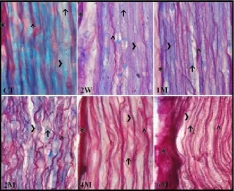
Histopathology: The peripheral nerve of all groups exhibited bundles of myelinated nerve fibers. All nerves fibers, nerve bundles and the entire nerve were surrounded by connective tissue of different thickness. In Luxol fast blue and Picro-Sirus red stained section, one could observe the epineurium, which covered the entire peripheral nerve. In 2W and 1M diabetic groups, the amount of dense connective tissue in epineurium looked minimal but with the progression of the duration of hyperglycemia, the amount of epineurial connective tissues increased. Compared with age-matched control the long-standing diabetic groups of 2, 4 and 6M exhibited numerous thickened epineurial collagen fibers around the entire nerve (Figure 1 and 2). Thin perineurial connective tissue was thin and uniform in 2W, 1M and 2M diabetic groups. However, in 4M and 6M diabetic groups quite thick perineurial collagen was observed as compared to age-matched control groups (Figure 1). Individual nerve fibers were surrounded by thin endoneurial connective tissues. Hematoxyline and eosin stained sections showed large nuclei of Schwann cells over the myelin sheath as well as small nuclei of fibrocytes in the endoneurium. In 2, 4 and 6M diabetic groups the collagen fibers in the endoneurium were thicker than in age-matched control groups. In 2W and 1M diabetic groups, only slight changes were observed (Figure 3). In LFB and PSR stain, 4 and 6M diabetic groups the myelin took faint staining or remained unstained but in 2W, 1 and 2M group myelin stained very lightly as compared with age-matched control groups (Figure 1). In 2W and 1M diabetic groups, the connective tissue around the perineurial blood vessel had few collagen fibers. And the same was observed in epineurium also. In 2, 4 and 6M diabetic groups similar finding were also observed but there was a slight difference in the collagen fiber thickness around blood vessels (Figure 4).
Figure 1. Longitudinal sections of the sciatic nerve from control and all diabetic groups. Note the varying sized myelinated fibers in different groups of the sciatic nerve (↑), endoneurium (>), perineurium (˄), and epineurium (*). Luxol fast blue and Picrosirius red. X1000
Figure 2. Transverse sections of the sciatic nerve from control and all diabetic groups. Note: myelinated nerve fibers (↑) blood capillaries, (↑↑). Collagen fibers (red) along the nerve fibers and around the blood vessels in 1, 2, 4 and 6M diabetic groups are
thicker and numerous. Picrosirius red with Luxol fast blue. X1000
Table 1. Quantitative changes in the myelinated axons of the sciatic nerve in diabetic animals as compared with age-matched control groups (number ± standard deviation; N = 6 for each group).
Group Number of myelinated fibers Number of small myelinated fibers (˂ 7 µm) Number of large myelinated fibers (> 7 µm)
2W-Control 412.13 ± 18.92 195.01 ± 09.47 217.13 ± 07.89
2W-Diabetic 402.25 ± 24.92 183.12 ± 14.43 219.13 ± 06.44
1M-Control 408.25 ± 22.52 202.38 ± 06.52 205.88 ± 09.20
1M-Diabetic 401.63 ± 20.19 174.25 ± 18.05 227.38 ± 09.41
2M-Control 418.38 ± 23.69 188.25 ± 18.14 230.13 ± 07.53
2M-Diabetic 385.13 ± 18.98 149.25 ± 17.29 235.88 ± 05.79
4M-Control 421.88 ± 29.47 182.75 ± 33.81 239.13 ± 08.16
4M-Diabetic 351.13 ± 17.13 106.88 ± 16.05 244.25 ± 10.26
6M-Control 423.75 ± 26.30 178.63 ± 30.93 245.13 ± 08.39
[image:3.595.43.544.646.791.2]Biochemical analysis: Serum creatinine levels were significantly increased (P<0.05) in 1, 2, 4 and 6M diabetic groups as compared to age-matched control groups. Conversely, serum total protein levels significantly decreased (P<0.05) in all diabetic groups as compared to age-matched control groups.
DISCUSSION
Diabetes mellitus is the most common disorder associated with impairment in the metabolism of carbohydrates, lipids and proteins (American Diabetes Association, 2017) in which tissues fail to react properly to insulin in the form of glucose resistance and glucose intolerance, resulting in hyperglycemia (American Diabetes Association, 2015). Hyperglycemia-induced oxidative stress and altered antioxidant levels may lead to potentially severe secondary complications in a number of organ systems and development of peripheral and central
nervous systems abnormalities (Vincent et al., 2004; Faizal et
al., 2017). In the present study, the general changes observed in diabetic animals were the same as previously reported
(Faizal et al., 2017). There was a reduction of body weight in
all diabetic groups which could be attributed to muscle wasting and loss of tissue proteins as a result of lack of insulin, causing
increased glycolysis and gluconeogenesis (Air et al., 2002;
Jain, 2014). These findings were also in agreement with
previous related studies (Doddigarla et al., 2016; Faizal et al.,
2017). Arrangement and direction of peripheral nerve fibers in the present study appeared similar to other related studies
(Malak et al., 2015). However, long-standing
hyperglycemia-induced structural alteration in the peripheral nerve e.g., in 4M and 6M diabetic groups there was poor staining of myelin sheath due to subtle demyelination of nerve fibers. One of the earlier studies also showed similar results in STZ-induced
diabetic rats (Lee et al., 2017) concluding that it might be due
[image:4.595.96.521.68.279.2]to irreversible damage to the peripheral nerve.
Figure 3. Longitudinal sections of the sciatic nerve. Note: myelinated nerve fibers (↑) Schwann cell nucleus, (>) and nucleus of fibrocytes (˄). Inset shows aggregation of Schwann cell nuclei. H &E stain. X1000
Figure 4. Transverse sections of the sciatic nerve showing blood vessels, (↑↑) RBC (>) and myelinated nerve fibers (↑) and collagen fibers (red) along the nerve fibers and in the adventitia of the blood vessels. The adventitia is of comparable thickness among control, 2 weeks, 1 and 2 months
[image:4.595.112.503.330.557.2]Connective tissues provide structural support to nerve bundles
and blood vessels at all level of organization (Faizal et al.,
2018). It has been noted that prolonged hyperglycemia initiates inflammation, associated fibrosis and excess deposition of extracellular matrix in the perivascular connective tissue, followed by a vascular occlusion, ischemia and cell atrophy
(Kundalic et al., 2014). Some researchers have demonstrated
thicker collagen fibers in the connective tissue of endoneurium, perineurium, and epineurium in diabetic groups
than in controls (Kundalic et al., 2014; Elgayar et al., 2017).
In the current study, the control and 2W diabetic groups showed the presence of connective tissue with thin collagen fibers in endoneurium, perineurium, and epineurium and also around the blood vessels inside the peripheral nerve. However, 2M, 4M and 6M diabetic groups showed progressively increasing the thickness of collagen fibers in the endoneurium, perineurium, and epineurium of the peripheral nerve. Earlier studies demonstrated advanced glycation end products (AGEs) are concerned with the advancement of submesothelial fibrosis
and neoangiogenesis (De Vriese et al., 2003). Progressive
fibrosis in a diabetic heart by PKC-β and p38
mitogen-activated protein kinase expression (Olubunmi et al., 2016),
and excess collagen accumulation in endoneurial compartment adversely affects the regeneration and growth of nerve fibers supported by Schwann cells (Bradley, 2000). In diabetic neuropathy, epineurial vessels are most prone to macrovascular changes like occlusion of blood vessels and thrombosis.
These changes also impair nerve blood flow and cause hypoperfusion, basement membrane thickening, and tunica
intima cells proliferation (Llewelyn et al., 1998; Ibrahim et al.,
1999; Tesfaye et al., 2005), Epineurial arteriolar attenuation
with venous tortuosity and distension also results in the
reduction of endoneurial blood flow (Tesfaye et al., 1993). In
the chronic hyperglycemic state, endothelial aldose reductase increases polyol pathway activity as well as vasoconstriction in peripheral nerve producing ischemic pathogenesis resulting in
neuropathy (Dyck et al., 1985). Advanced glycation end
products are formed from proteins like laminin, fibronectin and
collagens, mainly I, III, IV and VI (Nukada et al., 1996; Goh et
al., 2008) and neurofilaments in the nerve axonal fibers and myelin protein in Schwann cells may also be modified by AGEs in nerve fibers influencing the repair mechanism of
damaged nerves (Wada et al., 2005). In the current study,
fibrosis was observed around the blood vessels in prolonged hyperglycemia groups due to deposition of collagen fibers. Comparison of the current result and previous reports on perineurial and endoneurial fibrosis and thickening of collagen
fibers around the adventitia of nerve capillaries (Faizal et al.,
2017) suggests that hyperglycemia accelerates fibrosis. The vascular concept of peripheral neuropathy implies that diabetes-induced endothelial dysfunction resultant decrease in nerve blood flow, vascular reactivity, and endoneurial hypoxia plays a key role in functional and morphological changes in the diabetic nerve. Fibrosis impairs the balance of endoneurial homeostasis and regenerating ability of the nerve fibers. Sciatic nerve, which is the thickest peripheral nerve containing
the sensory, motor, and autonomic nerve fibers (Gewandter et
al., 2017), was the object of the current study. The difference between large-size and small-size nerve fibers concerns
anatomy, transmitting pathways, neurogenesis, energy
utilization, ion distribution, relationship with glial cells and
sensitivity to differing neurotrophic factors (Zotova et al.,
2008). Small-diameter neurons degenerate in experimental
diabetic neuropathy (Beiswenger et al., 2008).
In the current study, the myelinated nerve fibers of the sciatic nerve at many locations appeared disorganized and degenerated with varying grades of defects in myelin sheath in addition to their focal variable thickness. Reduction of the size of myelinated nerve fiber in diabetic rats may be attributed to a loss of neurofilaments due to decreased protein synthesis in the
neuronal cell bodies (Yagihashi et al., 1990). The loss of
axonal cytoskeletal elements may aggravate the vulnerability of diabetic nerves to environmental factors followed by fiber degeneration and eventually fiber loss. In addition prolonged hyperglycemia, oxidative stress activates polyol pathway,
induces sorbitol accumulation and impairs nerve Na+-K+
-ATPase activity leading to nerve dysfunction in terms of
slowing of nerve conduction (Tarr et al., 2013) and structural
abnormalities of diabetic neuropathy (Sima et al., 1985). In the
current study long-standing hyperglycemic rats, myelinated nerve fibers in the sciatic nerve showed quantitative changes like reduced number of total myelinated fibers and loss of small-sized myelinated fibers resulting into the increased proportion of large myelinated fibers.
The serum creatinine formed from amino acid metabolism is known to be a significant marker of diabetic nephropathy
(Ceriello et al., 2000; Ronco et al., 2010). Our results showed
a high serum creatinine level in all diabetic groups parallel to the severity of hyperglycemia as compared with age-matched control. However, the serum total protein levels were reduced in all diabetic groups. Those results hint to a positive correlation of hyperglycemia and the development of diabetic
nephropathy (Sjoholm et al., 2006) with a low-grade
inflammatory process (Sjoholm et al., 2006). Similar
observations have been shown in the other related study (De
Almeida et al., 2012). Based on the findings of the present
study it is concluded that the prolonged hyperglycemic state leads to increased serum creatinine and reduced serum total protein levels and overall decrease in small-sized nerve fibers as well as heaping up of collagen around the nerve and blood vessels, which may constitute important contributing factors in development of diabetic peripheral neuropathy.
Conflict of interest
The authors declare to have no conflict of interest.
Acknowledgements
The author’s gratefully acknowledge the colleagues and authorities of the Department of Anatomy, JN Medical College, and Aligarh Muslim University for strong support.
REFERENCE
ADA (American Diabetes Association) Classification and diagnosis of diabetes. Diabetes Care. 2015; 38: 8-16. ADA (American Diabetes Association). Classification and
diagnosis of diabetes. Diabetes Care. 2017; 40: 11-24. Air EL., Strowski MZ., Benoit SC., Conarellos L., Salituro
GM., Guan XM., Liu K., Woods SC., Zhang BB. 2002. Small molecule insulin mimetics reduce food intake and
body weight and prevent development of obesity. Nat
Med., 8: 179-183.
Beiswenger KK., Calcutt NA., Mizisin AP. 2008. Epidermal nerve fiber quantification in the assessment of diabetic
Biessels GJ., Kappelle AC., Bravenboer B., Erkelens DW., Gispen WH. 1994. Cerebral function in diabetes mellitus.
Diabetologia. 37: 643-650.
Bischhoffshausen VS., Ivulic D., Alvarez P., Schuffeneger VC., Idiaquez J., Fuentes C., Morande P., Fuentes I., Palisson F., Bennett DL., Calvo M. 2017. Recessive dystrophic epidermolysis bullosa results in painful small fibre neuropathy. Brain. 140: 1238-1251.
Boulton A.J., Vinik A.I., Arezzo J.C., Bril V., Feldman E.L., Freeman R., Malik R.A., Maser R.E., Sosenko J.M., Ziegler D. Diabetic neuropathies. Diabetes Care. 2005; 28: 956-962.
Bradley JL., King RH., Thomas P. 2000. The extracellular
matrix of peripheral nerve in diabetic polyneuropathy. J
Peripher Nerv Syst., 5: 243-244.
Ceriello A., Morocutti A., Franceschina M., Quagliaro L., Moro M., Damante G. 2000. Defective intracellular antioxidant enzyme production in type 1 diabetic patients
with nephropathy. Diabetes. 49: 2170-2177.
De Almeida DAT., Camila PB., Ethel LBN. 2012. Ana Angelica HF. Evaluation of Lipid Profile and Oxidative Stress in STZ Induced Rats Treated with Antioxidant
Vitamin. Braz Arch Biol Technol., 55: 527-536.
De Vriese AS., Flyvbjerg A., Mortier S., Tilton RG., Lameire NH. 2003. Inhibition of the interaction of AGE-RAGE prevents hyperglycemia-induced fibrosis of the peritoneal
membrane. J Am Soc Nephrol., 14: 2109-2118.
Doddigarla Z., Parwez I., Abidi S., Jamal A. 2016. Effect of chromium picolinate and melatonin either in single or in a
combination in alloxan induced male Wistar rats. J Biomed
Sci., 6: 1-7.
Dyck PJ., Giannini C. 1996. Pathologic alterations in the
diabetic neuropathies of humans: a review. J Neuropathol
Exp Neurol., 55: 1181-1193.
Dyck PJ., Hansen S., Karnes J., O'Brien P., Yasuda H., Windebank A., Zimmerman B. 1985. Capillary number and
percentage closed in human diabetic sural nerve. Proc Natl
Acad Sci., 82: 2513-2517.
Elgayar SA., Eltony SA., Sayed AA., Abbas AY. 2017. Protective effect of vitamin B com-plex in diabetic
peripheral neuropathy - Histopathological study. Eur J
Anat., 21:173-187.
Faizal M., Khan AA. 2018. A Histomorphometric Study on the Neurohypophysis of STZ-induced Diabetic Albino
Rats. Int J Neuro Res., 4: 371-378.
Faizal M., Khan AA. 2017. Effect of streptozotocin-induced
diabetes on the autonomic ganglia of albino rats. Anatomy.,
11: 51-60.
Faizal MP., Khan AA., Elsy B. 2017. Effect of experimental
hyperglycemia on the trigeminal ganglia of albino rats. Int
J Health Sci Res., 7: 191-198.
Faizal PA., Khan AA. 2017. Impact of experimental hyperglycemia on the lumbosacral dorsal root ganglia of
albino rats. Int J Med Health Sci., 6:158-164.
Gewandter JS., Burke L., Cavaletti G., Dworkin RH., Gibbons C., Gover TD., Herrmann DN., Mcarthur JC., McDermott MP., Rappaport BA., Reeve BB. 2017. Content validity of symptom based measures for diabetic, chemotherapy, and
HIV peripheral neuropathy. Muscle Nerve. 55: 366-372.
Goh SY., Cooper ME. 2008. The role of advanced glycation end products in progression and complications of diabetes.
J Clin Endocrinol Metab., 93: 1143-1152.
Ibrahim S., Harris ND., Radatz M., Selmi F., Rajbhandari S., Brady L., Jakubowski J., Ward JD. 1999. A new minimally
invasive technique to show nerve ischaemia in diabetic
neuropathy. Diabetologia. 42: 737-742.
Jain D., Bansal MK., Dalvi R., Upganlawar A., Somani R. 2014. Protective effect of diosmin against diabetic
neuropathy in experimental rats. J Integr Med., 12: 35-41.
Kundalic B., Ugrenovic S., Jovanovic, Stefanovic N., Petrovic V., Kundalic J., Stojanovic V., Zivkovic V., Antic V. 2014. Morphometric analysis of connective tissue sheaths of sural
nerve in diabetic and nondiabetic patients. Bio Med. Res
Int., 24: 1-7.
Lee EC., Kim MO., Roh GH., Hong SE. 2017. Effects of exercise on neuropathy in streptozotocin-induced diabetic
rats. Ann Rehabil Med., 41: 402-412.
Llewelyn JG., Thomas PK., King RH. 1998. Epineurial
microvasculitis in proximal diabetic neuropathy. J Neurol.,
245: 159-165.
Low PA., Vernino S., Suarez G. 2003. Autonomic dysfunction
in peripheral nerve disease. Muscle Nerve., 27: 646-661.
Malak HW., Saleh SI., Salah El., Din RA., Hamid AHF. 2015. Histological and immunohistochemical study on the consequences of acute glycemic level alteration on the dorsal root ganglia and sciatic nerve integrity in neonatal
albino rats. Egyptian J Histol., 38: 332-345.
Malik RA., Tesfaye S., Newrick PG., Walker D., Rajbhandari SM., Siddique I., Sharma AK., Boulton AJ., King RH., Thomas PK., Ward JD. 2005. Sural nerve pathology in diabetic patients with minimal but progressive neuropathy. Diabetologia. 48: 578-585.
Maser RE., Lenhard MJ., Pohlig RT., Balagopal PB. 2017. Osteopontin and clusterin levels in type 2 diabetes mellitus: differential association with peripheral autonomic nerve
function. Neurol Sci., 21: 1-6.
Mohseni S., Badii M., Kylhammar A., Thomsen NO., Eriksson KF., Malik R.A., Rosen I., Dahlin LB. 2017. Longitudinal study of neuropathy, microangiopathy, and autophagy in
sural nerve: Implications for diabetic neuropathy. Brain
Behav., 7: 1-9.
Naik NS, Lamani S, Devarmani SS. 2015. The role of serum
magnesium level in type 2 diabetes mellitus. Int J Res Med
Sci., 3: 556-559.
Nukada H., van RIJ AM., Packer SG., McMorran PD. 1996. Pathology of acute and chronic ischaemic neuropathy in atherosclerotic peripheral vascular disease. Brain. 119: 1449-1450.
Olubunmi AA., Oluwafeyisetan OA., Peter MO., Owira. 2016.
Naringin Reduces Hyperglycemia-Induced Cardiac
Fibrosis by Relieving Oxidative Stress. Plos One., 11:
1-15.
Pek SL., Sum CF., Yeoh LY., Lee SB., Tang WE., Lim SC., Tavintharan S. 2017. Association of apolipoprotein-CIII
(apoC-III), endothelium-dependent vasodilation and
peripheral neuropathy in a multi-ethnic population with
type 2 diabetes. Metabolism. 72: 75-82.
Prior R., Helleputte VL., Benoy V., Bosch DVL. 2017. Defective axonal transport: A common pathological
mechanism in inherited and acquired peripheral
neuropathies. Neurobiol Dis., 24: 1-65.
Rolim LC., da Silva EM., De Sa JR., Dib SA. 2017. A systematic review of treatment of painful diabetic neuropathy by pain phenotype versus treatment based on
medical comorbidities. Front Neurol., 8: 1-6.
Ronco C., Grammaticopoulos S., Rosner M., Decal M., Soni S., Lentini P. 2010. Oliguria, creatinine and other
biomarkers of acute kidney injury. Contrib Nephrol., 164:
Sengupta P. 2013. The Laboratory Rat: Relating Its Age With
Human's. Int J Prev Med., 4: 624–630
Sima AA., Sugimoto K. 1999. Experimental diabetic
neuropathy: an update. Diabetologia., 42: 773-788.
Sima AAF., Brismar T. 1985. Reversible diabetic nerve dysfunction: structural correlates to electrophysiological
abnormalities. Ann Neurol., 18: 21-29.
Sjoholm A., Nystrom T. 2006. Inflammation and the etiology
of type 2 diabetes. Diabetes Metab Res Rev., 22: 4-10.
Swaminathan A., Cros P., Seddon JA., Mirgayosieva S., Asladdin R., Dusmatova Z. 2017. Peripheral neuropathy in a diabetic child treated with linezolid for multidrug-resistant tuberculosis: a case report and review of the
literature. BMC Infect Dis., 17: 417-422.
Tarr JM., Kaul K., Chopra M., Kohner EM., Chibber R. 2013.
Pathophysiology of diabetic retinopathy. ISRN
Ophthalmol. 15: 1-13.
Tesfaye S., Chaturvedi N., Eaton SE., Ward JD., Manes C., Ionescu-Tirgoviste C., Witte DR., Fuller JH. 2005.
Vascular risk factors and diabetic neuropathy. N Engl J
Med., 352: 341-350.
Tesfaye S., Harris N., Jakubowski JJ., Mody C., Wilson RM., Rennie IG., Ward JD. 1993. Impaired blood flow and arterio-venous shunting in human diabetic neuropathy: a novel technique of nerve photography and fluorescein
angiography. Diabetologia. 36: 1266-1274.
Tomlinson DR., Gardiner NJ. 2008. Glucose neurotoxicity.
Nat Rev Neurosci., 9: 36-45.
Ugwu ET. 2017. Diabetic neuropathic cachexia with profound autonomic dysfunction in a young female with type 1
diabetes. Acta Diabetol., 13: 1-4.
Vincent AM., Russell JW., Low P., Feldman EL. 2004. Oxidative stress in the pathogenesis of diabetic neuropathy.
Endocr Rev., 25: 612-628.
Wada R., Yagihashi S. 2005. Role of advanced glycation end products and their receptors in development of diabetic
neuropathy. Ann N Y Acad Sci., 1043: 598-604.
Yagihashi S., Kamijo M., Watanabe K. 1990. Reduced myelinated fiber size correlates with loss of axonal neurofilaments in peripheral nerve of chronically
streptozotocin diabetic rats. Am J Pathol., 136:1365-1373.
Ziegler D. 2017. Diabetic peripheral and autonomic
neuropathy. Textbook of Diabetes., pp. 580-608.
Zochodne DW., Sun HS., Cheng C., Eyer J. 2004. Accelerated diabetic neuropathy in axons without neurofilaments. Brain. 12: 2193-2200.
Zotova EG., Schaumburg HH., Raine CS., Cannella B., Tar M., Melman A., Arezzo JC. 2008. Effects of hyperglycemia on rat cavernous nerve axons: a functional
and ultra structural study. Exp Neurol., 213: 439-447.