research communications
Acta Cryst.(2019). E75, 163–166 https://doi.org/10.1107/S2056989018018376
163
Received 3 December 2018 Accepted 28 December 2018
Edited by J. Jasinsk, Keene State College, USA
Keywords:crystal structure; semicarbazone; phenylhydrazinecarboxamide.
CCDC reference:1887613
Supporting information:this article has supporting information at journals.iucr.org/e
Crystal structure of 2-[(2
E
)-2-methyl-3-phenylprop-2-en-1-ylidene]-
N
-phenylhydrazinecarboxamide
S. R. Saritha,aL. Anitha,aS. R. Layana,aM. Sithambaresanb* and M. R. Sudarsanakumara
aDepartment of Chemistry, Mahatma Gandhi College, University of Kerala, Thiruvananthapuram 695 004, Kerala, India, andbDepartment of Chemistry, Faculty of Science, Eastern University, Sri Lanka, Chenkalady, Sri Lanka.
*Correspondence e-mail: msithambaresan@gmail.com
The title compound, C17H17N3O, crystallizes with two independent molecules in
the asymmetric unit. The semicarbazone moieties of these independent molecules (I and II) are essentially planar [maximum deviation of 0.042 (1) A˚ in molecule I and 0.041 (1) A˚ in molecule II], with the terminal phenyl rings twisted away from the mean plane of the semicarbazone moiety, making dihedral angles of 60.26 (8) and 28.76 (9) in molecule I and 31.07 (9) and
35.45 (8)in molecule II. The molecules both exhibit an Econfiguration with
respect to the C C and azomethine C N bonds. In the crystal, two classical N—H O hydrogen-bonding interactions are present between the two molecules, forming a centrosymmetric dimer, while a weak C—H O non-classical hydrogen-bonding interaction, with a donor–acceptor distance of 3.476 (2) A˚ , interconnects two neighbouring centrosymmetric dimers to form a cage-like structure. These cage structures are interconnected by weak C—H
interactions with an H distance of 2.790 A˚ , forming supramolecular chains along thec-axis direction.
1. Chemical context
Semicarbazones are oxygen and nitrogen contributor ligands whose significance lies in their versatility of molecular sequence, which allows diverse geometries to be obtained. Semicarbazones exhibit amido–iminol tautomerism in solu-tion due to the interacsolu-tion of solvent molecules, but generally exist in the amido form in the solid state. The FT–IR and NMR spectra of semicarbazones indicate the existence of a keto form in the solid state that can be confirmed by single crystal X-ray diffraction analysis (Kurupet al., 2011; Sreekanth
et al., 2004). Biological properties linked to antimicrobial (Siji
et al., 2010) and antiparasitic (Soareset al., 2011) effects make semicarbazones important ligands in coordination chemistry. Compared to Gentamycin, a commonly used antibiotic,
N4-phenylsemicarbazone derivatives exhibit moderate
anti-bacterial activity at higher concentrations and also show DNA cleavage properties (Layanaet al., 2016). Semicarbazones can function as brilliant ligands in a variety of metal ions (Kalaet al., 2007) and co-ordinate to metal ions either in neutral (Sijiet al., 2011) or in anionic forms (Reenaet al., 2008). Structural studies of many semicarbazones and N4 -phenylsemicarb-azones have been reported and some of them adopt an E
configuration with respect to the azomethine double bond along with both inter- and intramolecular hydrogen-bonding interactions (Reena et al., 2010; Layana et al., 2014, 2018). Semicarbazones form complexes with a variety of structural
features such as monomer, dimer and one-dimensional poly-mers (Kunnathet al., 2016).-Methyl-trans-cinnamaldehyde, a precursor for the synthesis of-methyl-trans
-cinnamaldehyde-N4-phenylsemicarbazone, has significant antifungal activity and can self-couple and form complexes with some transition metals (Shreazet al., 2011). The diverse structural features and substantial biological applications have prompted us to synthesize a new semicarbazone derived from-methyl-trans -cinnamaldehyde andN4-phenylsemicarbazide.
2. Structural commentary
The title compound crystallizes in the triclinic space groupP1 symmetry with two independent molecules, I and II, in the asymmetric unit (Fig. 1). The semicarbazone units in I and II are essentially planar, with maximum deviations from the least-squares plane of 0.042 (1) A˚ for N2 in molecule I and 0.041 (1) A˚ for N4 in molecule II. The terminal phenyl rings in both two molecules are twisted away from the semicarbazone mean plane, making dihedral angles of 60.26 (8) and 28.76 (9)
in molecule I and 31.07 (9) and 35.45 (8)in molecule II. Both
molecules exist in anEconfiguration with respect to the C C and azomethine C N bonds. The azomethine C N and keto C O bond lengths [1.273 (2) and 1.2269 (17) A˚ , respectively] in molecule I are shorter than those for molecule II [1.2766 (19) A˚ and 1.2302 (18) A˚ respectively]. In contrast, the C N and C O bond lengths bond lengths reported for the two independent molecules of 2-benzoylpyridine semi-carbazone are 1.294 (2) and 1.295 (2) A˚ and 1.2360 (19) and 1.2390 (19) A˚ respectively (de Limaet al., 2008).
3. Supramolecular features
In the crystal, two classical and one non-classical hydrogen-bonding interactions are observed. Molecules I and II are linked into centrosymmetric dimers through N2—H20 O2
and N5—H50 O1 hydrogen bonds withD Adistances of
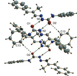
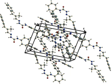
2.808 (2) A˚ , and 2.8639 (19) A˚, respectively (Fig. 2, Table 1), while C13—H13 O2 interactions with a D A distance of 3.476 (2) A˚ , interconnect adjacent dimers, creating cage-like structures that are linked by weak C—H interactions into supramolecular chains along the c-axis direction (Fig. 3). No significant–interactions occur. The packing viewed along thebaxis is shown in Fig. 4.
3.1. Database survey
The structure of the title compound has not previously been reported (CSD version 5.39, update of August 2018; Groomet al., 2016). All geometric parameters in the title compound
164
Sarithaet al. C17H17N3O Acta Cryst.(2019). E75, 163–166
[image:2.610.311.567.108.185.2] [image:2.610.55.288.193.279.2]research communications
Figure 2
N—H O hydrogen bonds and weak C—H O intermolecular inter-actions (dashed lines) generating centrosymmetric dimers and a cage-like structure.
Table 1
Hydrogen-bond geometry (A˚ ,).
Cg1 is the centroid of the C1–C6 ring.
D—H A D—H H A D A D—H A
N5—H50
O1 0.88 (1) 1.99 (1) 2.8639 (19) 174 (2)
N2—H20
O2 0.88 (1) 1.93 (1) 2.808 (2) 176 (2)
N3—H30
N1 0.87 (1) 2.13 (2) 2.6146 (18) 115 (1)
N6—H60
N4 0.87 (1) 2.17 (2) 2.6261 (19) 112 (2)
C13—H13 O2i 0.93 2.64 3.476 (2) 149
C32—H32 Cg1ii 0.93 2.79 3.518 (2) 136
[image:2.610.301.561.449.707.2]Symmetry codes: (i)xþ1;y;zþ1; (ii)xþ1;y;zþ2.
Figure 1
[image:2.610.48.294.560.718.2]agree well with those reported in the literature with the C10— N1/C27—N4 [1.273 (2) and 1.2766 (19) A˚ ], N1— N2/N4—N5 [1.3691 (17) and 1.3679 (18) A˚ ] and C11—O1/C28—O2 [1.2269 (17) and 1.2302 (18) A˚ ] bond distances being comparable to those in benzaldehyde-N4
-phenylsemicarb-azone [1.273 (2), 1.369 (2) and 1.225 (2) A˚ ; Layanaet al., 2014] and vanillin-N-phenylthiosemicarbazone [1.2726 (17), 1.3801 (15) and 1.2404 (15) A˚ ; Layanaet al., 2016]
4. Synthesis and crystallization
Hot ethanolic solutions ofN4-phenylsemicarbazide (0.1512 g, 1 mmol) and -methyl-trans-cinnamaldehyde (0.14 ml, 1 mmol) were mixed and refluxed for about 4 h. Colourless block-shaped crystals of the title compound (yield 83%) were separated by filtration, washed with ethanol and dried over P4O10in vacuo. Single crystals (m.p. 4632 K) were obtained
by slow evaporation of a 1:1 mixture of ethyl acetate and ethanol.
Analysis calculated: C, 73.03; H, 6.09, N, 15.04%. Found: C, 72.66; H, 6.32; N, 15.29%. Spectrometric data. FT–IR max
(KBr, cm1): The spectrum of the title compound shows characteristic absorption bands of the main functional groups at IR (max, cm
1
): 3379 (4NH), 3192 (2NH), 1685 (C O) 3072, 2960 (C—H aromatic), 1591 (C N), 1029 (N—N). FT– Raman (cm1) 3055 (N—H), 1613 (C O), 1577 (C N), 1137 (N—N).1H NMR (400 MHz) (DMSO-d6, ppm):H2.2 (s, 3H,
methyl), 7–7.5 (m, 10H, Ar—H), 6.7 (s, 1H, methine), 7.7 (s, 1H, azomethine), 8.6 (s, 2H, amine), 10.6 (s, 1H, iminol H).
13
C NMR (400 MHz) (DMSO-d6, ppm):C135.2 (C6), 129.12
(C1 and C5), 128.4 (C2 and C4), 119.6 ppm (C3), 152.9 (C7), 146.4 (C8), 138.9 (C9), 136.5 (C10), 12.9 (C17), 134.3 (C11), 128.5 ppm (C12 and C16), 127.4 ppm (C13 and C15) and 122.4 ppm (C14). UV–visible (200–1000, nm): 268 (–*), 342 (n–
*).
research communications
Acta Cryst.(2019). E75, 163–166 Sarithaet al. C
[image:3.610.46.296.70.325.2]17H17N3O
165
Figure 4
[image:3.610.121.499.457.740.2]The packing viewed along thebaxis. Figure 3
5. Refinement
Crystal data, data collection and structure refinement details are summarized in Table 2. Reflections (111) and (001) were omitted due to bad agreement. All hydrogen atoms bound to carbon atoms were positioned geometrically with C—H distances of 0.93–0.96 A˚ and refined as riding, withUiso(H) =
1.2Ueq(C) or 1.5Ueq(C-methyl). The NH hydrogen atoms were
located in a difference-Fourier map and refined with N—H restrained to 0.880.01 A˚ .
Acknowledgements
The authors are thankful to the SAIF, STIC, Cochin Univer-sity of Science and Technology, Kochi, Kerala, India for providing the instrumental facilities for single-crystal X-ray diffraction, elemental analysis, FT–IR, NMR and UV–Vis
spectroscopic studies and to the SAIF IIT Madras, Chennai, India for the FT–Raman spectroscopic data. They are grateful to Dr. M. R. Prathapachandra Kurup, Department of Applied Chemistry, Cochin University of Science & Technology, Kochi-22, India, for the use of theDIAMONDsoftware.
Funding information
Funding for this research was provided by: University of Kerala, Thiruvananthapuram, India.
References
Brandenburg, K. (2010).DIAMOND. Crystal Impact GbR, Bonn, Germany.
Bruker (2004). APEX2, SAINT and SADABS. Bruker AXS Inc., Madison, Wisconsin, USA.
Farrugia, L. J. (2012).J. Appl. Cryst.45, 849–854.
Groom, C. R., Bruno, I. J., Lightfoot, M. P. & Ward, S. C. (2016).Acta Cryst.B72, 171–179.
Kala, U. L., Suma, S., Kurup, M. R. P., Krishnan, S. & John, R. P. (2007).Polyhedron,26, 1427–1435.
Kunnath, R. J., Sithambaresan, M., Aravindakshan, A. A., Natarajan, A. & Kurup, M. R. P. (2016).Polyhedron,113, 73–80.
Kurup, M. R. P., Varghese, B., Sithambaresan, M., Krishnan, S., Sheeja, S. R. & Suresh, E. (2011).Polyhedron,30, 70–78. Layana, S. R., Saritha, S. R., Anitha, L., Sithambaresan, M.,
Sudarsanakumar, M. R. & Suma, S. (2018).J. Mol. Struct. 1157, 579–586.
Layana, S. R., Siji, V. L., Sudarsanakumar, M. R., Suma, S., Kurup, M. R. P. & Sikha, T. S. (2016).J. Indian Chem. Soc.93, 577–586. Layana, S. R., Sithambaresan, M., Siji, V. L., Sudarsanakumar, M. R.
& Suma, S. (2014).Acta Cryst.E70, o591.
Lima, D. F. de, Pe´rez-Rebolledo, A., Ellena, J. & Beraldo, H. (2008).
Acta Cryst.E64, o177.
Reena, T. A. & Kurup, M. R. P. (2010).J. Chem. Crystallogr.40, 927– 932.
Reena, T. A., Seena, E. B. & Kurup, M. R. P. (2008).Polyhedron,27, 1825–1831.
Sheldrick, G. M. (2008).Acta Cryst.A64, 112–122. Sheldrick, G. M. (2015).Acta Cryst.C71, 3–8.
Shreaz, S., Sheikh, R. A., Bhatia, R., Neelofar, K., Imran, S., Hashmi, A. A., Manzoor, N., Basir, S. F. & Khan, L. A. (2011).Biometals,24, 923–933.
Siji, V. L., Kumar, M. R. S., Suma, S. & Kurup, M. R. P. (2010).
Spectrochim. Acta A,76, 22–28.
Siji, V. L., Sudarsanakumar, M. R., Suma, S., George, A. & Thomas, P. V. (2011).Indian J. Chem. Sect. A,50, 793–797.
Soares, R. O. A., Echevarria, A., Bellieny, M. S. S., Pinho, R. T., de Leo, R. M. M., Seguins, W. S., Machado, E. M., Canto-Cavalheiro, M. M. & Leon, L. L. (2011).Exp. Parasitol.129, 381–387. Sreekanth, A., Kala, U. L., Nayar, C. R. & Kurup, M. R. P. (2004).
Polyhedron,23, 41–47.
Westrip, S. P. (2010).J. Appl. Cryst.43, 920–925.
166
Sarithaet al. C17H17N3O Acta Cryst.(2019). E75, 163–166
[image:4.610.44.291.92.395.2]research communications
Table 2
Experimental details.
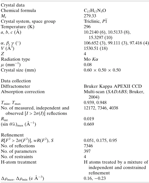
Crystal data
Chemical formula C17H17N3O
Mr 279.33
Crystal system, space group Triclinic,P1
Temperature (K) 296
a,b,c(A˚ ) 10.2140 (6), 10.5133 (8),
15.3297 (10)
,,(
) 106.652 (3), 99.111 (3), 97.416 (4)
V(A˚3) 1530.51 (18)
Z 4
Radiation type MoK
(mm1) 0.08
Crystal size (mm) 0.600.500.50
Data collection
Diffractometer Bruker Kappa APEXII CCD
Absorption correction Multi-scan (SADABS; Bruker,
2004)
Tmin,Tmax 0.939, 0.948
No. of measured, independent and observed [I> 2 (I)] reflections
12172, 7346, 4038
Rint 0.019
(sin/)max(A˚1) 0.669
Refinement
R[F2> 2 (F2)],wR(F2),S 0.051, 0.175, 0.95
No. of reflections 7346
No. of parameters 397
No. of restraints 4
H-atom treatment H atoms treated by a mixture of
independent and constrained refinement
max,min(e A˚3) 0.16,0.23
Computer programs: APEX2, SAINT and XPREP (Bruker, 2004), SHELXL2014
supporting information
sup-1
Acta Cryst. (2019). E75, 163-166
supporting information
Acta Cryst. (2019). E75, 163-166 [https://doi.org/10.1107/S2056989018018376]
Crystal structure of 2-[(2
E
)-2-methyl-3-phenylprop-2-en-1-ylidene]-
N
-phenyl-hydrazinecarboxamide
S. R. Saritha, L. Anitha, S. R. Layana, M. Sithambaresan and M. R. Sudarsanakumar
Computing details
Data collection: APEX2 (Bruker, 2004); cell refinement: APEX2 and SAINT (Bruker, 2004); data reduction: SAINT and
XPREP (Bruker, 2004); program(s) used to solve structure: SHELXL2014 (Sheldrick, 2015); program(s) used to refine structure: SHELXS97 (Sheldrick, 2008); molecular graphics: ORTEP-3 for Windows (Farrugia, 2012) and DIAMOND
(Brandenburg, 2010); software used to prepare material for publication: publCIF (Westrip, 2010).
2-[(2E)-2-Methyl-3-phenylprop-2-en-1-ylidene]-N-phenylhydrazinecarboxamide
Crystal data
C17H17N3O
Mr = 279.33
Triclinic, P1
a = 10.2140 (6) Å
b = 10.5133 (8) Å
c = 15.3297 (10) Å
α = 106.652 (3)°
β = 99.111 (3)°
γ = 97.416 (4)°
V = 1530.51 (18) Å3
Z = 4
F(000) = 592
Dx = 1.212 Mg m−3
Mo Kα radiation, λ = 0.71073 Å Cell parameters from 3309 reflections
θ = 2.6–27.7°
µ = 0.08 mm−1
T = 296 K Block, colorless 0.60 × 0.50 × 0.50 mm
Data collection
Bruker Kappa APEXII CCD diffractometer
Radiation source: fine-focus sealed tube Graphite monochromator
ω and φ scan
Absorption correction: multi-scan (SADABS; Bruker, 2004)
Tmin = 0.939, Tmax = 0.948
12172 measured reflections 7346 independent reflections 4038 reflections with I > 2σ(I)
Rint = 0.019
θmax = 28.4°, θmin = 2.2°
h = −13→12
k = −13→13
l = −20→15
Refinement
Refinement on F2 Least-squares matrix: full
R[F2 > 2σ(F2)] = 0.051
wR(F2) = 0.175
S = 0.95 7346 reflections 397 parameters 4 restraints
Hydrogen site location: mixed
H atoms treated by a mixture of independent and constrained refinement
w = 1/[σ2(F
o2) + (0.1017P)2] where P = (Fo2 + 2Fc2)/3 (Δ/σ)max < 0.001
supporting information
sup-2
Acta Cryst. (2019). E75, 163-166
Special details
Geometry. All esds (except the esd in the dihedral angle between two l.s. planes) are estimated using the full covariance
matrix. The cell esds are taken into account individually in the estimation of esds in distances, angles and torsion angles; correlations between esds in cell parameters are only used when they are defined by crystal symmetry. An approximate (isotropic) treatment of cell esds is used for estimating esds involving l.s. planes.
Fractional atomic coordinates and isotropic or equivalent isotropic displacement parameters (Å2)
x y z Uiso*/Ueq
supporting information
sup-3
Acta Cryst. (2019). E75, 163-166
H21 −0.3953 0.5993 0.3110 0.092* C22 −0.2836 (2) 0.4633 (2) 0.33250 (14) 0.0643 (5) H22 −0.2347 0.5174 0.3908 0.077* C23 −0.25859 (16) 0.3344 (2) 0.29541 (12) 0.0507 (4) C24 −0.15906 (16) 0.27357 (19) 0.34168 (12) 0.0498 (4) H24 −0.1223 0.2101 0.3017 0.060* C25 −0.11244 (15) 0.29476 (17) 0.43239 (11) 0.0431 (4) C26 −0.15538 (17) 0.38927 (19) 0.51113 (12) 0.0516 (4) H26A −0.0976 0.4761 0.5297 0.077* H26B −0.1493 0.3543 0.5627 0.077* H26C −0.2469 0.3982 0.4916 0.077* C27 −0.01521 (15) 0.21447 (17) 0.45568 (11) 0.0451 (4) H27 0.0122 0.1537 0.4079 0.054* C28 0.17376 (15) 0.13993 (19) 0.63940 (12) 0.0474 (4) C29 0.17182 (15) 0.24127 (19) 0.80442 (12) 0.0482 (4) C30 0.1829 (2) 0.3675 (2) 0.86669 (14) 0.0740 (6) H30 0.1683 0.4397 0.8452 0.089* C31 0.2159 (2) 0.3874 (3) 0.96110 (15) 0.0878 (7) H31 0.2219 0.4729 1.0029 0.105* C32 0.23933 (19) 0.2836 (3) 0.99329 (15) 0.0733 (6) H32 0.2616 0.2974 1.0569 0.088* C33 0.2302 (2) 0.1594 (3) 0.93208 (15) 0.0766 (7) H33 0.2476 0.0882 0.9540 0.092* C34 0.1954 (2) 0.1371 (2) 0.83737 (14) 0.0675 (5) H34 0.1880 0.0510 0.7961 0.081* N1 0.44999 (12) −0.16681 (14) 0.51975 (9) 0.0447 (3) N2 0.34882 (14) −0.09748 (16) 0.50394 (10) 0.0485 (4) N3 0.35427 (14) −0.17255 (17) 0.34979 (10) 0.0536 (4) N4 0.03382 (12) 0.22481 (14) 0.53988 (9) 0.0455 (3) N5 0.12237 (14) 0.14139 (16) 0.55249 (10) 0.0516 (4) N6 0.13105 (14) 0.22397 (17) 0.70885 (10) 0.0538 (4) O1 0.20160 (11) −0.04300 (13) 0.40141 (8) 0.0525 (3) O2 0.25451 (13) 0.06620 (15) 0.65140 (8) 0.0648 (4) H3′ 0.4260 (12) −0.1964 (17) 0.3742 (11) 0.049 (5)* H5′ 0.1507 (17) 0.0898 (17) 0.5059 (9) 0.060 (6)* H6′ 0.080 (2) 0.276 (2) 0.6905 (15) 0.087 (7)* H2′ 0.3176 (16) −0.0501 (17) 0.5509 (9) 0.055 (5)*
Atomic displacement parameters (Å2)
U11 U22 U33 U12 U13 U23
supporting information
sup-4
Acta Cryst. (2019). E75, 163-166
C8 0.0415 (8) 0.0493 (10) 0.0467 (10) 0.0124 (7) 0.0108 (7) 0.0198 (8) C9 0.0615 (11) 0.0734 (14) 0.0521 (11) 0.0290 (10) 0.0109 (8) 0.0186 (10) C10 0.0455 (8) 0.0494 (11) 0.0446 (9) 0.0136 (7) 0.0116 (7) 0.0192 (8) C11 0.0416 (8) 0.0477 (10) 0.0457 (9) 0.0108 (7) 0.0081 (7) 0.0197 (8) C12 0.0453 (9) 0.0457 (10) 0.0419 (9) 0.0117 (7) 0.0044 (7) 0.0128 (8) C13 0.0478 (10) 0.0906 (16) 0.0518 (11) 0.0219 (10) 0.0054 (8) 0.0141 (11) C14 0.0620 (12) 0.138 (2) 0.0514 (12) 0.0239 (13) 0.0164 (10) 0.0196 (14) C15 0.0725 (13) 0.113 (2) 0.0420 (11) 0.0223 (12) 0.0025 (10) 0.0188 (12) C16 0.0545 (10) 0.0749 (15) 0.0532 (11) 0.0158 (9) −0.0066 (9) 0.0134 (10) C17 0.0432 (9) 0.0663 (13) 0.0519 (11) 0.0134 (8) 0.0044 (8) 0.0185 (9) C18 0.0621 (11) 0.0881 (17) 0.0470 (11) 0.0220 (10) 0.0057 (9) 0.0169 (11) C19 0.0634 (12) 0.120 (2) 0.0508 (12) 0.0199 (13) −0.0005 (9) 0.0295 (13) C20 0.0599 (12) 0.116 (2) 0.0783 (16) 0.0254 (13) 0.0060 (11) 0.0555 (16) C21 0.0739 (13) 0.0754 (16) 0.0897 (16) 0.0222 (11) 0.0032 (12) 0.0422 (14) C22 0.0677 (12) 0.0597 (14) 0.0642 (12) 0.0129 (9) −0.0033 (9) 0.0257 (11) C23 0.0463 (9) 0.0648 (13) 0.0449 (10) 0.0096 (8) 0.0081 (7) 0.0240 (9) C24 0.0504 (9) 0.0522 (11) 0.0458 (10) 0.0135 (8) 0.0078 (7) 0.0133 (8) C25 0.0403 (8) 0.0432 (10) 0.0439 (9) 0.0064 (7) 0.0046 (7) 0.0137 (8) C26 0.0530 (9) 0.0535 (12) 0.0508 (10) 0.0179 (8) 0.0109 (8) 0.0163 (9) C27 0.0445 (8) 0.0455 (10) 0.0431 (9) 0.0114 (7) 0.0069 (7) 0.0103 (8) C28 0.0397 (8) 0.0527 (11) 0.0461 (10) 0.0114 (7) 0.0022 (7) 0.0120 (8) C29 0.0404 (8) 0.0587 (12) 0.0454 (9) 0.0135 (7) 0.0081 (7) 0.0147 (9) C30 0.0992 (15) 0.0679 (15) 0.0585 (13) 0.0323 (12) 0.0211 (11) 0.0154 (11) C31 0.1139 (19) 0.0819 (18) 0.0553 (14) 0.0197 (14) 0.0196 (13) 0.0005 (13) C32 0.0642 (12) 0.104 (2) 0.0472 (12) 0.0100 (12) 0.0097 (9) 0.0199 (13) C33 0.0860 (15) 0.0902 (19) 0.0625 (14) 0.0214 (13) 0.0101 (11) 0.0380 (14) C34 0.0845 (13) 0.0593 (14) 0.0558 (12) 0.0146 (10) 0.0066 (10) 0.0174 (10) N1 0.0417 (7) 0.0494 (9) 0.0485 (8) 0.0147 (6) 0.0088 (6) 0.0214 (7) N2 0.0490 (8) 0.0593 (10) 0.0444 (8) 0.0231 (7) 0.0120 (6) 0.0207 (7) N3 0.0499 (8) 0.0735 (11) 0.0438 (8) 0.0295 (7) 0.0071 (6) 0.0216 (8) N4 0.0417 (7) 0.0472 (9) 0.0465 (8) 0.0127 (6) 0.0045 (6) 0.0136 (7) N5 0.0512 (8) 0.0588 (10) 0.0441 (9) 0.0247 (7) 0.0042 (7) 0.0119 (8) N6 0.0547 (8) 0.0642 (11) 0.0449 (8) 0.0263 (7) 0.0072 (7) 0.0159 (8) O1 0.0492 (6) 0.0641 (9) 0.0501 (7) 0.0244 (6) 0.0089 (5) 0.0218 (6) O2 0.0679 (8) 0.0774 (10) 0.0485 (7) 0.0406 (7) 0.0013 (6) 0.0124 (7)
Geometric parameters (Å, º)
supporting information
sup-5
Acta Cryst. (2019). E75, 163-166
C5—H5 0.9300 C24—H24 0.9300 C6—C7 1.469 (2) C25—C27 1.450 (2) C7—C8 1.334 (2) C25—C26 1.493 (2) C7—H7 0.9300 C26—H26A 0.9600 C8—C10 1.453 (2) C26—H26B 0.9600 C8—C9 1.487 (2) C26—H26C 0.9600 C9—H9A 0.9600 C27—N4 1.2766 (19) C9—H9B 0.9600 C27—H27 0.9300 C9—H9C 0.9600 C28—O2 1.2302 (18) C10—N1 1.273 (2) C28—N6 1.346 (2) C10—H10 0.9300 C28—N5 1.358 (2) C11—O1 1.2269 (17) C29—C34 1.363 (3) C11—N2 1.355 (2) C29—C30 1.373 (3) C11—N3 1.356 (2) C29—N6 1.410 (2) C12—C13 1.373 (2) C30—C31 1.380 (3) C12—C17 1.378 (2) C30—H30 0.9300 C12—N3 1.406 (2) C31—C32 1.354 (3) C13—C14 1.368 (3) C31—H31 0.9300 C13—H13 0.9300 C32—C33 1.353 (3) C14—C15 1.374 (3) C32—H32 0.9300 C14—H14 0.9300 C33—C34 1.382 (3) C15—C16 1.359 (3) C33—H33 0.9300 C15—H15 0.9300 C34—H34 0.9300 C16—C17 1.375 (3) N1—N2 1.3691 (17) C16—H16 0.9300 N2—H2′ 0.881 (9) C17—H17 0.9300 N3—H3′ 0.868 (9) C18—C19 1.383 (3) N4—N5 1.3679 (18) C18—C23 1.390 (3) N5—H5′ 0.879 (9) C18—H18 0.9300 N6—H6′ 0.872 (9)
supporting information
sup-6
Acta Cryst. (2019). E75, 163-166
C8—C7—C6 127.81 (16) C25—C26—H26B 109.5 C8—C7—H7 116.1 H26A—C26—H26B 109.5 C6—C7—H7 116.1 C25—C26—H26C 109.5 C7—C8—C10 118.17 (15) H26A—C26—H26C 109.5 C7—C8—C9 124.61 (15) H26B—C26—H26C 109.5 C10—C8—C9 117.15 (14) N4—C27—C25 121.85 (15) C8—C9—H9A 109.5 N4—C27—H27 119.1 C8—C9—H9B 109.5 C25—C27—H27 119.1 H9A—C9—H9B 109.5 O2—C28—N6 123.87 (15) C8—C9—H9C 109.5 O2—C28—N5 120.66 (16) H9A—C9—H9C 109.5 N6—C28—N5 115.48 (14) H9B—C9—H9C 109.5 C34—C29—C30 118.95 (17) N1—C10—C8 120.96 (15) C34—C29—N6 122.71 (17) N1—C10—H10 119.5 C30—C29—N6 118.29 (17) C8—C10—H10 119.5 C29—C30—C31 120.2 (2) O1—C11—N2 120.94 (15) C29—C30—H30 119.9 O1—C11—N3 124.61 (14) C31—C30—H30 119.9 N2—C11—N3 114.45 (13) C32—C31—C30 120.5 (2) C13—C12—C17 119.33 (16) C32—C31—H31 119.8 C13—C12—N3 117.83 (13) C30—C31—H31 119.8 C17—C12—N3 122.81 (15) C33—C32—C31 119.5 (2) C14—C13—C12 120.14 (16) C33—C32—H32 120.3 C14—C13—H13 119.9 C31—C32—H32 120.3 C12—C13—H13 119.9 C32—C33—C34 120.8 (2) C13—C14—C15 120.64 (19) C32—C33—H33 119.6 C13—C14—H14 119.7 C34—C33—H33 119.6 C15—C14—H14 119.7 C29—C34—C33 120.1 (2) C16—C15—C14 119.16 (18) C29—C34—H34 120.0 C16—C15—H15 120.4 C33—C34—H34 120.0 C14—C15—H15 120.4 C10—N1—N2 116.30 (14) C15—C16—C17 120.91 (16) C11—N2—N1 120.63 (14) C15—C16—H16 119.5 C11—N2—H2′ 119.3 (11) C17—C16—H16 119.5 N1—N2—H2′ 120.1 (11) C16—C17—C12 119.81 (17) C11—N3—C12 127.30 (13) C16—C17—H17 120.1 C11—N3—H3′ 111.3 (11) C12—C17—H17 120.1 C12—N3—H3′ 120.4 (11) C19—C18—C23 121.3 (2) C27—N4—N5 116.14 (14) C19—C18—H18 119.4 C28—N5—N4 120.26 (15) C23—C18—H18 119.4 C28—N5—H5′ 117.5 (12) C20—C19—C18 119.9 (2) N4—N5—H5′ 122.2 (12) C20—C19—H19 120.0 C28—N6—C29 125.66 (14) C18—C19—H19 120.0 C28—N6—H6′ 113.7 (15) C21—C20—C19 119.53 (19) C29—N6—H6′ 120.3 (15)
supporting information
sup-7
Acta Cryst. (2019). E75, 163-166
C4—C5—C6—C1 −0.2 (3) C24—C25—C27—N4 −178.73 (16) C4—C5—C6—C7 −177.99 (19) C26—C25—C27—N4 −1.0 (2) C2—C1—C6—C5 0.8 (3) C34—C29—C30—C31 −0.8 (3) C2—C1—C6—C7 178.68 (18) N6—C29—C30—C31 176.69 (19) C5—C6—C7—C8 −47.8 (3) C29—C30—C31—C32 0.9 (4) C1—C6—C7—C8 134.4 (2) C30—C31—C32—C33 −0.1 (4) C6—C7—C8—C10 −179.13 (17) C31—C32—C33—C34 −1.0 (3) C6—C7—C8—C9 −2.4 (3) C30—C29—C34—C33 −0.2 (3) C7—C8—C10—N1 171.39 (16) N6—C29—C34—C33 −177.57 (17) C9—C8—C10—N1 −5.6 (3) C32—C33—C34—C29 1.1 (3) C17—C12—C13—C14 0.6 (3) C8—C10—N1—N2 −179.88 (14) N3—C12—C13—C14 178.7 (2) O1—C11—N2—N1 177.56 (15) C12—C13—C14—C15 −0.1 (4) N3—C11—N2—N1 −2.8 (2) C13—C14—C15—C16 −0.4 (4) C10—N1—N2—C11 177.09 (15) C14—C15—C16—C17 0.6 (4) O1—C11—N3—C12 3.3 (3) C15—C16—C17—C12 −0.1 (3) N2—C11—N3—C12 −176.34 (17) C13—C12—C17—C16 −0.4 (3) C13—C12—N3—C11 149.8 (2) N3—C12—C17—C16 −178.43 (18) C17—C12—N3—C11 −32.2 (3) C23—C18—C19—C20 −0.4 (3) C25—C27—N4—N5 178.64 (15) C18—C19—C20—C21 1.2 (4) O2—C28—N5—N4 −179.26 (16) C19—C20—C21—C22 −0.7 (4) N6—C28—N5—N4 0.2 (2) C20—C21—C22—C23 −0.7 (3) C27—N4—N5—C28 −176.31 (16) C21—C22—C23—C18 1.4 (3) O2—C28—N6—C29 0.5 (3) C21—C22—C23—C24 −179.73 (19) N5—C28—N6—C29 −179.02 (16) C19—C18—C23—C22 −0.9 (3) C34—C29—N6—C28 −37.1 (3) C19—C18—C23—C24 −179.82 (19) C30—C29—N6—C28 145.5 (2)
Hydrogen-bond geometry (Å, º)
Cg1 is the centroid of the C1–C6 ring.
D—H···A D—H H···A D···A D—H···A
N5—H5′···O1 0.88 (1) 1.99 (1) 2.8639 (19) 174 (2) N2—H2′···O2 0.88 (1) 1.93 (1) 2.808 (2) 176 (2) N3—H3′···N1 0.87 (1) 2.13 (2) 2.6146 (18) 115 (1) N6—H6′···N4 0.87 (1) 2.17 (2) 2.6261 (19) 112 (2) C13—H13···O2i 0.93 2.64 3.476 (2) 149 C32—H32···Cg1ii 0.93 2.79 3.518 (2) 136