Plants and microorganisms are able to generate all 20 amino acids necessary for the protein syn-thesis and they can even synsyn-thesise many more amino acids. For example, more than 700 of non-protein amino acids have been isolated from plants as secondary metabolites. Some of these amino acids appear to play an important role as interme-diates in the biosynthesis of the plant signalling molecules, vitamins, and other constituents (e.g. alkaloids, bile salts, and pigments). They repre-sent storage forms of nitrogen or sulfur, some of them play a role in the enhancement of tolerance against abiotic stress, some act in the plant chemi-cal defence system against a variety of organisms as they manifest antimetabolic effects in viruses, bacteria, fungi, as well as in lower and higher plants and animals. Many of these amino acids are, however, considered to be only biochemical oddities. Some of these amino acids also show
beneficial properties as they exhibit significant antioxidative and anticarcinogenic activities in animals. In animals, some non-protein amino acids act in intracellular metabolic processes (e.g. in the metabolism of lipids), as macroergic substrates, and neurotransmitters, and intermediates in the synthesis of some biologically active compounds (e.g. hormones) and stimulants.
1 3-AMINO ACIDS AND 4-AMINO ACIDS
β-Alanine (3-aminopropanoic acid) is involved in the biosynthesis of pantothenic acid, coen-zyme A (HS-CoA), and some histidine dipeptides (VELÍŠEK et al. 2006). The principal pathway of
β-alanine formation (KEGG) is the decarboxyla-tion of L-aspartic acid catalysed by the pyridoxal 5´-phosphate protein aspartate-1-decarboxylase (EC 4.1.1.11) (Figure 1).
Partly supported by the Ministry of Education, Youth and Sports of the Czech Republic, Project No. MSM 6046137305.
Biosynthesis of Food Constituents: Amino Acids:
4. Non-protein Amino Acids – a Review
JAN VELÍŠEK, ROMAN KUBEC and KAREL CEJPEK
Department of Food Chemistry and Analysis, Faculty of Food and Biochemical
Technology, Institute of Chemical Technology Prague, Prague, Czech Republic
Abstract
VELÍŠEK J., KUBEC R., CEJPEK K. (2006): Biosynthesis of food constituents: Amino acids: 4. Non-protein amino acids – a review. Czech J. Food Sci., 24: 93–109.
This review article gives a brief survey of the principal pathways that lead to the biosynthesis of the most important non-protein amino acids occurring in foods and feeds. These amino acids have been divided into the following groups: 3-amino acids and 4-amino acids, N-substituted amino acids, alicyclic amino acids, hydroxyamino acids, sulfur-con-taining amino acids, basic amino acids, and taurine.
γ-Aminobutanoic acid (GABA, 4-aminobuta-noic acid) acts as a chemical messenger in the cell communication. It predominantly occurs in the brain tissue (but also in plant and microbial cells) where it acts as a neurotransmitter inhibi-tor. It is also a constituent of some peptides, e.g. nisine. γ-Aminobutanoic acid is predominantly formed by decarboxylation of L-glutamic acid catalysed by the pyridoxal 5´-phosphate protein, i.e. glutamate decarboxylase (EC 4.1.1.15) (Figure 2) (KEGG).
2 N-SUBSTITUTED AMINO ACIDS 2.1 Sarcosine and glycine betaine
Glycine N-methyltransferase (EC 2.1.1.20)1
ca-talyses the transfer of the methyl group of S-ade- nosyl-methionine (SAM or AdoMet) to glycine to form S-adenosylhomocysteine (AdoHcy) and
N-methylglycine (sarcosine, Figure 3). The enzyme is unique among methyltransferases since the methylated product has no physiological activity. It has been suggested that the major role of glycine
N-methyltransferase is to regulate the S
-adenosyl-methionine/S-adenosylhomocysteine ratio rather than to synthesise sarcosine2 (KEGG).
Glycine betaine (N,N,N-trimethylglycine, tri-methylammoniumacetobetaine) is produced by certain higher plants and bacteria. It is synthesised at elevated rates in response to various types of environmental stress via two distinct pathways: oxidation of choline, or N-methylation of glycine (CHEN & MURATA 2002). For almost all biological systems, including most animals, plants (e.g. plants of the family Chenopodiaceae, such as spinach), and microorganisms, glycine betaine biosynthesis is accomplished in chloroplasts by the first pathway, conversion of choline via the unstable interme-diate betaine aldehyde (JAIN & SELVARAJ 2000). The first step of choline oxidation is catalysed by ferredoxin-dependent choline monooxygenase (EC 1.14.15.7), and the second one by NAD(P)-dependent betaine aldehyde dehydrogenase (EC 1.2.1.8)3 (Figure 4).
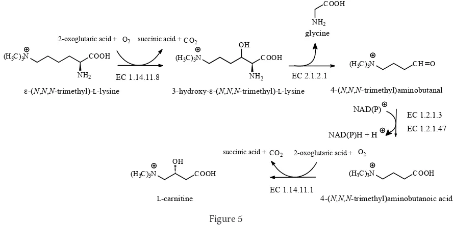
2.2 Carnitine
L-Carnitine [3-hydroxy-4-(N,N,N-trimethyl)- aminobutyric acid] is required for the transfer of
ac-1In addition to its enzymatic role, glycine N-methyltransferase has been shown to be a major folate binding protein in the rat liver cytosol, to be a polycyclic aromatic hydrocarbon-binding protein and a mediator of cytochrome P4501A1 gene expression.
2Sarcosine is also formed as a product of choline and creatine catabolism. Both compounds have several metabolic fates. Choline may be converted into acetylcholine, to phosphatidylcholine or to N,N-dimethylglycine, which is then converted to sarcosine and ultimately to glycine. Dimethylglycine dehydrogenase (EC 1.5.99.2) is an enzyme cataly-sing the oxidative demethylation of N,N-dimethylglycine to form sarcosine. Subsequently, sarcosine dehydrogenase (EC 1.5.99.1) converts sarcosine into glycine. Hydrolysis of creatine by creatinase (EC 3.5.3.3) yields sarcosine and urea. The product of creatinine hydrolysis by creatinine deamidase (EC 3.5.4.21), N-methylhydantoin, yields N -car-bamoylsarcosine by the catalysis of N-methylhydantoinase (ATP-hydrolysing) (EC 3.5.2.14).
3Bacteria Escherichia coli also form glycine betaine from choline but do not use choline monooxygenase (EC 1.14.15.7) as they have choline dehydrogenase (EC 1.1.99.1) instead that generates hydrogen peroxide. Glycine betaine bio-synthesis in the microorganisms Arthrobacter globiformis requires only one enzyme choline oxidase (EC 1.1.3.17). Recently, a novel pathway of glycine betaine biosynthesis from glycine via sarcosine (glycine N-methyltransferase, EC 2.1.1.20) and N,N-dimethylglycine was found in two extremely halophilic microorganisms Ascinopolyspora halo-philia and Ectothiorhodospira halochloris.
HOOC
NH2
COOH
CO2
HOOC NH2 EC 4.1.1.11
[image:2.595.68.532.86.183.2]L-aspartic acid �-alanine
Figure 1 Figure 2
CO2
EC 4.1.1.15
L-glutamic acid
HOOC NH2
COOH HOOC
NH2
tivated acyl groups across intracellular membranes. Many organisms, ranging from bacteria to mam-mals, are able to synthesise carnitine from lysine and methionine. Lysine becomes available in the form of ε-(N,N,N-trimethyl)lysine after lysosomal hydrolysis of proteins that contain this amino acid as a result of the post-translational methylation of lysine residues by lysine N-methyltransferase (EC 2.1.1.43), using SAM as the methyl donor.
In the first step of the carnitine biosynthe-sis, ε-(N,N,N-trimethyl)lysine is hydroxylated to β-hydroxy-ε-(N,N,N-trimethyl)lysine [3-hy-droxy-ε-(N,N,N-trimethyl)lysine] by trimethyl-lysine dioxygenase (EC 1.14.11.8). Subsequently,
β-hydroxy-ε-(N,N,N-trimethyl)lysine is cleaved by retroaldolisation into glycine and 4-(N,N,N -trimethyl)aminobutanal [γ-(N,N,N-trimethyl)amin
obutyraldehyde] by the pyridoxal 5´-phosphoric acid protein β-hydroxy-ε-(N-trimethyl)lysine aldolase (EC 2.1.2.1). 4-(N,N,N-trimethyl)aminobutanal is subsequently oxidised by NAD(P)-dependent 4-trimethylaminobutyraldehyde dehydrogenase (EC 1.2.1.47) or aldehyde dehydrogenase (NAD+) (EC
1.2.1.3) to form 4-(N,N,N-trimethyl)aminobutanoic acid (γ-butyrobetaine). Finally, 4-(N,N,N -tri-methylaminobutanoic acid is hydroxylated at the C-3 position by γ-butyrobetaine hydroxylase (EC 1.14.11.1) to form carnitine (Figure 5). Like lysyl hydroxylase (EC 1.14.11.4, VELÍŠEK & CEJPEK 2006), trimethyllysine dioxygenase and 4-trimethyl-aminobutyraldehyde dehydrogenase are non-heme ferrous-iron dioxygenases that require 2-oxoglu-taric acid and molecular oxygen as substrates, and Fe2+ and L-ascorbic acid (for regeneration of Fe2+
Figure 3
(H3C)3N CH2OH (H3C)3N CH O (H3C)3N COOH
choline betaine aldehyde glycine betaine
ferredoxin (reduced) + O2 ferredoxin (oxidized) + 2 H2O NAD(P) + H2O NAD(P)H + H
EC 1.14.15.7 EC 1.2.1.8
Figure 4
NAD(P)H + H NAD(P)
(H3C)3N COOH
NH2
(H3C)3N COOH
NH2
OH
NH2
COOH
(H3C)3N CH O
(H3C)3N COOH
(H3C)3N COOH
OH 2-oxoglutaric acid + O2
2-oxoglutaric acid + O2
succinic acid + CO2
succinic acid + CO2
glycine
�-(N,N,N-trimethyl)-L-lysine 3-hydroxy-�-(N,N,N-trimethyl)-L-lysine 4-(N,N,N-trimethyl)aminobutanal
4-(N,N,N-trimethyl)aminobutanoic acid
L-carnitine
EC 1.14.11.8 EC 2.1.2.1
EC 1.14.11.1
[image:3.595.76.521.537.758.2]EC 1.2.1.3 EC 1.2.1.47
Figure 5 EC 2.1.1.20
+ +
COOH S
NH2 H3C
Ad
S-adenosyl-L-methionine
+ H
COOH S
NH2
Ad
S-adenosyl-L-homocysteine
glycine NH2
COOH
sarcosine N
COOH
ions) as cofactors (VAZ et al. 1998; SWIEGERS et al. 2002).
3 ALICYCLIC AMINO ACIDS 3.1 1-Aminocyclopropane-1-carboxylic acid
Ethylene is a gaseous, plant signalling molecule (hormone) that regulates many processes of seed germination, plant growth and development, flow-ering, fruit ripening, abscission, and senescence. Plant cells can also synthesise ethylene when they encounter various stress factors, such as physical wounding, pathogen attack, flooding, chilling injury, or the presence of heavy metals. The induc-tion of ethylene synthesis also plays a crucial role in a certain herbicide mode of action.
The biosynthesis of ethylene in higher plants proceeds in several steps. The immediate ethylene precursor is 1-aminocyclopropane-1-carboxylic acid that is synthesised from methionine via SAM (VELÍŠEK & CEJPEK 2006). The formation of 1-ami-nocyclopropane-1-carboxylic acid is catalysed by the pyridoxal 5´-phosphate protein, 1-aminocy-clopropane-1-carboxylate synthase (EC 4.4.1.14). The enzymatic conversion of this acid into ethylene
proceeds via unstable N -hydroxy-1-aminocyclo-propane-1-carboxylic acid, which breaks down into ethylene (derived from the C-2 and C-3 carbons of 1-aminocyclopropane-1-carboxylic acid) and cyanoformic acid. The latter compound is further decomposed into hydrogen cyanide and CO2 as shown in Figure 6. The enzyme involved in this process is 1-aminocyclopropane-1-carboxylic acid oxidase (EC 1.14.17.4), a member of the ferrous-dependent family of non-heme oxygenases. Like other members of the family, it requires ferrous ions and utilises molecular oxygen and ascorbic acid. It also requires bicarbonate ions or CO2 as activators (BARLOW et al. 1997; CHARNG et al.
2001).
3.2 Hypoglycin and related amino acids The unripe fruits of the Jamaican ackee tree (Blighia sapida, Sapindaceae) contain an unusual free amino acid called hypoglycin, i.e. (2S,4S )-3-(methylenecyclopropyl)alanine, also known as L-β-(methylenecyclopropyl)alanine, L-3-(methyl enecyclopropyl)alanine or 2-amino-4,5-methano-hex-5-enoic acid. The free amino acid (hypogly-cin A) is found in the arils and seeds of the fruit
Figure 6 hydrogen cyanide
+ H
EC 4.4.1.14
N
N N
N NH2
CH2
OH OH
O H3C S
5´-methylthioadenosine
COOH NH OH N
C HOOC
S-adenosyl-L-methionine 1-aminocyclopropane-1-carboxylic acid
N-hydroxy-1-aminocyclopropane-1-carboxylic acid cyanoformic acid
H3C
COOH
S
NH2
N
N N
N NH2
CH2
OH OH
O
EC 1.14.17.4
COOH
NH2
1 2
3
L-dehydroascorbic acid L-ascorbic acid
+ +
H2O O2
CO2
H2O +
H C N +
and its content significantly decreases in the arils with ripeness (from 1000–1110 mg/kg to less than 100 mg/kg)4. γ-Glutamyl hypoglycin (hypoglycin B)
is found only in the ackee seeds. Traces of the lower homologue of hypoglycin A with hypoglycaemic activity, i.e. 2-(methylenecyclopropyl)glycine, its
γ-glutamyl dipeptide, and (2S,1´S,2´S)-2-(2´-car- boxycyclopropyl)glycine (Figure 7) are present in the immature seeds (SHERRATT 1986; KEAN 1989; NATALINI et al. 2000).
Significant amounts of hypoglycin A and hy-poglycin B occur also in the seeds of Billia
hip-pocastanum (Hippocastanaceae) from Costa Rica and Acer pseudoplatanus (Aceraceae), a common sycamore of temperate zones. The lower homologue of hypoglycin, L-α-(methylenecyclopropyl)glycine, was found in the seeds of Litchi sinensis ( Sapin-daceae) (KEAN 1989).
Symptoms of ackee poisoning (commonly known as Jamaican vomiting sickness or more accu-rately the toxic hypoglycaemic syndrome) oc-cur 6–48 h after the ingestion of unripe arils and include nausea, vomiting, drowsiness, muscular and mental exhaustion, and hypoglycaemia. The
H COOH
NH2
H2C
H
COOH
N H2C
H
COOH O
NH2
H
hypoglycin A hypoglycin B
COOH
NH2
HOOC H
H
2-(2´-carboxycyclopropyl)glycine COOH
NH2
H2C
2-(methylenecyclopropyl)glycine
Figure 8 Figure 7
4The Food and Drug Administration and Health Canada set the limit of 100 mg/kg (BLAKE et al. 2004). HN
H2C
COOH
O
COOH NH2
hypoglycin B
GSH
L-Cys-Gly
H3C COOH
O
COOH
O HO
C1
C1
COOH
O H2C
COOH
O H2C
H2O
2 H 2-oxobutanoic acid 5-hydroxy-2-oxopentanoic acid 2-oxopent-4-enoic acid
2-oxopent-3,4-dienoic acid 2-oxo-3,4-methanopent-4-enoic acid
2-(methylenecyclopropyl)glycine COOH H2C
NH2
COOH H2C
O
3-hydroxy-3-carboxy-4,5-methanohex-4-enoic acid 2-hydroxy-3-carboxy-4,5-methanohex-4-enoic acid
2-oxo-4,5-methanohex-5-enoic acid hypoglycin A
COOH H2C
NH2
COOH
H3C COOH
OH
COOH
H3C COOH
OH
COOH H2C
mechanism of action is in that the toxin follows a similar metabolic pathway as branched chained amino acids, thus producing the active metabolite methylenecyclopropylacetyl-CoA. It is known that this metabolite then irreversibly binds to FAD and thereby inactivates medium chain acylde-hydrogenases that are essential for the complete
β-oxidation of fatty acids (HENRY et al. 1998; BLAKE
et al. 2004).
Only a few reports exist on the biosynthesis of hypoglycin, which links to the lower homologue, L-α-(methylenecyclopropyl)glycine. It is inferred that the first step is analogous to isoleucine bio-synthesis as it starts with the dehydration and deamination of threonine to yield 2-oxobutyric (2-oxobutanoic) acid. The addition of the C1 unit from methionine (probably from SAM) to 2-oxo- butanoic acid is supposed to give 5-hydroxy-2-oxopentanoic acid which eliminates water to yield 2-oxopent-4-enoic acid. 2-Oxopent-4-enoic acid is oxidised to allenic acid (2-oxopent-3,4-dienoic acid) to which methionine adds the second C1 unit giving rise to 2-oxo-3,4-methanopent-4-enoic acid. The formation of α-(methylenecyclopropyl)glycine is then accomplished by transamination (Figure 8) (KEAN & LEWIS 1981; KEAN 1989).
Acetyl-CoA is subsequently added to 2-oxo-3,4-methanopent-4-enoic acid by the same mechanism by which (S)-2-isopropylmalic acid is formed in the leucine biosynthetic pathway (the relevant enzymes of the leucine pathway appear not to be absolutely specific). The product, 2-hydroxy-2-(2-methylcycloprop-1-en-1-yl)butane-1,4-dioic acid (3-hydroxy-3-carboxy-4,5-methanohex-4-enoic acid), then exactly follows the remaining steps of that pathway, the sequence of which
being isomerisation to 2-hydroxy-3-(2-methyl-cycloprop-1-en-1-yl)butane-1,4-dioic acid (2-hy- droxy-3-carboxy-4,5-methanohex-4-enoic acid) and dehydrogenation with decarboxylation yield-ing 2-oxo-4,5-methanohex-5-enoic acid. Finally, transamination of the latter compound, β-(methyle necyclopropyl)pyruvic acid, leads to hypoglycin A. The enzyme γ-glutamyl transpeptidase (EC 2.3.2.2) catalyses the reaction in which glutathione (GSH), acting as donor, forms γ-glutamyl peptide (i.e. hypoglycin B) with acceptor hypoglycin A (Fig-ure 8).
4 HYDROXYAMINO ACIDS 4.1 Dihydroxyphenylalanine
A relatively small number of tyrosine molecules are directly hydroxylated to give 3,4-dihydroxy- L-phenylalanine (L-DOPA, levodopa)5. This
re-action is catalysed by (6R)-5,6,7,8-tetrahydro- L-biopterin (L-erythro -5,6,7,8-tetrahydrobiopt-erin, BH4)-dependent monooxygenase enzyme (tyrosine hydroxylase, EC 1.14.16.2) (Figure 9) (IUBMB 2003).
5 SULFUR-CONTAINING AMINO ACIDS 5.1 S-Alk(en)ylcysteines and their sulfoxides
S-Alk(en)ylcysteines and their sulfoxides belong to the most common non-protein amino acids. Although their occurrence was throught to be associated almost exclusively with Allium species, their distribution in the plant kingdom appears to be much broader. They commonly occur in many
5DOPA can undergo various biochemical fates in all living organisms. In animals, it is involved in melanogenesis and neurotoxicity as oxidation reactions catalysed by tyrosinase (EC 1.14.18.1) convert DOPA into a heterogenous polymer melanin, the main pigment in the mammalian skin, hair and eyes, other reactions can lead to formation of catecholamines, e.g. the neurotransmitter noradrenaline (norepinephrine) and the hormone adrenaline (epine-phrine) (VELÍŠEK 2002). Its metabolism in plants is of particular importance as it leads to formation of some food constituents, such as alkaloids (e.g. salsalinol in bananas is a product from dopamine and acetaldehyde combining via the Pictet-Spengler reaction), certain pigments (betalains), and undesirable bitter substances (i.e. verbascoside and oleuropein in olives).
Figure 9
O2 H2O
HO
HO
COOH
NH2
HO
COOH
NH2
EC 1.14.16.2
3,4-dihydroxy-L-phenylalanine
5,6,7,8-tetrahydrobiopterin 7,8-dihydrobiopterin L-tyrosine
N N
NH
N NH2
O H3C
OH
OH H
H N
N N
N NH2
O H3C
OH OH
H
other plants (e.g. genera Brassica, Vigna, Petiveria,
Tulbaghia, Scorodocarpus and Acacia, among many others) as well as in several mushrooms (e.g. genera
Marasmius, Collybia, Lentinus) and marine algae (e.g. genera Chondria and Undaria).
Garlic (Allium sativum), onion (A. cepa), and oth-er memboth-ers of the genus Allium (Liliaceae) typically contain 1–5% dry weight of S-alk(en)ylcysteines sulfoxides. The pool generally consists of vary-ing relative proportions of four major derivatives – (RC,SS)-S-allyl-, (RC,SS)-S-methyl-, (RC,SS)-S -propyl- and (RC,RS)-(E)-S-(prop-1-en-1-yl)cysteine sulfoxides (alliin, methiin, propiin and isoalliin) (Figure 10). The presence of two other derivatives, namely (RC,SS)-S-ethyl- and (RC,SS)-S-butylcysteine sulfoxides, was also reported (KUBEC et al. 2000, 2002). However, due to their very low abundance, the contribution of the latter two compounds to the aroma formation is rather limited in the most common edible Allium species.
The typical flavour of Allium vegetables is formed by the enzymatic cleavage of odourless
S-alk(en)ylcysteine sulfoxides when the cellular tissue is disrupted6. In the intact tissue, alliin and
the other S-alk(en)ylcysteine sulfoxides are located in the cytoplasm and the C−S lyase enzyme (al-liinase, EC 4.4.1.4) in the vacuole. Disruption of the plant tissue (by cutting, slicing, chopping etc.)
results in the release of alliinase and subsequent
α,β-elimination of S-alk(en)ylcysteine sulfoxides, affording the corresponding alk(en)ylsulfenic acids and α-aminoacrylic acid. The latter compound spontaneously decomposes to yield ammonia and pyruvic acid (via α-iminoacrylic acid). Conden-sation of the arising alk(en)ylsulfenic acids leads to the formation of thiosulfinates, the flavour principles of freshly disrupted Allium vegetables (Figure 11). The most typical amino acid of onion,
S-(prop-1-en-1-yl)cysteine sulfoxide (isoalliin), enzymatically decomposes yielding prop-1-en-1-ylsulfenic acid (Figure 12). This sulfenic acid can be either spontaneously transformed into prop-1-en-1-yl-containing thiosulfinates or, by the action of the lachrymatory factor synthase, it yields the irritating lachrymatory factor of onion, (Z)-propanethial S-oxide (IMAI et al. 2002). The arising S-alk(en)yl alkanethiosulfinates can par-ticipate in an astonishing variety of subsequent reactions which strongly depend on the conditions (particularly on the polarity of medium and tem-perature), and which afford miscellaneous types of organosulfur compounds, such as sulfides, vi-nyldithiins, ajoenes, etc. (BLOCK 1992) (Figure 13). These compounds exhibit a broad spectrum of health-promoting activities, e.g. hypolipidemic, antithrombotic, antioxidant, hypocholesterolemic, cancer-preventive and anticancer effects.
S-Alk(en)ylcysteine sulfoxides are rather inert metabolites and probably are not active inter-mediates of the main metabolic chains. They are generally considered to serve as storage, waste/ Figure 10
Figure 11
R
S COOH
NH2 O
S-alk(en)yl-L-cysteine sulfoxides
H2C COOH
NH2
2-aminoacrylic acid
H2O S-alk(en)yl-L-cysteine sulfoxide
R
S COOH
NH2
O EC 4.4.1.4
H2O
H3C COOH
O NH3
+
R S S R O R
S H O
alk(en)yl sulfenic acid
pyruvic acid dialk(en)yl thiosulfinate
R S OH
R S OH
end products or chemical defence agents against predators. Several biosynthetic pathways for the formation of S-alk(en)ylcysteine sulfoxides have been proposed. The generally accepted mecha-nism involves sulfate assimilation into cysteine, which subsequently enters the glutathione cy-cle. Michael addition of γ-glutamylcysteine to methacrylic acid (originating from valine) af-fords γ-glutamyl-S-(2-carboxypropyl)cysteine, which undergoes sequential decarboxylation to
γ-glutamyl-S-(1-propenyl)cysteine and oxidation to γ-glutamyl-S-(1-propenyl)cysteine sulfoxide (PARRY & SOOD 1985). The latter is cleaved by
γ-glutamyltranspeptidase (EC 2.3.2.1) to isoalliin. Parallel processes probably consist of Michael addition of glutathione to methacrylic acid, giv-ing S-(2-carboxypropyl)glutathione (VELÍŠEK et
al. 2006), which is subsequently converted into
γ-glutamyl-S-(2-carboxypropyl)cysteine. Meth-ylation of glutathione yields S-methylglutathione, conversion of the latter compound leads to the formation of γ-glutamyl-S-methylcysteine. The situation with alliin biogenesis is less clear. Per-haps, alliin is also formed by decarboxylation of
γ-glutamyl-S-(2-carboxypropyl)cysteine, i.e. by a process of a different regiospecifity than the formation of isoalliin (LANCASTER & SHAW 1989; JONES et al. 2004).
5.2 S-Methylmethionine
Apart from its incorporation into proteins and other functions, methionine is the precursor of
S-methylmethionine, a compound that serves Figure 12
H3C
S O
H2N COOH
S N
O
H3C COOH
H
H3C
S OH
isoalliin
cycloalliin
(E)-prop-1-en-1-yl sulfenic acid
(Z)-propanethialS-oxide
(E,E)-di(prop-1-en-1-yl) thiosulfinate EC 4.4.1.4
S O
H3C
S S O H3C
CH3
lachrymatory factor synthase
o
Figure 13 EC 4.4.1.4
H2C
S OH
allylsulfenic acid alliin
H2C
S S O
CH2
S CH2
S S
CH2
S S
CH2 diallyl disulfide diallyl trisulfide
thioacrolein
(E)-ajoene (Z)-ajoene
2-vinyl-4H-1,3-dithiin
3-vinyl-4H-1,2-dithiin allicin
S COOH
NH2
H2C
O
CH2
S S O
S H2C
O S
H2C S
S CH2
H2C
as the storage form of “labile” methyl groups in plants and/or plays a role in preventing SAM ac-cumulation (GAKIERE et al. 2002). S -Methylme-thionine (also called vitamin U or cabbigen) has been shown to prevent formation of duodenum and stomach ulcers and hyperlipidaemia. High levels of S-methylmethionine occur in vegetables belonging to the Brassicaceae family. It is found in higher levels in kohlrabi (90 mg/kg) and in cab-bage (75 mg/kg). The levels in other vegetables are lower and those found in fruits are about 1 mg/kg (VELÍŠEK 2002).
S-Methylation is the only reaction step in the biochemical production of S-methylmethionine from methionine in plants (STEFELS 2000). This reaction, catalysed by methionine S -methyltrans-ferase (EC 2.1.1.12), is dependent on SAM and requires Zn2+ or Mn2+ ions (KEGG) (Figure 14).
6 BASIC AMINO ACIDS AND RELATED AMINO COMPOUNDS
6.1 Creatine and phosphocreatine
The biosynthesis of creatine from glycine, which reacts with the guanidyl group of arginine to form
guanidinoacetic acid, is catalysed by glycine amidi-notransferase (EC 2.1.4.1). Under the catalysis of guanidinoacetate N-methyltransferase (EC 2.1.1.2), SAM is the source of the N-methyl group to form creatine, which is phosphorylated by ATP by the action of creatine kinase (EC 2.7.32). Spontane-ous slow cyclisation of phosphocreatine (creatine phosphate) yields creatinine (Figure 15), which is also formed by thermal cyclisation of creatine (IUBMB 2003).
6.2 Pipecolic acid
The higher homologue of proline, pipecolic acid (L-2-piperidine carboxylic acid), occurs frequently in higher plants. Pipecolic acid also occurs in ani-mal tissues (e.g. in brain) where it acts as a stimu-lant of γ-aminobutanoic acid (GABA) receptors. It appears to be universally derived from lysine through oxidative deamination of the α-amino group catalysed by lysine oxidase (EC 1.4.3.14). The reaction product, 6-amino-2-oxohexanoic acid, spontaneously forms ∆1-piperideine-2-carboxylic
acid, which is reduced by either NAD(P)-dependent
∆1-piperideine-2-carboxylate reductase (EC 1.5.1.21)
or ∆1-pyrroline-2-carboxylate reductase (EC 1.5.1.1,
AdoMet AdoHcy
H3C
S COOH
NH2
L-methionine
EC 2.1.1.12
H3C
S COOH
NH2 CH3
S-methyl-L-methionine
Figure 14
N COOH
L-pipecolic acid
H2N COOH
NH2
L-lysine
H2N COOH
O N COOH
O2+H2O H2O2+NH3 H2O
6-amino-2-oxohexanoic acid �1-piperideine-2-carboxylic acid
NAD(P)H+H NAD(P)
EC 1.4.3.14 EC 1.5.1.1
EC 1.5.1.21 H
Figure 16 Figure 15
N
N
HN O
H3C
H NH2
COOH
COOH
NH
NH2 HN
COOH
N
NH2
HN
CH3
COOH
N
NH HN
CH3
P
Arg Orn AdoMet AdoHcy
glycine guanidinoacetic acid creatine phosphocreatine
EC 2.1.4.1 EC 2.1.1.2 EC 2.7.3.2
ATP ADP
MEYER & GROBBELAAR 1986) (Figure 16). Mono-hydroxypipecolic acids (occurring, e.g., in Inga spe-cies, Mimosaceae) are synthesised from pipecolic acid, whereas dihydroxypipecolic acids are derived from monohydroxypipecolic acids with the same absolute stereospecificity (MORTON 1998).
6.3 Substituted alanines
About 2000 plant species are known to produce cyanide (mainly in the form of cyanogenic glyco-sides) as a part of their chemical defence systems. Furthermore, all vascular plants and numerous fungi and algae produce cyanide as a by-product in the synthesis of the plant hormone ethylene and during early germination of many seeds. Therefore, plants had to evolve effective detoxifying strategies (LARSEN et al. 2004). All vascular plants possess the enzyme β-cyanoalanine synthase (EC 4.4.1.9) which couples hydrogen cyanide with cysteine to produce β-cyanoalanine7. β-Cyanoalanine can be
then converted into γ-glutamyl-β-cyanoalanine by
γ-glutamyltransferase (EC 2.3.2.2). In the next meta-bolic step, the enzyme β-cyanoalanine hydratase (EC 4.2.1.65) produces asparagine (Figure 17), an amino acid important for nitrogen storage (VELÍŠEK & CEJPEK 2006), which can be further metabolised to aspartic acid and ammonia by the action of the enzyme asparaginase (EC 3.5.1.1).
β-Cyanoalanine synthase enzymes (EC 4.4.1.9) reported in plants and bacteria can also catalyse some other reactions. For example, they act as
O-acetylserine(thiol)lyases (EC 2.5.1.47). Vice versa, O-acetylserine(thiol)lyase enzymes usually catalyse the insertion of hydrogen sulfide into
O-acetylserine to yield cysteine and acetic acid (VELÍŠEK & CEJPEK 2006), and they are also capable of cyanide detoxification via cysteine consumption,
resulting in the formation of L-3-cyanoalanine (β-cyanoalanine) and hydrogen sulfide.
Moreover, O-acetylserine(thiol)lyases-like pro-teins function (they possess additional catalytic activity) in plant secondary metabolism through synthesis of various β-substituted heterocyclic alanines in the presence of suitable precursors.
O-Acetylserine serves as the donor of the alanyl moiety. Some of these amino acids are toxic to, or physiologically active in, organisms in which they do not normally occur. β-Cyanoalanine itself is a neurotoxic amino acid occurring in higher quan-tities in Vicia and Lathyrus species (Fabaceae).
β-(Isoxazolin-5-on-2-yl)alanine or 2-(2-amino-2-carboxyethyl)isoxazolin-5-one from Lathyrus,
Vicia, Pisum, and Lens species (Fabaceae) shows antimycotic activity towards the yeast Saccharomy-ces cerevisiae. Being a structural analogue (antago-nist) of L-glutamic acid, this amino acid exhibits neuroexcitatory activity and can act as a precursor of 3-aminopropionitrile, L-2,4-diaminobutanoic acid and the neurolathyrogenic 3-(N -oxalyl)-L-2,3-diaminopropionic acid (β-N-oxalyl-L-alanine) in some Vicia and Lathyrus species. L-Quisqualic acid (Figure 18) present in Quisqualis species ( Com-bretaceae) has neuroexcitatory (it is a stimulant of L-glutamic acid receptors) activity and is used as a vermicide in Chinese medicine (Figure 18) (KUO & LAMBEIN 1991; IKEGAMI et al. 1992; YIGZAW
et al. 2001).
By contrast, some other β-substituted heterocy-clic alanines are synthesised by specific synthases from O-acetylserine and suitable heterocyclic precursors (IKEGAMI & MURAKOSHI 1999). The formation of β-(pyrazol-1-yl)alanine from pyrazole (Citrullus species) is catalysed by pyrazolealanine synthase (EC 2.5.1.51), while L-mimosine synthase (EC 2.5.1.52) acts on 3,4-dihydroxypyridine and
L-cysteine
HS COOH
NH2
C COOH
N
NH2
H2N COOH
NH2
O
L-asparagine L-3-cyanoalanine
EC 4.4.1.9 EC 4.2.1.65
HCN H2S H2O
Figure 17
yields L-mimosine present in Mimosa and Leucaena
species (Leguminosae). L-Willardiine in Pisum,
[image:11.595.69.523.506.708.2]Acacia, and Fagus species and isomeric L-isowil- lardiine from Pisum and Crotalaria species are formed from uracil by the action of willardiine synthase (isowillardiine synthase, EC 2.5.1.53) (Figure 19). L-Lupinic acid is produced from (E)-zeatin under the catalysis of lupinic acid syn-thase (EC 2.5.1.50) in Lupinum species. Mimosine is a thyrotoxic amino acid which shows depila-tory activity (causes the loss of hair in growing animals). Uracilylalanine L-willardiine in Pisum,
Acacia, and Fagus species is a neuroactive amino acid. Lupinic acid is a metabolite of the cytokinin (E)-zeatin, which is also present in plants as a riboside8 (Figure 19).
6.4 Substituted aminobutanoic acids
The higher homologues of heterocyclic β -sub-stituted alanines with one more carbon in the side chain can be considered either as a group of homoserine derivatives or as a group of γ
-substi-tuted 2-aminobutanoic acids (KLAIR et al. 1998).
N -(3-Amino-3-carboxypropyl)azetidine-2-car-boxylic acid from the beechnut seeds (Fagus syl-vatica), nicotianamine from the beechnut seeds and tobacco (Nicotiana tabacum), mugineic acid from rice (Oriza sativa), the higher homologue of β-(isoxazolin-5-on-2-yl)alanine derived from 2-aminobutanoic acid (VELÍŠEK & CEJPEK 2006), e.g. α-amino-γ-(isoxazolin-5-on-1-yl)butanoic acid or 2-(3-amino-3-carboxypropyl)isoxazolin-5-on from the grass pea (Lathyrus sativus) seeds, L-canavanine and L-canaline occurring in the jack beans (Canavalia ensiformis) and sword beans (C. gladiata) are examples of some other non-protein amino acids bearing the 3-amino-3-car-boxypropyl moiety (Figure 20).
N -(3-Amino-3-carboxypropyl)azetidine-2-car-boxylic acid, nicotianamine, mugineic acid and its analogues act as phytosiderophores (iron-chelat-ing amino acids) produced in higher plants that promote the uptake of iron from the soil (MORI 1994). All these amino acids (similarly to 1-amino-cyclopropane-1-carboxylic acid) are derived from SAM as the donor of the 3-amino-3-carboxypropyl side chain. For example, the biosynthesis of nico-tianamine catalysed by niconico-tianamine synthase (EC 2.5.1.43) requires three molecules of SAM (Figure 21).
2-(3-Amino-3-carboxypropyl)isoxazolin-5-on has been shown to cause neurotoxic symptoms similar to neurolathyrogenic
L-2,4-diaminobuta-Figure 19 Figure 18
8The function of cytokinins is reflected by changes in the levels of the individual cytokinin forms. For example, cis/ trans isomerization, O-glucosylation, formation of lupinic acid, and degradation by cytokinin oxidase (EC 1.4.3.6) can destroy their activity.
2-(2-amino-2-carboxyethyl)isoxazolin-5-one COOH
NH2 O N
O
COOH
NH2 HN ON
O O
L-quisqualic acid
2-(2-amino-2-carboxyethyl)isoxazolin-5-one COOH
NH2 O N
O
COOH
NH2 HN
O N
O O
L-quisqualic acid
(E)-zeatin
H
COOH NH2 CH3
OH N
N N
N N
L-lupinic acid
N NH
O
O
COOH
NH2 N
O
HO COOH
NH2
L-mimosine L-willardine
N
N COOH
NH2
�-(pyrazol-1-yl)-L-alanine
N
N COOH
NH2 O
O H
L-isowillardine
H OH
CH3 H
N
N N
noic acid as it acts as its precursor. It has also been linked to the biosynthesis of 2-cyanoethylisoxa-zolin-5-one present in the shoots and seedlings of L. sativus, which can cause osteolathyrism in animals. The proposed biosynthetic pathways for the neurotoxic amino acids in Lathyrus species are given in Figure 22. The formation of 2-(2-amino-2-carboxyethyl)isoxazol-5-one from O-acetylserine is catalysed by cysteine synthase (EC 2.5.1.47). 3-Aminopropionitrile, L-2,4-diaminobutanoic acid and 3-(N-oxalyl)-L-2,3-diaminopropionic acid can also form from L-3-cyanoalanine.
Canavanine, L-2-amino-4-(guanidinooxy)bu- tanoic acid, an analogue of arginine, is a potent
arginine antagonist being able to manifest an-timetabolic effects in bacteria and fungi, as well as in higher plants and animals. This amino acid can replace arginine as a substrate in most meta-bolic reactions. Canavanine-containing plants employ this amino acid in their chemical defence systems (as a protective allelochemical), as its storage creates an effective protective barrier to herbivore predation and various diseases. Cana-vanine is known to possess cytotoxicity to tumour cells in culture and experimental tumours in vivo
(ROSENTHAL 1991; JANG et al. 2002).
Canaline, L-2-amino-4-(aminooxy)butanoic acid, a structural analogue of ornithine, is a lysine an-Figure 20
2-(3-amino-3-carboxypropyl)isoxazolin-5-one COOH
NH2
N COOH
N COOH
N COOH
COOH
NH2
H
COOH
NH2
O N NH
H2N
H
COOH
NH2
O H2N
N COOH
N COOH
COOH
OH OH
H
N-(3-amino-3-carboxypropyl)azetidine-2-carboxylic acid nicotianamine mugineic acid
L-canavanine L-canaline
COOH
NH2
O
N O
Figure 21
N COOH
N COOH
COOH OH OH
H
mugineic acid 3´´-deamino-3´´-oxonicotianamine
H N
COOH
N COOH
COOH O H
N COOH
N COOH
COOH OH
2 ´-deoxymugineic acid
H2N
+
S-adenosyl-L-methionine
3 H3C
COOH
S
N N N
N NH2
CH2
OH OH
O
EC 2.5.1.43
3 H
3
N N N
N NH2
CH2
OH OH
O H3C S
5´-methylthioadenosine nicotianamine
H N
COOH
N COOH
COOH
NH2
EC 2.6.1.80
2-oxoglutaric acid + O2
L-glutamic acid
EC 1.1.1.285 EC 1.14.11.24
2-oxoglutaric acid + O2
tagonist. It is unique by being the only naturally occurring amino acid possessing an aminooxy moiety. It is a highly toxic compound that repre-sents a protective allelochemical against insects and predators. It reacts with the pyridoxal 5´-phosphate moiety of many aminotransferases and decarboxylases to form a covalently bound oxime that inactivates, often irreversibly, the enzyme (ROSENTHAL 1997; LEE & KWON 2000).
It was proposed that canaline originates from as-partic acid but the precise mechanism by which this amino acid is formed needs to be elucidated.
Ca-navanine is biosynthesised from canaline through the urea (ornithine) cycle via O -uredohomoser-ine (ornith-uredohomoser-ine carbamoyltransferase, EC 2.1.3.3) and canavaninosuccinic acid (argininosuccinate synthase, EC 6.3.4.5), which splits off succinic acid (probably by the action of argininosuccinate lyase, EC 4.3.2.1) to yield canavanine (Figure 23) (NATELSON et al. 1977; LEE et al. 1997).
Canavanine is catabolised to canaline and urea by arginase (EC 3.5.3.1). The transamination re-action then possibly leads to the formation of 4-aminooxy-2-propanoic acid. Its decarboxyla-Figure 22
Figure 23
HOOC COOH
NH2
L-aspartic acid
COOH
NH2
O N NH
H2N
H
COOH
NH2
O H2N
L-canavanine
L-canaline O-uredohomoserine
N O COOH
NH2
O
H2N
H
L-canavaninosuccinic acid
O COOH
NH2
N N
HN
COOH COOH
H H P
H2N
O
P
carbamoyl phosphate
EC 2.1.3.3
EC 6.3.4.5
HOOC
COOH succinic acid
EC 4.3.2.1 H3C COOH
COOH
NH2
O N
O
O N
O H
isoxazolin-5-one
2-(2-amino-2-carboxyethy)isoxazolin-5-one
COOH
NH2
O
N O
2-(3-amino-3-carboxypropyl)-isoxazolin-5-one
EC 2.5.1.47
COOH
NH2
O N
O HO
COOH
NH2
H2N
L-2,3-diaminopropanoic acid
COOH
NH2
N O HOOC
H
COOH
NH2
N O
HOOC H O-acetyl-L-serine
H2O
3-(N-oxalyl)-L
-2,3-diaminopropionic acid oxalyl-CoA
HS-CoA
C O
N O
N
2-cyanoethylisoxazolin-5-one
H2N COOH
NH2
L-2,4-diaminobutanoic acid
H2N
C N
3-aminopropionitrile
in L. sativus
[image:13.595.103.493.518.749.2]tion and oxidation of the aldehyde formed finally yield 3-isoxazolidone that occurs in relatively high levels in the Jack been seedlings (Figure 24) (SUGII et al. 1981).
7 TAURINE
In mammalian tissues, taurine (1-aminoethane-2-sulfonic acid) is a ubiquitous semi-essential amino acid occurring as a free compound, although its concentrations in different tissues and fluids vary widely. Good sources of taurine include meat (0.02–0.1% fresh weight) and some types of sea-food, the latter being particularly rich sources. In mammalian tissues, taurine is the most abundant amino acid in the skeletal muscle, heart, retina,
brain, and leukocytes. It also occurs in insect tissues and is particularly abundant in the flight muscle and eye. On the other hand, taurine is completely absent from plants.
Taurine is essential for many biological processes, such as bile salt synthesis (the major pathway of taurine metabolism), development of the brain and eyes, reproduction, osmoregulation as well as the anti-inflammatory activity of leukocytes (LOMBARDINI 1991; PARK et al. 2002). It also plays a significant role as an antioxidant preventing the oxidative damage that occurs during the aging process (EPPER & DAWSON 2001).
Recent studies provide an evidence that taurine is also a constituent of some biological macromol-ecules. For example, taurine-containing modified
L-cysteine
HS COOH
NH2
S COOH
O HO
NH2
S COOH
O NH2
O HO
S O
HO NH2
S NH2
O O HO
HS NH2
L-cysteine sulfinic acid
L-cysteic acid taurine
hypotaurine L-cysteamine
O2
CO2
CO2 O2
H2O + NAD
H + NADH EC 1.13.11.20
EC 4.4.1.10
EC 4.1.1.29
EC 4.1.1.29
EC 1.13.11.19
H2SO3
H2S
EC 1.8.1.3
Figure 25
4-aminooxy-2-oxopropanoic acid
3-aminooxypropanal 3-aminooxypropanoic acid
COOH
NH2
O N NH
H2N
H
COOH
NH2
O H2N
L-canavanine L-canaline
EC 3.5.3.1 H2O
H2N NH2
O urea
3-isoxazolidone N
O O
H
COOH O
H2N
O
H O
H2N
O O
H2N COOH
NH3
CO2
1/2 O2
H2O
1/2 O2
uridines have been found in the human mitochon-dria. Taurine combined with higher fatty acids, such as 2-(octadecanoylamino)ethanesulfonic acid, occurs in the lipotaurine fraction of some protozoa cells (e.g. Tetrahymena thermophila) (KAYA & SANO 1991).
The estimated mean daily intake of taurine is around 58 mg. Taurine-containing health drinks, usually containing 4 g/l of taurine, are marketed worldwide for the treatment of various physi-ological conditions, for the improvement of the athletic performance, and for the general well being (SCHULLER-LEVIS & PARK 2003). Taurine is biosynthesised from cysteine by two distinct path-ways (Figure 25). The so-called cysteine sulfinate pathway is the major route for taurine biosynthesis in the liver and also in the brain. It includes oxi-dation of the thiol group of cysteine by cysteine dioxygenase (EC 1.13.11.20), which requires Fe2+ and NAD(P)H to form cysteine sulfinic acid
(3-sulfinoalanine). This enzyme is important in the regulation of amino acid levels, such as the level of cysteine and methionine, and the peptide glutath-ione. The decarboxylation of cysteine sulfinic acid by cysteine sulfinic acid decarboxylase (EC 4.1.1.29) generates hypotaurine (1-aminoethane-2-sulfinic acid). Subsequent oxidation of the latter compound by hypotaurine dehydrogenase (EC 1.8.1.3) yields taurine. Hypotaurine can also be formed by oxi-dation of cysteamine by the action of cysteamine dioxygenase (EC 1.13.11.19).
Cysteine sulfinic acid decarboxylase is the rate-limiting enzyme in taurine biosynthesis. It is a pyridoxal 5´-phosphate-dependent enzyme and its activity can be repressed by several factors, such as thyroid and steroid (estrogen) hormones, and high-protein diets (KAISAKIA et al. 1995; EPPER & DAWSON 2001).
Cysteine sulfinic acid decarboxylase is also re-sponsible for the direct transformation (decar-boxylation) of cysteic acid (cysteine sulfonic acid) to taurine. Cysteic acid is formed from cysteine by the action of cysteine lyase (EC 4.4.1.10) (DO & TAPPAZ 1996).
EC (Enzyme Commission) numbers and some common abbreviations
EC (Enzyme Commission) numbers, assigned by IUPAC-IUBMB, were taken from KEGG: Kyoto Ency-clopedia of Genes and Genomes, http://www.biologie. uni-hamburg.de. In many structures, the abbreviation
P is used to represent the phosphate group and PP the diphosphate group. At physiological pH, these and some other groups will be ionised, but in pictures the unionised forms are depicted to simplify the structures, to eliminate the need for counter-ions, and to avoid mechanistic confusion.
SAH S-adenosyl-L-homocysteine (AdoHcy) SAM S-adenosyl-L-methionine (AdoMet) ADP adenosine 5´-diphosphate
ATP adenosine 5´-triphosphate
CoA coenzyme A as a part of a thioester DOPA 3,4-dihydroxy-L-phenylalanine FAD flavine adenine dinucleotide GSH glutathione (reduced)
NADH nicotinamide adenine dinucleotide
NADPH nicotinamide adenine dinucleotide phosphate P phosphoric acid
References
BARLOW J.N., ZHANG Z., JOHN P., BALDWIN J.E., SCHO- FIELD C.J. (1997): Inactivation of 1-aminocyclopropane-1-carboxylate oxidase involves oxidative modifications. Biochemistry, 36: 3563–3569.
BLAKE O.A., JACKSON J.C., JACKSON M.A., GORDON C.L.A. (2004): Assessment of dietary exposure to the natural toxin hypoglycin in ackee (Blighia sapida) in Jamaican consumers. Food Research International, 37: 833–838.
BLOCK E. (1992): The organosulfur chemistry of the genus Allium-implications for the organic chemistry of sulfur. Angewandte Chemie, Int. Ed. Engl., 31: 1135–1178. CHARNG Y.-Y., CHOU S.-J., JIAANG W.-T., CHEN S.-T.,
YANG S.F. (2001): The catalytic mechanism of 1-ami-nocyclopropane-1-carboxylic acid oxidase. Archives of Biochemistry and Biophysics, 385: 179–185.
CHEN T.H.H., MURATA N. (2002): Enhancement of to-lerance of abiotic stress by metabolic engineering of betaines and other compatible solutes. Current Opinion in Plant Biology, 5: 250–257.
DO K.Q., TAPPAZ M.L. (1996): Specificity of cysteine sulfi-nate decarboxylase (CSD) for sulphur-containing amino- acids. Neurochemistry International, 28: 363–371. EPPER B., DAWSON R., Jr. (2001): Dietary taurine
mani-pulations in aged male Fischer 344 rat tissue: taurine concentration, taurine biosynthesis, and oxidative mar-kers. Biochemical Pharmacology, 62: 29–39.
HENRY S.H., PAGE S.W., BOLGER P.M. (1998): Hazard assessment of ackee fruit (Blighia sapida). Human and Ecological Risk Assessment, 4: 1175–1187.
IKEGAMI F., HORIUCHI S., KOBORI M., MORISHIGE I., MURAKOSHI I. (1992): Biosynthesis of neuroactive amino acids by cysteine synthases in Lathyrus latifolius. Phytochemistry, 31: 1991–1996.
IKEGAMI F., MURAKOSHI I. (1999): Enzymic synthesis of non-protein β-substituted alanines and some higher ho-mologues in plants. Phytochemistry, 35: 1089–1104. IMAI S., TSUGE N., TOMOTAKE M., NAGATOME Y., SAWADA
H., NAGATA T., KUMAGAI H. (2002): An onion enzyme that makes the eyes water. Nature, 419: 685.
IUBMB (2003): http://www.chem.qmul.ac.uk/iubmb/ enzyme/reaction/.
JAIN R.K., SELVARAJ G. (2000): Choline import into chlo-roplasts limits glycine betaine synthesis in tobacco: analysis of plants engineered with a chloroplastic or a cy-tosolic pathway. Metabolic Engineering, 2: 300–311. JANG M.H., JUN D.Y., RUE S.W., HAN K.H., PARK W., KIM
Y.H. (2002): Arginine antimetabolite L-canavanine induces apoptotic cell death in human Jurkat T cells via caspase-3 activation regulated by Bcl-2 or Bcl-xL. Biochemical and Biophysical Research Communicati-ons, 295: 283–288.
JONES M.G., HUGHES J., TREGOVA A., MILNE J., TOMSETT A.B., COLLIN H.A. (2004): Biosynthesis of the flavour precursors in onion and garlic. Journal of Experimental Botany, 55: 1903–1918.
KAISAKIA P.A., JERKINS A.A., GOODSPEED D.C., STEELE R.D. (1995): Cloning and characterization of rat cysteine sulfinic acid decarboxylase. Biochimica et Biophysica Acta, 1262: 79–82.
KAYA K., SANO T. (1991): Definition of total biosynthesis pathway of taurolipids in Tetrahymena cells. Biochimica et Biophysica Acta, 1084: 101–104.
KEAN E.A. (1989): Hypoglycin. In Toxicants of plant ori-gin. Vol. III. In: CHEEKE P.R. (ed.): Proteins and Amino Acids. CRC Press, Inc., Boca Raton: 229–262. KEAN E.A., LEWIS C.E. (1981): Biosynthesis of L-β
-(me-thylenecyclopropyl)-alanine (hypoglycin) in Blighia sapida. Phytochemistry, 20: 2161–2164.
KEGG: Kyoto Encyclopedia of Genes and Genomes, http://www.biologie.uni-hamburg.de.
KLAIR S.S., MOHAN H.R., KITAHARA T. (1998): A novel synthetic approach towards phytosiderophores: expedi-tious synthesis of nicotianamine and 2´-deoxymugineic acid. Tetrahedron Letters, 39: 89–92.
KUBEC R., KIM S., MCKEON D.M., MUSAH R.A. (2002): Isolation of S-n-butylcysteine sulfoxide and six n-bu- tyl-containing thiosulfinates from Allium siculum. Journal of Natural Products, 65: 960–964.
KUBEC R., SVOBODOVÁ M., VELÍŠEK J. (2000): Distri-bution of S-alk(en)ylcysteine sulfoxides in some Al- lium species. Identification of a new flavor precursor: S-ethylcysteine sulfoxide (ethiin). Journal of Agricul-tural and Food Chemistry, 48: 428–433.
KUO Y.-H., LAMBEIN F. (1991): Biosynthesis of the neu-rotoxin β-N-oxalyl-α,β-diaminopropionic acid in cal-lus tissue of Lathyrus sativus. Phytochemistry, 30: 3241–3244.
LANCASTER J.E., SHAW M.L. (1989): γ-Glutamyl peptides in the biosynthesis of S-alk(en)yl-L-cysteine sulfoxides (flavour precursors) in Allium. Phytochemistry, 28: 455–460.
LARSEN M., TRAPP S., PIRANDELLO A. (2004): Removal of cyanide by woody plants. Chemosphere, 54, 325–333. Cited in: MEYER J.J.M., GROBBELAAR N. (eds.) (1986): Biosynthesis of pipecolic acid and 4-hydroxypipecolic acid. Phytochemistry, 25: 1469–1470.
LEE Y., KWON Y.M. (2000): Identification of an isoform of ornithine carbamoyltransferase that can effectively utilize canaline as a substrate from the leaves of Cana-valia lineata. Plant Science, 151: 145–151.
LEE Y., LEE C.B., KIM S.-G., KWON Y.M. (1997): Purifi-cation and characterization of ornithine carbamoyl-transferase from the chloroplasts of Canavalia lineata leaves. Plant Science, 122: 217–224.
LOMBARDINI J.B. (1991): Taurine: retinal function. Brain Research Reviews, 16: 151–169.
MEYER J.J.M., GROBBELAAR N. (eds.) (1986): Biosyn-thesis of pipecolic acid and 4-hydroxypipecolic acid. Phytochemistry, 25: 1469–1470.
MORI S. (1994): Iron acquisition by plants. Current Opi-nion in Plant Biology, 2, 250–253.
MORTON T.C. (1998): Chemotaxonomic significance of hydroxylated pipecolic acids in Central American Inga (Fabaceae: Mimosoideae: Ingeae). Biochemical Systematics and Ecology, 26: 379–401.
NATALINI B., CAPODIFERRO V., DE LUCA C., ESPINAL R. (2000): Isolation of pure (2S,1´S,2´S )-2(2´-carboxy-cyclopropyl)glycine from Blighia sapida (akee). Journal of Chromatography A, 873: 283–286.
NATELSON S., KOLLER A., TSENG H.Y., DODS R.F. (1977): Canaline carbamoyltransferase in human liver as part of a metabolic cycle in which guanidino compounds are formed. Clinical Chemistry, 23: 960–966.
PARK E., PARK S.Y., WANG C., XU J., LAFAUCI G., SCHUL-LER-LEVIS G. (2002): Cloning of murine cysteine sul-finic acid decarboxylase and its mRNA expression in murine tissues. Biochimica et Biophysica Acta, 1574: 403–406.
sulfo-xide in onions (Allium cepa). Journal of the American Chemical Society, 111: 4514–4515.
ROSENTHAL G.A. (1991): The biochemical basis for the deleterious effects of L-canavanine. Phytochemistry, 30: 1055–1058.
ROSENTHAL G.A. (1997): L-Canaline: a potent antimeta-bolite and anti-cancer agent from leguminous plants. Life Science, 60: 1635–1641.
SCHULLER-LEVIS G.B., PARK E. (2003): Taurine: new im-plications for an old amino acid. FEMS Microbiological Letters, 226: 195–202.
SHERRATT H.S.A. (1986): Hypoglycin, the famous toxin of the unripe Jamaican ackee fruit. Trends Pharmaco-logical Science, 7: 186–191.
STEFELS J. (2000): Physiological aspects of the production and conversion of DMSP in marine algae and higher plants. Journal of Sea Research, 43: 183–197.
SUGII M., MIURA H., NAGATA K. (1981): 3-Isoxazolidone from Jack been seedlings. Phytochemistry, 20: 451–453. SWIEGERS J.H., VAZ F.M., PRETORIUS I.S., WANDERS
R.J.A., BAUER F.F. (2002): Carnitine biosynthesis in Neurospora crassa: identification of a cDNA coding for ε-N-trimethyllysine hydroxylase and its functional expression in Saccharomycescerevisiae. FEMS Micro-biological Letters, 210: 19–23.
VAZ F.M., VAN GOOL S., OFMAN R., IJLST L., WANDERS R.J.A. (1998): Carnitine biosynthesis: identification of the cDNA encoding human γ-butyrobetaine hydroxylase. Biochemical and Biophysical Research Communicati-ons, 250: 506–510.
VELÍŠEK J. (2002): Chemie potravin. 2. vydání. Ossis, Tábor.
VELÍŠEK J., CEJPEK K. (2006): Biosynthesis of food consti-tuents: Amino acids: 1. The glutamic acid and aspartic acid group – a review. Czech Journal of Food Sciences, 24: 1–10.
VELÍŠEK J., KUBEC R., CEJPEK K. (2006): Biosynthesis of food constituents: Peptides – a review. Czech Journal of Food Sciences, 24: in press.
YIGZAW Y., LARRSON N., GORTON L., RUZGAS T., SOLO-MON T. (2001): Liquid chromatographic determination of total and β-N-oxalyl-L-α,β-diaminopropionic acid in Lathyrus sativus seeds using both refractive index and bioelectrochemical detection. Journal of Chroma-tography A, 929: 13–21.
Received for publication June 23, 2005 Accepted after corrections September 26, 2005
Corresponding author:
Prof. Ing. JAN VELÍŠEK, DrSc., Vysoká škola chemicko-technologická v Praze, Fakulta potravinářské a biochemické technologie, Ústav chemie a analýzy potravin, Technická 5, 166 28 Praha 6, Česká republika