http://go.warwick.ac.uk/lib-publications
Original citation:Westacott, Daniel J. and Cooke, Stephen J. . (2012) Functional outcome following direct repair or intervertebral fusion for adolescent spondylolysis: a systematic review. Journal of Pediatric Orthopaedics B, Vol. 21 (No. 6). pp. 596-601. ISSN 1473-5865
Permanent WRAP url:
http://wrap.warwick.ac.uk/51988
Copyright and reuse:
The Warwick Research Archive Portal (WRAP) makes the work of researchers of the University of Warwick available open access under the following conditions. Copyright © and all moral rights to the version of the paper presented here belong to the individual author(s) and/or other copyright owners. To the extent reasonable and practicable the material made available in WRAP has been checked for eligibility before being made available.
Copies of full items can be used for personal research or study, educational, or not-for-profit purposes without prior permission or charge. Provided that the authors, title and full bibliographic details are credited, a hyperlink and/or URL is given for the original metadata page and the content is not changed in any way.
Publisher’s statement:
© 2012 Lippincott Williams & Wilkins, Inc. A note on versions:
The version presented here may differ from the published version or, version of record, if you wish to cite this item you are advised to consult the publisher’s version. Please see the ‘permanent WRAP url’ above for details on accessing the published version and note that access may require a subscription.
Functional outcome following direct repair or intervertebral fusion for
adolescent spondylolysis: a systematic review
Daniel J WESTACOTT, Stephen J COOKE
Department of Orthopaedics, University Hospital of Coventry and Warwickshire, UK
Corresponding Author: Mr Daniel Westacott c/o Julie Moran
Paediatric Trauma & Orthopaedics
Office 4 (ABS10011) Ward 16 (Children's Unit) University Hospital
Clifford Bridge Road Coventry
CV2 2DX
Abstract
A systematic review of the literature was performed to establish if direct repair of the pars
defect or intervertebral fusion achieves better Oswestry Disability Index scores in
adolescent spondylolysis or low grade spondylisthesis. Nine studies met the inclusion
criteria, reporting a minimum total of 80 direct repairs and 108 fusions due to presumed
replication of data between studies. Little statistically or clinically significant difference
could be established between the two interventions. The only comparative study showed
improved long term outcome with fusion. Further well-designed prospective comparative
studies are required to establish the optimum treatment for this condition.
Key Words
Spondylolysis
Spondylolisthesis
Pediatric
Outcome Assessment
Oswestry Disability Index
Introduction
Spondylolysis is a condition affecting the lumbar spine in which there is a unilateral or
bilateral defect of the pars interarticularis. Such defects have been identified in 11.5% of
adult Caucasians and the majority remain asymptomatic [1,2]. However, the condition can
cause pain and progress to spondylolisthesis. Symptoms are more likely to occur in
children and adolescents undertaking sports that involve repetitive forced hyperextension
of the lower back, such as gymnastics [3]. Spondylolysis is considered to be a fatigue
fracture due to the high stresses put through the lumbar spine, particularly the L5 pars
interarticularis, as a consequence of our bipedal gait.
Treatment is usually conservative and the majority of cases will settle with abstinence
from sport and physiotherapy [4]. For persistent pain, or in cases of neurological
compromise, surgery may be indicated. This traditionally involved posterior or
posterolateral fusion of the affected segment [5,6]. However, it has been proposed that
this might cause unnecessary stiffness and next level disc degeneration due to the loss of a
spinal motion segment. Therefore, attention has turned more recently to direct repair of
the defect. Various techniques have been described, involving internal fixation of the
defect, either with a screw or cerclage wiring [7-9]. Healing may be augmented by
autologous iliac crest bone graft. While this has the benefit of being a smaller operation,
non-union and pseudarthrosis rates of up to 25% have been reported [10]. This is higher
than following fusion [11]. Furthermore, Seitsalo et al [12] demonstrated that fusion does
a group of patients treated surgically for symptomatic spondylolysis. Fusion remains the
treatment of choice for high grade spondylolisthesis and any slip associated with spina
bifida, degenerative disc or facet disease, dysplastic bony changes or segmental instability
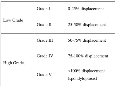
[13]. The grading system for spondylolisthesis, as described by Meyerding, is detailed in
Table 1.
Due to the lack of clarity as to the optimum surgical intervention, this review aims to
establish if there is a difference in functional outcome between direct repair of the defect
and intervertebral fusion for adolescent spondylolysis or low grade spondylolisthesis.
Methodology
For this review, functional outcome will be evaluated using the Oswestry Disability Index
(ODI). This patient-reported score is among the leading disease-specific outcome
measures for lumbar disorders [14]. It has been validated for a number of different lumbar
conditions [15-17] and has been used as a bench mark to validate numerous other
instruments [14]. 0-19% is considered as minimal disability, 20-39% moderate, 40-59%
severe, and over 60% as crippled [18]. For lumbar surgical procedures, the minimum
clinically important difference in ODI score has been calculated as 12.8 points [19]. The
Literature Search and Study Selection
A search of the Medline database (1945-present) was performed using OvidSP on 6th
February 2012. The search strategy is detailed in Table 2. As recommended by the
Cochrane Handbook of Systematic Reviews [20], a variety of search terms were
employed, combining index terms and free-text terms, to identify papers dealing with
patient-reported outcomes. Abstracts were assessed for relevance and full-texts were
reviewed of those that met the inclusion criteria on initial assessment. The reference
sections of these papers were scrutinised for further relevant articles.
Studies were included if a patient group with average age of less than 21 years received
direct repair or intervertebral fusion for spondylolysis or low grade spondylolisthesis
(Meyerding Grade I and II) and was assessed post-operatively with the Oswestry
Disability Index. All levels of evidence were included but case reports and case series
with fewer than five patients were excluded.
Results
127 studies were identified by the Medline search strategy. The process by which articles
were selected is detailed in Figure 1. 14 were selected for full-text assessment on the basis
of their abstract. Nine studies met the inclusion criteria and are detailed in Table 3. Two
were excluded because the average age was over 21 [11,21], two because the ODI was not
in the appropriate age group [24]. All the included studies were level III or IV evidence.
Formal evaluation of methodological quality was not performed but the key weaknesses
of the studies are detailed in Table 4. Pooled statistical analysis was not attempted due to
the variability of surgical procedures performed in each group and the apparent likelihood
of patient duplication between studies, as detailed in Table 4.
Four studies reported outcome following direct repair of the defect. Altaf et al reported a
prospective case series of 20 patients with L5 spondylolysis, pain lasting more than 12
months despite conservative treatment and a normal disc on MRI. They were treated with
direct stabilization of the pars defect using a pair of pedicle screws connected by a
U-shaped modular link passing beneath the spinous process [25]. The defect was filled with
autologous bone graft and compression achieved by tightening the link to the screws.
Average ODI decreased from 54 pre-operatively to 8 post-operatively. The authors
concluded that 90% of patients had an excellent clinical outcome and state that the
strength of this construct removes the need for post-operative immobilization, although
the non-union rate was 20%, with half of these being symptomatic.
Debnath et al performed direct repair of a spondylolytic defect without associated
spondylolisthesis on 22 young athletes [26] who had failed conservative treatment,
although the term ‘athlete’ was not defined. 19 patients underwent a Buck’s repair
(passing a cortical lag screw across a grafted defect), modified to include bone grafting of
the lamina and the transverse process. Three patients received Scott’s wiring, using an
figure-of-eight over the spinous process. Mean ODI dropped from 39.5 to 10.7. Two
patients that underwent Scott’s wiring required revision to posterolateral fusion for
non-union. The authors conclude that a modified Buck’s fusion results in a significant
improvement in ODI for professional sportsmen and women.
Debnath et al also reported the outcome in 42 patients with unilateral pars defects [27].
The majority responded to conservative treatment with activity modification, bracing and
physical therapy but eight remained symptomatic and underwent modified Buck’s fusion
as previously described, following a positive response to local anaesthetic pars
infiltration. They were treated contemporaneously at the same centre with those reported
in Debnath’s earlier work, so there may be some duplication of patients between the two
studies. Mean ODI dropped from 39.4 to 6.4, the best outcome of any repair, although the
lesions were unilateral and therefore not associated with a spondylolisthesis. One patient
with spina bifida suffered a symptomatic non-union, requiring posterior fusion.
Koptan et al treated ten patients who had developed spondylolysis following correction of
painless idiopathic scoliosis [28]. Local anaesthetic infiltration of the pars defect was used
to confirm the cause of pain. In five cases, a 1mm double cable was looped between a
pedicle screw and the spinous process bilaterally. Five cases received a modular construct
similar to that used by Altaf et al. All had iliac crest bone graft placed in the defects.
Three studies were identified that reported functional outcome after posterior or
posterolateral fusion. It appears that these studies report the same patient group from
different perspectives. Remes et al compared outcome with abnormal MRI findings [29],
Lamberg et al looked at functional and radiological outcomes [30], while Helenius et al
compared ODI to the Scoliosis Research Council questionnaire [31]. Post-operative mean
ODI ranged from 6.3 for posterolateral fusions to 11.3 for posterior fusions.
One study, published in two papers reporting early and long term follow up, compared
direct repair with intervertebral fusion [32,33]. Schlenzka et al compared outcome in 28
patients who underwent Scott’s wiring with 28 who received posterolateral segmental
fusion without instrumentation. At mean follow-up of 54 months there was no significant
difference in ODI between repair and fusion. However, the fusion group did significantly
Discussion
On the basis of the data included in this review of the literature, it is very difficult to
recommend one intervention over the other in terms of outcome, particularly when
considering the lack of quality comparative data from well-designed studies. While a
number of case series describing various methods of direct repair suggest good results
[25-28], these were comparable to those of fusion in the largest studies [29-31].
The only study drawing direct comparisons between the two treatments suggested fusion
to provide better outcome in the long term [33]. While this was statistically significant,
the difference between an ODI score of 4 and 11 may not be clinically significant, with
both groups sitting within the bracket of ‘minimal disability’. There were important
methodological weaknesses of the study, including treatment allocation, differing
post-operative protocols and differing pathologies in terms of defect level and degree of slip.
It should also be noted that the direct repairs in the comparative study were performed
using Scott’s wiring. Kip et al found that screw fixation has greater biomechanical
strength than wiring and is therefore more likely to lead to union of the defect [34]. Two
of the three patients that underwent Scott’s wiring in the study by Debnath et al required
revision for non-union [26].
One of the proposed advantages of direct repair is preservation of the motion segment and
and abnormal MRI findings [29]. Schlenzka et al found no difference in the MRI signal
intensity of the disc above the operated segment between repair and fusion [32], bringing
into question the theoretical benefit of direct repair. They also observed increased
operative time, blood loss and re-operations in the direct repair group. Furthermore, given
the non-union rate of up to 20% with direct repair [25,26], the case in its favour becomes
harder to argue. That said, these studies were not able to link non-union with worse
functional outcome. Direct repair may be considered less invasive but most still use iliac
crest bone graft which has significant donor site morbidity [35]. Due to pseudarthrosis
and progression of slip, secondary fusion becomes necessary in up to 57% of direct
repairs, depending on technique [36].
The level of the defect should be considered when deciding treatment method. A
posterolateral fusion has been suggested to be the gold standard for L5 spondylolysis [37],
with repair reserved for more cephalad defects. The majority of the fusions in this review
were performed at L5. However, many repairs were also performed at this level
(including all the cases in the study by Altaf et al). Degree of slip may impact upon the
preferred treatment. Schlenzka et al demonstrated better outcome with fusion than direct
repair. The patients in the repair group had a greater degree of slip on average than both
the patients in the fusion group and the patients in the other studies that reported on direct
repair. It may be that direct repair should be reserved for cases with minimal or no slip.
The total number of patients in this review is comparatively small, with a maximum of 88
that there is duplication of patients between the two studies by Debnath et al as they were
performed at the same centre and the study periods coincide. Furthermore, it seems very
likely that Helenius et al, Remes et al and Lamberg et al all report the same population. It
is unclear when the fusions reported by Schlenzka et al were performed. This replication
means that there may be as few as 80 direct repairs and 108 fusions included in this
review, with all of the fusions having been performed at the same centre.
It must be remembered that the ODI has not been validated in adolescent spondylolysis.
The SRC questionnaire may be a more appropriate instrument in adolescents as it includes
questions on cosmetic appearance and does not feature questions on sexual function.
Helenius et al showed it to correlate well with the ODI in patients undergoing fusion for
adolescent spondylolysis [31]. Furthermore, the ODI had not been validated to be used in
Finnish at the time these studies were presented, although this has now been performed
[38]. There were also inconsistencies in the use of the ODI between studies. Schlenzka et
al modified the tool, removing the question on sexual function [32].
Due to the apparent long term advantage of fusion and the lack of clarity as to the
theoretical advantages of direct repair, a randomised controlled trial is necessary to
establish the optimum mode of treatment for this condition. The issues of level of disease
and degree of slip would have to be addressed, either through inclusion criteria or
randomisation. Subgroup analysis may be required to separate the efficacy of the various
interventions described. The outcome measures should include clinician-based evaluation
degeneration), and patient-reported outcomes, including the ODI and SRC questionnaires.
A long time period will be required for recruitment and follow-up for an RCT in this
condition, so in the meantime the authors look forward to the long term outcomes from
the studies of direct repair featured in this review.
In conclusion, this systematic review does not demonstrate a clinically significant
difference in functional outcome, as measured by the ODI, between direct repair and
fusion for paediatric spondylolysis or low grade spondylolisthesis. Further well-designed
References
1. Kalichman L, Kim DH, Li L, Guermazi A, Berkin V, Hunter DJ. Spondylolysis and spondylolisthesis: prevalence and association with low back pain in the adult community-based population. Spine 2009; 34(2):199-205.
2. Beutler WJ, Fredrickson BE, Murtland A, Sweeney CA, Grant WD, Baker D. The natural history of spondylolysis and spondylolisthesis: 45-year follow-up
evaluation. Spine 2003; 28(10):1027-35.
3. Rossi F, Dragoni S. Lumbar spondylolysis: occurrence in competitive athletes. Updated achievements in a series of 390 cases. J Sports Med Phys Fitness 1990; 30(4):450-2.
4. Pizzutillo PD, Hummer CD 3rd. Nonoperative treatment for painful adolescent spondylolysis or spondylolisthesis. J Pediatr Orthop 1989; 9(5):538-40.
5. Sherman FC, Rosenthal RK, Hall JE. Spine fusion for spondylolysis and spondylolisthesis in children. Spine 1976; 4(1):59-66.
6. Wiltse LL, Jackson DW. Treatment of spondylolisthesis and spondylolysis in children. Clin Orthop Relat Res 1976; 117:92-100.
7. Rajasekaran S, Kamath V, Avadhani A. Bucks fusion. Eur Spine J 2010; 19(2):343-4.
8. Nicol RO, Scott JH. Lytic spondylolysis. Repair by wiring. Spine 1986; 11(10):1027-30.
9. Sales de Gauzy J, Vadier F, Cahuzac JP. Repair of lumbar spondylolysis using Morscher material: 14 children followed for 1-5 years. Acta Orthop Scand. 2000; 71(3):292-6.
10. Pai VS, Hodgson B, Pai V. Repair of spondylolytic defect with a cable screw reconstruction. Int Orthop. 2008; 32(1):121-5.
11. Ming-li F, Hui-liang S, Yi-min Y, Huai-jian H, Qing-ming Z, Cao-Li. Analysis of factors related to prognosis and curative effect for posterolateral fusion of lumbar low-grade isthmic spondylolisthesis. Int Orthop. 2009; 33(5):1335-40.
13. Tsirikos AI, Garrido EG. Spondylolysis and spondylolisthesis in children and adolescents. J Bone Joint Surg Br 2010; 92(6):751-759.
14. Fairbank JC, Pynsent PB. The Oswestry Disability Index. Spine 2000; 25(22):2940-52.
15. Ferrari R. Responsiveness of the short-form 36 and oswestry disability
questionnaire in chronic nonspecific low back and lower limb pain treated with customized foot orthotics. J Manipulative Physiol Ther 2007; 30(6):456-8.
16. O'Shea FD, Riarh R, Anton A, Inman RD. Assessing back pain: does the Oswestry Disability Questionnaire accurately measure function in ankylosing spondylitis? J Rheumatol 2010; 37(6):1211-3.
17. Tomkins-Lane CC, Battié MC. Validity and reproducibility of self-report
measures of walking capacity in lumbar spinal stenosis. Spine 2010; 35(23):2097-102.
18. Fairbank JC, Couper J, Davies JB, O'Brien JP. The Oswestry low back pain disability questionnaire. Physiotherapy 1980; 66(8):271-3.
19. Copay AG, Glassman SD, Subach BR, Berven S, Schuler TC, Carreon LY. Minimum clinically important difference in lumbar spine surgery patients: a choice of methods using the Oswestry Disability Index, Medical Outcomes Study questionnaire Short Form 36, and pain scales. Spine J 2008; 8(6):968-74.
20. http://www.cochrane-handbook.org - last accessed 15th July 2011
21. Lauber S, Schulte TL, Liljenqvist U, Halm H, Hackenberg L. Clinical and radiologic 2-4-year results of transforaminal lumbar interbody fusion in degenerative and isthmic spondylolisthesis grades 1 and 2. Spine 2006; 31(15):1693-8.
22. Gehrchen PM, Dahl B, Katonis P, Blyme P, Tøndevold E, Kiaer T. No difference in clinical outcome after posterolateral lumbar fusion between patients with isthmic spondylolisthesis and those with degenerative disc disease using pedicle screw instrumentation: a comparative study of 112 patients with 4 years of follow-up. Eur Spine J. 2002; 11(5):423-7.
23. Bernicker JP, Kohl HW 3rd, Sahni I, Esses SI. Long-term functional and radiographic follow-up of surgically treated isthmic spondylolisthesis. Am J Orthop 1999; 28(11):631-6.
25. Altaf F, Osei NA, Garrido E, Al-Mukhtar M, Natali C, Sivaraman A, Noordeen HH. Repair of spondylolysis using compression with a modular link and screws. J Bone Joint Surg Br 2011;93(1):73-7.
26. Debnath UK, Freeman BJ, Gregory P, de la Harpe D, Kerslake RW, Webb JK. Clinical outcome and return to sport after the surgical treatment of spondylolysis in young athletes. J Bone Joint Surg Br 2003; 85(2):244-9.
27. Debnath UK, Freeman BJ, Grevitt MP, Sithole J, Scammell BE, Webb JK. Clinical outcome of symptomatic unilateral stress injuries of the lumbar pars interarticularis. Spine 2007; 32(9):995-1000.
28. Koptan WM, ElMiligui YH, ElSharkawi MM. Direct repair of spondylolysis presenting after correction of adolescent idiopathic scoliosis. Spine J. 2011; 11(2):133-8.
29. Remes V, Lamberg T, Tervahartiala P, Helenius I, Schlenzka D, Yrjönen T et al Long-term outcome after posterolateral, anterior, and circumferential fusion for high-grade isthmic spondylolisthesis in children and adolescents: magnetic resonance imaging findings after average of 17-year follow-up. Spine 2006; 31(21):2491-9.
30. Lamberg TS, Remes VM, Helenius IJ et al Long-term clinical, functional and radiological outcome 21 years after posterior or posterolateral fusion in childhood and adolescence isthmic spondylolisthesis. Eur Spine J 2005; 14(7):639-44.
31. Helenius I, Lamberg T, Osterman K et al Scoliosis research society outcome instrument in evaluation of long-term surgical results in spondylolysis and low-grade isthmic spondylolisthesis in young patients. Spine 2005; 30(3):336-41.
32. Schlenzka D, Seitsalo S, Poussa M, Osterman K. Operative treatment of
symptomatic lumbar spondylolysis and mild isthmic spondylolisthesis in young patients: direct repair of the defect or segmental spinal fusion? Eur Spine J 1993; 2(2):104-12.
33. Schlenzka D, Remes V, Helenius I et al Direct repair for treatment of symptomatic spondylolysis and low-grade isthmic spondylolisthesis in young patients: no benefit in comparison to segmental fusion after a mean follow-up of 14.8 years. Eur Spine J 2006; 15(10):1437-47.
34. Kip PC, Esses SI, Doherty BI, Alexander JW, Crawford MJ. Biomechanical testing of pars defect repairs. Spine 1994; 19(23):2692-7.
36. Giudici F, Minoia L, Archetti M, Corriero AS, Zagra A. Long-term results of the
direct repair of spondylolisthesis. European Spine Journal 2011; 20 Suppl 1:115-20.
37. Cheung EV, Herman MJ, Cavalier R, Pizzutillo PD. Spondylolyisis and
spondylolisthesis in children and adolescents. II. Surgical management. J Am Acad Orthop Surg 2006; 14:488-98.
38. Pekkanen L, Kautiainen H, Ylinen J, Salo P, Häkkinen A. Reliability and validity study of the Finnish version 2.0 of the oswestry disability index. Spine 2011; 36(4):332-8.
Figure Legends
Figure 1. Flowchart demonstrating the process of article selection
Medline Search
n = 127
Original studies n = 110
Relevant title n = 61
Met inclusion criteria following full-text
assessment n = 9 Relevant abstract
n = 14
Reviews, case reports, duplicates
n = 17
Title not relevant n = 49
Excluded following abstract review
n = 47
Excluded following full-text assessment
Low Grade
Grade I 0-25% displacement
Grade II 25-50% displacement
High Grade
Grade III 50-75% displacement
Grade IV 75-100% displacement
Grade V >100% displacement (spondyloptosis)
1 Spondylolysis/su [Surgery] 251
2 Spondylolisthesis/su [Surgery] 1604
3 1 or 2 1750
4 limit 3 to (english language and humans and "all child (0 to 18 years)") 415
5
Treatment Outcome/ or Disability Evaluation/ or Pain Measurement/ or
oswestry.mp. or Questionnaires/ 791423
6 "Outcome Assessment (Health Care)"/ 40525
7 5 or 6 823138
8 4 and 7 127
Population Direct Repair Fusion Mean ODI score as %
(Range) ± SD Altaf
et al, 2011 [25]
20 patients with L5 splY (9 with grade I splI)
Modular link between pedicle screws
Pre-op: 54 (42-78) Post-op: 8 (0-42)
Mean follow-up: 4 years Mean age: 13.9
Debnath et al, 2003 [26]
22 young athletes with uni- or bilateral splY at L3, L4 or L5
19 modified Buck’s repair 3 Scott’s wiring
Pre-op: 39.5 ± 8.7 Post-op: 10.7 ± 12.9
Follow-up at 2 years Mean age: 20.2 Debnath
et al, 2007 [27]
8 patients with unilateral splY
Modified Buck’s repair at L3, L4 or L5
Pre-op: 39.4 ± 3.6 Post-op: 6.4 ± 5.2
Follow-up at 2 years Mean age: 20 Helenius
et al, 2005 [31]
108 patients with splY or low grade splI, 95% at L5 level
29 posterior 79 postero-lateral
8.2 (0-68)
Mean follow-up: 20.9 years Mean age: 15.9
Koptan et al, 2011 [28]
10 patients with splY at L3, L4 or L5 (3 with grade I splI)
Looped wire or modular link between pedicle screws
Pre-op: 52 (46–74) Post-op: 11 (0–34)
Mean follow-up: 4.5 years Mean age: 16
Lamberg et al, 2005 [30]
107 patients with splY or low grade splI, 95% at L5 level
29 posterior 79 postero-lateral
8.2 (0-68)
Mean follow-up: 20.9 years Mean age: 15.3
Remes et al, 2006 [29]
103 patients with splY or low grade splI, 95% at L5 level
29 posterior 79 postero-lateral
Posterior: 11.3 (0–68) Posterolateral: 6.3(0–48) Mean follow-up: 21.0 years Mean age: 15.9
Schlenzka et al, 1993 [32]
Adolescent splY +/- low grade splI
28 patients, L3, L4 or L5: Scott’s wiring with autologous bone graft
28 patients, all L5 level: posterolateral fusion
Repair group: 7.6 ± 8.9 Fusion group: 8.6 ± 11.8
Mean follow-up: 54 months Mean age: 18.2 for repair, 16.2 for fusion
Schlenzka et al, 2006 [33]
Longer term follow-up of previous study group
As above As above Repair group: 11.4 (0-52) Fusion group: 4.3 (0-16) P = 0.02
Minimum 11 year follow-up
Study Design Level Weaknesses
Altaf et al, 2011 [25]
Prospective case series
IV Unclear patient selection and post-operative protocol No independent observer
Debnath et al, 2003 [26]
Prospective case series
IV Unclear inclusion criteria Unclear treatment allocation
Likely patient overlap with following study Debnath et
al, 2007 [27]
Prospective case series
IV Unclear patient selection Small sample size
Likely patient overlap with previous study Helenius et
al, 2005 [31]
Retrospective case series
IV Inconsistency of fusion technique and level Unclear treatment allocation
Attrition bias
Likely patient overlap with studies by Schlenzka et al Koptan et
al, 2011 [28]
Prospective case series
IV Small sample size
Other spinal pathology present Inconsistency of treatment Lamberg
et al, 2005 [30]
Retrospective case series
IV Same patient group as Helenius et al, one unexplained missing patient Remes et al, 2006 [29] Retrospective case series
IV Same patient group as Helenius et al, without 5 claustrophobic patients who could not undergo MRI
Schlenzka et al, 1993 [32]
Non-randomised comparative trial
III No pre-operative scoring
Treatment group decided by surgeon’s preference
Difference between groups in disease level and degree of slip Attrition bias
Schlenzka et al, 2006 [33]
III