http://www.scirp.org/journal/as ISSN Online: 2156-8561
ISSN Print: 2156-8553
Utilization of Pyrosequencing to Monitor the
Microbiome Dynamics of Probiotic Treated
Poultry (Gallus gallus domesticus) during
Downstream Poultry Processing
Vamsy Priya Guttala1, Enrique G. Medrano2, Joey Bray1, Beatrice Clack1* 1Stephen F. Austin State University, Department of Biology, Nacogdoches, USA
2United States Department of Agriculture, Agricultural Research Service, College Station, USA
Abstract
Antibiotic growth promoters that have been historically employed to control pathogens and increase the rate of animal development for human consump-tion are currently banned in many countries. Probiotics have been proposed as an alternative to control pathogenic bacteria. Traditional culture methods typically used to monitor probiotic effects on pathogens possess significant limitations such as a lack in sensitivity to detect fastidious and non-culturable bacteria, and are both time consuming and costly. Here, we tested next gener-ation pyrosequencing technology as a streamline and economical method to monitor the effects of a probiotic on microbial communities in juvenile poul-try (Gallus gallus domesticus) after exposure to several microbiological chal-lenges and litter conditions. Seven days and repeated again at 39 days follow-ing hatchfollow-ing, chicks were challenged with either Salmonella enterica serovar Enteritidis, Campylobacter jejuni, or no bacteria in the presence of, or without a probiotic (i.e., Bacillus subtilis) added to the feed. Three days following each of two challenges (i.e., days 10 and 42, respectively) the microbiome distribu-tions of the poultry caecum were characterized based on 16S rDNA analysis. Generated PCR products were analyzed by automated identification of the samples after pooling, multiplexing and sequencing. A bioinformatics pipeline was then employed to identify microbial distributions at the phylum and ge-nus level for the treatments. In conclusion, our results demonstrated that py-rosequencing technology is a rapid, efficient and cost-effective method to monitor the effects of probiotics on the microbiome of poultry propagated in an agricultural setting.
Keywords
Poultry Production, Probiotics, Pyrosequencing, Mutiplex Identifier (MID), How to cite this paper: Guttala, V.P.,
Me-drano, E.G., Bray, J. and Clack, B. (2017) Uti- lization of Pyrosequencing to Monitor the Microbiome Dynamics of Probiotic Treated Poultry (Gallus gallus domesticus) during Downstream Poultry Processing. Agricultur-al Sciences, 8, 675-691.
https://doi.org/10.4236/as.2017.87051
Received: May 23, 2017 Accepted: July 25, 2017 Published: July 28, 2017
Copyright © 2017 by authors and Scientific Research Publishing Inc. This work is licensed under the Creative Commons Attribution International License (CC BY 4.0).
http://creativecommons.org/licenses/by/4.0/
Microbiota
1. Introduction
Since 1995, the Poultry industry has become one of the globes’ largest and fastest growing segments of animal food production [1]. The United States is one of the largest producers of poultry with the broiler sector playing a major role [2]. Crowded poultry housing conditions are stressful to the birds leading to an ele-vated disease potential. Additionally, packed houses cause deterioration of envi-ronmental conditions providing a situation that is conducive to the spread of disease and thus increase the possibility of transmission to humans [3]. Out-breaks of campylobacteriosis and salmonellosis infections due to the consump-tion of contaminated poultry or derived products have occurred in human pop-ulations throughout the world and are thus a major concern [4][5][6][7][8].
Antimicrobial growth promoters (AGPs) consist of antibiotics that are added to the feed of animals to enhance their growth rate and production performance [9]. Unfortunately, the large quantities of AGPs that have been used in poultry production provided a source for development of antibiotic resistant bacteria [10]. For example, Campylobacter was found to be increasingly resistant to anti-biotics such as fluoroquinolones and macrolides that are used as antimicrobials for the treatment of campylobacteriosis [11]. Additionally, the development of resistance to antibiotics by Salmonella has also been reported [12]. Hence, there is an increased necessity not only to minimize AGP use but also to develop novel non-antibiotic-based alternative treatments. Probiotics are being considered to fill this gap with utilization in certain farms instead of antibiotics [13][14]. The most common probiotic additives used in the broiler industry include Aspergil-lus, BacilAspergil-lus, Bifidiobacterium, Candida, Lactobacillus and Sterptomyces [15][16] [17].
tech-nologies provide more nucleotides to characterize from a given DNA sample when compared with conventional approaches [20].
Here, we compared microbe populations present in chick cecum following feeding probiotics consisting of Bacillus subtilis with those fed a normal diet (feed without probiotics). Additionally, litter microbiota present before and after composting the litter were analysed. Bacterial populations were classified based on 16S rDNA sequencing analysis. An understanding of the development of the normal bacterial community provided a method to detect disruption in the flora and determine the effects of food animal management changes. The success and precise assessment of the bacterial information using high throughput pyrose-quencing demonstrated in this study may allow for timely manipulation of the intestinal flora with the intention of enhancing intestinal health and feed con-version ratios.
2. Materials and Methods
2.1. Probiotic Experiment
Poultry Rearing. A total of 450 male broilers (Gallus domesticus) were ob-tained from the Cobb-Vantress hatchery, Inc (Timpson, Texas, USA) imme-diately after hatching (i.e. zero days of age). At the hatchery, birds were vacci-nated for Marek’s disease, Newcastle’s disease and bronchitis using standard methods [21]. The broilers were divided among 28 floor pens (1.2 m 1.2 m) at the Stephen F. Austin State University (SFASU) Science Research Center, Na-cogdoches, TX, USA with 15 birds per pen on fresh litter. The facility is envi-ronmentally controlled with negative pressure rooms (i.e. air expelled from the room). To provide the birds a relatively stable thermal environment, ventilation and heat ranged from 32˚C daily to 21˚C nightly. All birds received the same basal diet formulated according to the Nutrient Requirements of Chickens [22]. Clean water and feed were provided ad libitum throughout the study via Lubing Feather Soft Nipple Drinkers and then Tube Feeders (QC Supply, Schuyler, NE, USA).
Preparation of Inocula for Challenges. From glycerol stocks, 500 µL a of Salmonella enterica poultry isolate was added to 30 mL of DifcoTM Rappaport- Vassiliadis R10 (Becton, Dickinson and Company, Franklin Lakes, NJ, USA) broth amended with novobiocin at 25 µg/mL (Sigma, St. Louis, MO) and nali-dixic acid at 20 µg/mL (Sigma, St. Louis, MO), and incubated for 16 - 18 h in an Innova 4300 shaker (New Brunswick, Edfield, CT, USA) at 37˚C and 250 RPM. A Campylobacter jejuni poultry isolate was propagated by adding 500 µL of a frozen glycerol stock to 30 mL of Bolton broth base (Sigma-Aldrich, St. Louis, MO) amended with novobiocin at 25 µg/mL (Sigma-Aldrich, St. Louis, MO) and nalidixic acid (20 µg/mL, Sigma-Aldrich, St. Louis, MO) and incubated at 42˚C for 16 - 18 h without any agitation in the presence of 10.0% carbon dioxide, 4.9% oxygen and 8% nitrogen obtained as a compressed gas (Gibson Laboratories, Lexington, KY, USA).
250 RPM in Innova 4300 shaker incubator. Similarly, 1 ml of C. jejuni was in-oculated in a 50 mL conical tube containing Bolton broth and incubated at 42˚C after the passage of compressed gas. Optical density of the culture was checked periodically until attaining an absorbance 0.45 at a wavelength of 625 nm. Once the cultures entered log phase, the bacteria were pelleted by centrifugation at 12,000 × g for 5 min. The supernatant was discarded and the cells were washed with sterile Phosphate Buffer Saline (PBS; 130 mM NaCl, 7 mM Na2HPO4, 3 mM NaH2PO4, pH 7.3) twice and then suspended in 40 mL of PBS for inocula-tions.
Final concentrations administered in the challenges were 3 × 10⁹ colony forming units (CFU)/mL for S.enterica and 1 × 10⁹ CFU/mL for C.jejuni. Birds were infected by oral gavage with S.enterica or C. jejuni in 0.2 mL of physiologi-cal saline 0.85% w/v on the 7th and 39th day of age as designated in Table 1. Control groups (Treatments 5 and 6) were provided PBS.
Caecum Samples. Previous studies by Barnes et al. (1972), and Wei et al. (2013) showed that a diverse microbiota was found primarily in the caecum. Therefore, this study focused on the ceca microbiome [23][24]. Three days fol-lowing each challenge, caecum samples (n = 56) were harvested (i.e. at the 10th day and 42nd). The cecal sacs were removed from two randomly selected birds per pen on the day of harvest. The caecum contents of both chicks from a pen were pooled for molecular analysis.
Genomic DNA Isolation. Caecum contents were aseptically scraped into ste-rile 50 mL tubes containing 10 mL of steste-rile PBS and mixed by vortexing for 3 min. Debris was removed by centrifugation at 700 × g for 1 min. The superna-tant was collected and centrifuged at 12,000 × g for 5 min to pellet bacteria that was then suspended in 2 mL PBS and centrifuged at 12,000 × g for 5 min. The PBS wash was repeated and the pellet was finally suspended in 2 ml PBS. Glyce-rol stocks were prepared by drawing 500 µL of the washed cells and flash freez-ing in liquid nitrogen immediately after the addition of 1 mL glycerol. The re-maining cells were stored at −20˚C for DNA extraction.
The bacterial genomic DNA was isolated using a Wizard Genomic DNA puri-fication Kit (Promega Corporation, Madison, USA) as per the manufacturer’s protocol. The DNA purity was checked spectrophotometrically using a Varian Cary 50 UV—Vis spectrophotometer equipped with a Hellma microcell tray for microliter sample volumes (Hellma Analytics, Mullheim, Germany).
2.2. Litter Compost Experiment
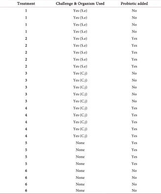
Table 1. Poultry experiment with probiotic treatments. Effect of the Probiotic (Bacillus
subtilis) on chick cecum microbial flora was determined after challenging birds with
Sal-monella enterica (S.e) serovar Enteritidis, Campylobacter jejuni (C.j), and appropriate controls. The experiment consisted of six treatments: Treatment 1 (S.e challenged and no Probiotic), Treatment 2 (S.e challenged + Probiotic), Treatment 3 (C.j challenged and no Probiotic), Treatment 4 (C.j challenged + Probiotic), Treatment 5 (no challenge + Probi-otic) and Treatment 6 (no challenge and no probiProbi-otic).
Treatment Challenge & Organism Used Probiotic added
1 Yes (S.e) No
1 Yes (S.e) No
1 Yes (S.e) No
1 Yes (S.e) No
2 Yes (S.e) Yes
2 Yes (S.e) Yes
2 Yes (S.e) Yes
2 Yes (S.e) Yes
2 Yes (S.e) Yes
3 Yes (C.j) No
3 Yes (C.j) No
3 Yes (C.j) No
3 Yes (C.j) No
3 Yes (C.j) No
4 Yes (C.j) Yes
4 Yes (C.j) Yes
4 Yes (C.j) Yes
4 Yes (C.j) Yes
4 Yes (C.j) Yes
5 None Yes
5 None Yes
5 None Yes
5 None Yes
6 None No
6 None No
6 None No
6 None No
and non-composted litter section. Each flock was reared for 49 days to an av-erage market weight of 2.4 kg/bird.
Litter Samplings. Each house (House 1 and House 2) was divided into four 38 m sections lengthwise. Using a 30 cm soil collection tube (Acorn Naturalists, Tustin, CA USA), six litter samples were collected per 38 m sections from each house. Samples were then pooled and homogenized to make four composted and four non-composted samples in sterile bags and stored at −20˚C. The bac-terial DNA from 2.5 g poultry litter was isolated using the ZR Soil microbe DNA midiprep kit (Zymoresearch, Irvine, USA) as per the manufacturer’s protocol.
2.3. Next-Generation Sequencing
16S rDNA Synthesis. Extracted DNA from the poultry probiotic experiment and litter compost studies were used as templates to amplify 16S rDNA se-quences using the polymerase chain reaction (PCR) in a MyCycler (BioRad La-boratories, Inc., USA). Reactions were performed in a 50 µL total volume with GoTaq Green Master Mix from Promega Corp. (Madison, USA). The forward primer 27F (5’–AGAGTTTGATCMTGGCTCAG–3’) is a 16S ribosomal DNA specific universal primer for prokaryotes that was previously employed by Lane et al. [25]. The universal reverse primer for prokaryotes called 519R (5’–GWA-
TTACCGCGGCKGCTG–3’) was used by Turner et al. [26].
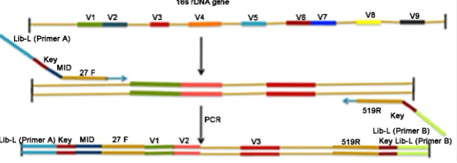
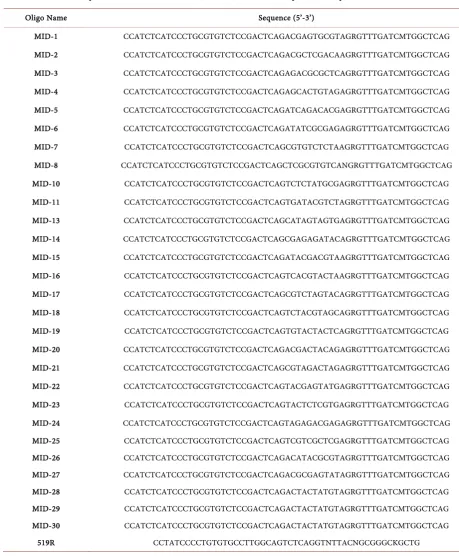
All the primers including Multiplex Identifiers (MIDs) listed in Table 2 were purchased from Sigma Genosys (a division of Sigma Aldrich). Figure 1 provides an illustration of the strategy used to combine the bacterial 16S ribosomal DNA primer set with the MIDs. The PCR conditions were the following: 97˚C for 5 minutes; 40 cycles of 60˚C for 1 minute, 72˚C for 1 minute 20 seconds and 95˚C for 30 seconds; followed by a 72˚C for 5 minutes and hold at 4˚C. The PCR am-plified product was analyzed using agarose gel electrophoresis.
[image:6.595.210.538.511.628.2]Pyrosequencing Application. After quantification of DNA, equal amounts of purified PCR products were pooled for Roche emPCR amplification that was performed as per the manufacturer’s protocol (454 Roche Life Sciences,
Table 2. Degenerate primers used for bacterial 16S rDNA amplification. Primers were designed with Roche 454 Lib–L forward primer (Primer A) at the 5’ end, the sequencing key in the middle, and with a Multiplex Identifier (MID) and universal primer at the 3’ end. The reverse primer with Roche 454 Lib-L (Primer B) and universal primer were positioned at the 3’ end.
Oligo Name Sequence (5’-3’)
MID-1 CCATCTCATCCCTGCGTGTCTCCGACTCAGACGAGTGCGTAGRGTTTGATCMTGGCTCAG
MID-2 CCATCTCATCCCTGCGTGTCTCCGACTCAGACGCTCGACAAGRGTTTGATCMTGGCTCAG
MID-3 CCATCTCATCCCTGCGTGTCTCCGACTCAGAGACGCGCTCAGRGTTTGATCMTGGCTCAG
MID-4 CCATCTCATCCCTGCGTGTCTCCGACTCAGAGCACTGTAGAGRGTTTGATCMTGGCTCAG
MID-5 CCATCTCATCCCTGCGTGTCTCCGACTCAGATCAGACACGAGRGTTTGATCMTGGCTCAG
MID-6 CCATCTCATCCCTGCGTGTCTCCGACTCAGATATCGCGAGAGRGTTTGATCMTGGCTCAG
MID-7 CCATCTCATCCCTGCGTGTCTCCGACTCAGCGTGTCTCTAAGRGTTTGATCMTGGCTCAG
MID-8 CCATCTCATCCCTGCGTGTCTCCGACTCAGCTCGCGTGTCANGRGTTTGATCMTGGCTCAG
MID-10 CCATCTCATCCCTGCGTGTCTCCGACTCAGTCTCTATGCGAGRGTTTGATCMTGGCTCAG
MID-11 CCATCTCATCCCTGCGTGTCTCCGACTCAGTGATACGTCTAGRGTTTGATCMTGGCTCAG
MID-13 CCATCTCATCCCTGCGTGTCTCCGACTCAGCATAGTAGTGAGRGTTTGATCMTGGCTCAG
MID-14 CCATCTCATCCCTGCGTGTCTCCGACTCAGCGAGAGATACAGRGTTTGATCMTGGCTCAG
MID-15 CCATCTCATCCCTGCGTGTCTCCGACTCAGATACGACGTAAGRGTTTGATCMTGGCTCAG
MID-16 CCATCTCATCCCTGCGTGTCTCCGACTCAGTCACGTACTAAGRGTTTGATCMTGGCTCAG
MID-17 CCATCTCATCCCTGCGTGTCTCCGACTCAGCGTCTAGTACAGRGTTTGATCMTGGCTCAG
MID-18 CCATCTCATCCCTGCGTGTCTCCGACTCAGTCTACGTAGCAGRGTTTGATCMTGGCTCAG
MID-19 CCATCTCATCCCTGCGTGTCTCCGACTCAGTGTACTACTCAGRGTTTGATCMTGGCTCAG
MID-20 CCATCTCATCCCTGCGTGTCTCCGACTCAGACGACTACAGAGRGTTTGATCMTGGCTCAG
MID-21 CCATCTCATCCCTGCGTGTCTCCGACTCAGCGTAGACTAGAGRGTTTGATCMTGGCTCAG
MID-22 CCATCTCATCCCTGCGTGTCTCCGACTCAGTACGAGTATGAGRGTTTGATCMTGGCTCAG
MID-23 CCATCTCATCCCTGCGTGTCTCCGACTCAGTACTCTCGTGAGRGTTTGATCMTGGCTCAG
MID-24 CCATCTCATCCCTGCGTGTCTCCGACTCAGTAGAGACGAGAGRGTTTGATCMTGGCTCAG
MID-25 CCATCTCATCCCTGCGTGTCTCCGACTCAGTCGTCGCTCGAGRGTTTGATCMTGGCTCAG
MID-26 CCATCTCATCCCTGCGTGTCTCCGACTCAGACATACGCGTAGRGTTTGATCMTGGCTCAG
MID-27 CCATCTCATCCCTGCGTGTCTCCGACTCAGACGCGAGTATAGRGTTTGATCMTGGCTCAG
MID-28 CCATCTCATCCCTGCGTGTCTCCGACTCAGACTACTATGTAGRGTTTGATCMTGGCTCAG
MID-29 CCATCTCATCCCTGCGTGTCTCCGACTCAGACTACTATGTAGRGTTTGATCMTGGCTCAG
MID-30 CCATCTCATCCCTGCGTGTCTCCGACTCAGACTACTATGTAGRGTTTGATCMTGGCTCAG
519R CCTATCCCCTGTGTGCCTTGGCAGTCTCAGGTNTTACNGCGGGCKGCTG
consensus sequences of the DNA libraries. The assembled contigs were used in a metagenomic analysis with the CAMERA database [27][28].
Statistical Analysis. Based upon the CAMERA BLASTn derived matches, the sequences were classified at the appropriate taxonomic levels based on Data Analysis Methodology offered by the Research and Testing Laboratory, Lubbock, TX, USA (http://rtlgenomics.com/). Additionally, RDP Naïve Bayesian rDNA classifier version 2.5 was used to organize the data into taxonomy groups with a
bootstrap cutoff of 80% (https://rdp.cme.msu.edu/). Two-Way ANOVA was
used with GraphPad Prism version 6.0.
(https://www.graphpad.com/scientific-software/prism/), in order to further ana-lyze the data and calculate the variance to observe the effect of treatments on the chick’s microbiome. A p-value <0.05 was considered statistically significant. The SAS based program JMP Genomics Version 5.1 was employed to organize the distribution of identified bacteria [29].
3. Results
3.1. Probiotic Poultry Experiment
Results of the 454 sequencing experiments performed on the samples collected on day 10 and 42 showed that the primers designed with the MIDs successfully amplified specific 16S rDNA regions of multiple bacteria. Pyrosequencing gen-erated 19.8 Mbp with average reads of 389 bp and 43.7 Mbp with average reads of 342 bp for the pooled chick caecum samples from the 10th and 42nd day collec-tion periods, respectively.
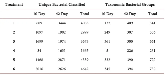
[image:8.595.207.541.586.737.2]The CAMERA BLASTn analysis using the collective data produced over 20,000 significant matches for all of the treatment samples (Table 3). The Two- Way ANOVA analysis using both the 10th and 42nd day samples from treatments
Table 3. Distribution of bacteria identified based on 16S ribosomal DNA sequence analy-sis from the caeca of chicks that had been provided a probiotic and challenged with either
Salmonella enterica (S.e) serovar Enteriditis or Campylobacter jejuni (C.j), and
appropri-ate controls. The experiment consisted of six treatments: Treatment 1 (S.e challenged and no Probiotic), Treatment 2 (S.e challenged + probiotic), Treatment 3 (C.jchallenged and no Probiotic), Treatment 4 (C.j challenged + Probiotic), Treatment 5 (no challenge + Probiotic) and Treatment 6 (no challenge and no probiotic). Genomic DNA was ex-tracted from poultry caeca at the 10th and 42nd day following each Treatment.
Treatment Unique Bacterial Classified Taxonomic Bacterial Groups
10 Day 42 Day Total 10 Day 42 Day Total
1 609 3444 4053 132 409 541
2 1097 1902 2999 249 307 556
3 1699 1974 3673 361 300 661
4 34 1631 1665 5 226 231
5 1468 2871 4339 332 390 722
that included exposure to S. enterica or C. jejuni along with the probiotic re-vealed a difference in the number of genera with respect to collection period. However, the probiotic treatment did not provide statistical evidence for a re-duction in pathogens detected (p = 0.1751). Overall, Firmicutes were predomi-nant in both days sampled from the six phyla identified (Figure 2(a)). Further, increased levels of beneficial genera such as Blautia, Eubacteria, Faecalibacteria, among others were detected from the 10th to the 42nd sample collections (Figure 2(b)). Clostridia that includes both pathogenic and non-pathogenic species were unaffected by treatment with or without the probiotic. Bacillus spp. were de-tected in all of the treatment samples (Figure 2(d)). A total of 6923 and 14,448 bacterial strains were identified for the 10th and 42nd day samples, respectively.
Figures 3-8 illustrate the distributions of the identified bacteria that
com-prised >1.5% of the population. However, the number of genera decreased as time increased irrespective of the treatment with or without the probiotic for both beneficial (Figure 2(b)) and pathogenic bacteria (Figure 2(c)). As Bacillus spp. are ubiquitous, expectantly they were detected in the entire sample analyzed
(Figure 2(d)).
(a) (b)
(c) (d)
Figure 2. Two-way ANOVA providing the distribution of significant (p < 0.05) phyla and genera based on 16S rDNA sequence analysis. Genomic DNA was extracted from poultry caeca at the 10th and 42nd day following a challenge (Chal+) or no challenge (Chal−) with a
bacterial pathogen (Salmonella enterica—S.e or Campylobacter jejuni—C.j), and/or pro-biotic (Bacillus subtilis) administration. Figure (a) illustrates the phylum distribution.
(a) (b)
Figure 3. Distribution of bacteria identified based on 16S ribosomal DNA sequence analysis from caeca of chicks that had been challenged with Salmonella enterica and not administered a probiotic. Genomic DNA was extracted from poultry caeca at the 10th
(Pie (a)) and 42nd (Pie (b)) day following the treatment.
[image:10.595.64.539.280.430.2](a) (b)
Figure 4. Distribution of bacteria identified based on 16S ribosomal DNA sequence analysis from caeca of chicks that had been challenged with Salmonella enterica and administered a probiotic. Genomic DNA was extracted from poultry caeca at the 10th
(Pie (a)) and 42nd (Pie (b)) day following the treatment.
(a) (b)
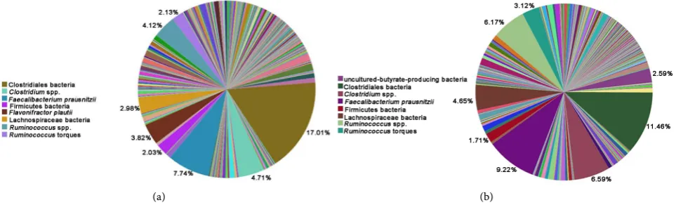
Figure 5. Distribution of bacteria identified based on 16S ribosomal DNA sequence analysis from caeca of chicks that had been challenged with Campylobacter jejuni and not administered a probiotic. Genomic DNA was extracted from poultry caeca at the 10th (Pie (a)) and 42nd (Pie (b)) day following the treatment.
3.2. Litter Compost Analysis
[image:10.595.63.537.488.636.2](a) (b)
Figure 6. Distribution of bacteria identified based on 16S ribosomal DNA sequence analysis from caeca of chicks that had been challenged Campylobacter jejuni and administered a probiotic. Genomic DNA was extracted from poultry caeca at the 10th (Pie
(a)) and 42nd (Pie (b)) day following the treatment.
[image:11.595.59.537.277.425.2](a) (b)
Figure 7. Distribution of bacteria identified based on 16S ribosomal DNA sequence analysis from caeca of chicks that had not been challenged with a bacterial pathogen and administered a probiotic. Genomic DNA was extracted from poultry caeca at the 10th (Pie (a)) and 42nd (Pie (b)) day following the treatment.
(a) (b)
Figure 8. Distribution of bacteria identified based on 16S ribosomal DNA sequence analysis from caeca of chicks that had not been challenged with a bacterial pathogen and not administered a probiotic. Genomic DNA was extracted from poultry caeca at the 10th (Pie (a)) and 42nd (Pie (b)) day following the mock a challenge with a bacterial pathogen and no probiotic administration.
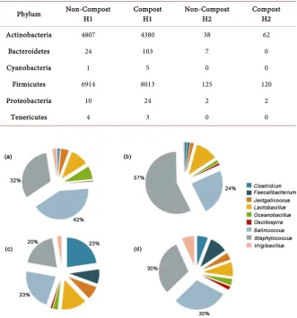
[image:11.595.58.540.487.633.2]irrespective of the treatments (Figure 9). Comparisons of genera from the phy-lum Actinobacteria showed that Bracybacterium was predominant in broiler lit-ter irrespective of the treatments (Figure 10).
4. Discussion
[image:12.595.208.540.272.628.2]The current study was intended to determine the proof of concept that next- generation sequencing technology could be applied to rapidly and efficiently
Table 4. Two poultry rearing facilities partitioned to house poultry pens (House 1 – H1) and House 2 - H2) that were bedded with either non-composted or composted wood shavings to assess prokaryotic composition differences between the litter. Extractions of DNA from litter samples were used to detect bacterial phyla composition based on 16S rDNA bacterial sequence analysis by employing the Ribosomal Database Project—Naive Bayesian rDNA classifier version 2.5 (https://rdp.cme.msu.edu/).
Phylum Non-Compost H1 Compost H1 Non-Compost H2 Compost H2
Actinobacteria 4807 4380 38 62
Bacteroidetes 24 103 7 0
Cyanobacteria 1 5 0 0
Firmicutes 6914 8013 125 120
Proteobacteria 10 24 2 2
Tenericutes 4 3 0 0
Figure 10. Distribution of genera from the phylum Firmicutes detected in com-posted and non-comcom-posted poultry litter. Extractions of DNA from litter sam-ples were used to determine bacterial phyla composition based on 16S rDNA bacterial sequence analysis by employing the Ribosomal Database Project (RDP)—Naive Bayesian rDNA classifier version 2.5 (https://rdp.cme.msu.edu/). Classifications from the RDP analysis of the sequences had an 80% cutoff. Charts (a) and (b) illustrate the 1st set of non-composted and composted litter genera. Charts (c) and (d) illustrate the 2nd set non-composted and composted litter genera.
assess the cecal microbiome of poultry treated with a probiotic. The MIDs provided a manner to pool samples for cost savings and sequencing efficiency yet differentiate between the sample sources. Effects of probiotic administration on the chick microbiome, specifically on two known pathogenic bacteria S. ente-rica and C. jejuni were the basis of the measurements. Additionally, the effect of windrow composting on microbial populations in reused poultry litter was also examined. Pyrosequencing results of pooled samples that reduced cost and processing time showed that the chick microbiome and poultry litter consists of numerous varieties of bacteria.
In accordance with previous work [30][31], Firmicutes were the predominant phylum identified. Representatives of Lachnospiraceae and Ruminococcaceae families were also detected as reported by Danzeisen et al. [32]. At genus level, the overall number of 16S rDNA sequence matches decreased from the first to second time interval sampled.
The probiotic employed in this study was found to not significantly reduce putative pathogen levels in the microbiome of the chick caecum. Though the chicks were challenged with S. enterica and C. jejuni, no significant difference was observed in their populations at day 10 or day 42 with or without the probi-otic. A possible explanation could be that study was conducted in an academic setting in contrast to an actual poultry production facility [32].
con-sidered possible alternatives to litter to examine the diversity of microbial com-munities. For example, Kim et al. [33], working on pigs showed that animal- to-animal variation could be negligible in genetically similar production animals where environment plays a critical role in determining animal gut microbiome. Kasier et al. [34] reported that colonization of S. enterica occurred on day seven post challenge. Interestingly, Wisner et al. [35], showed that the Salmonella was cleared after 3 and 4 days of post challenge which suggests that testing the feces could have helped in finding specific inoculated bacteria.
The poultry litter microbiota analysis showed an increased bacterial diversity in the poultry litter at the genus and species level after windrow composting (440 genera and 1700 species). Interestingly at the phylum level, (Table 4) reduced levels of Firmicutes, Proteobacteria and Tenericutes that constitute the majority of known pathogenic bacteria were measured. By the use of the RDP database, Firmicutes were the predominant phylum identified in reused litter irrespective of the treatments. Cressman et al. [36], showed that the pathogenic bacteria such as Campylobacter, Salmonella, Listeria and Yersinia species were not detectable by using traditional PCR platform screens. In contrast, the pyrosequencing me-thod detected these pathogenic genera. Chlortetracycline-resistant bacteria and tylosin-resistant bacteria were also observed in reused poultry litter that might enter birds’ gut that fed on this litter thus causing resistance to antibiotics. Sta-phylococcus and Clostridia were found to be the predominant genera in reused litter.
5. Conclusions
The results of this study showed that pyrosequencing is both a sensitive and po-werful tool to study the microbiome of chicks after treatments in poultry man-agement aimed to minimize downstream contamination. Also, the study showed that multiple samples can be sequenced simultaneously using MID’s thus, de-monstrating that next-generation sequencing is an economical platform in com-bination with freely available bioinformatic database tools for chick microbiome analysis. Additionally, the computational analysis provided a mechanism to identify novel and uncommon genera. There were over 400 genera and 800 spe-cies identified from the different treatments. Results from this study suggested that neither of the treatments (probiotic administration or in-house windrow composting) had caused significant reductions of pathogenic bacteria.
Acknowledgements
Michael Hume kindly provided the Salmonella and Campylobacter isolates. Scott Dowd generously contributed critical insight to the litter work. Thanks are due to Richard Hernandez for editorial support.
References
[1] Henry, R. and Rothwell, G. (1995) The World Poultry Industry, IFC Global Agribu-siness Series, World Bank, Washington DC. https://doi.org/10.1596/0-8213-3429-8 [2] United States Department of Agriculture, Economic Research Service (2005) US
and State Farm Cash Receipts 1924-2004. United States Department of Agriculture, Economic Research Service, Washington DC.
[3] Wolfe, N.D., Dunavan, C.P. and Diamond, J. (2007) Origins of Major Human In-fectious Diseases. Nature, 447, 279-283. https://doi.org/10.1038/nature05775 [4] Tauxe, R.V. (1992) Epidemiology of Campylobacter jejuni Infection in the United
States and Other Industrialized Nations. In:Nachamkin, I., Blaser, M.J. and Tomp-kins, L.S., Eds., Campylobacter jejuni: Current Status and Future Trends, American Association of Microbiologists, Washington DC, 9-19.
[5] Skirrow, M.B. (1998) Campylobacteriosis. In: Palmer, S.R., Soulsby, S.R.L. and Simpson, D.I.H., Eds., Zoonoses, Oxford University Press, New York, 37-46. [6] Corry, J.E.L. and Atabay, H.I. (2001) Poultry as a Source of Campylobacter and
Re-lated Organisms. Journal of Applied Microbiology, 90, 96S-114S. https://doi.org/10.1046/j.1365-2672.2001.01358.x
[7] Majowicz, S.E., Musto, J., Scallan, E., Angulo, F.J., Kirk, M., O’Brien, S.J., Jones, T.F., Fazil, A. and Hoekstra, R.M. (2010) The Global Burden of Nontyphoidal
Sal-monella gastroenteritis. Clinical Infectious Diseases, 50, 882-889.
https://doi.org/10.1086/650733
[8] Silva, J., Leite, D., Fernandes, M., Mena, C., Gibbs, P.A. and Teixeira, P. (2011)
Campylobacter Spp. As a Foodborne Pathogen: A Review. Frontiers in
Microbiolo-gy, 2, 200. https://doi.org/10.3389/fmicb.2011.00200
[9] Wegener, H.C., Aarestrup, F.M., Jensen, L.B., Hammerum, A.M. and Bager, F. (1999) Use of Antimicrobial Growth Promoters in Food Animals and Enterococcus
faecium Resistance to Therapeutic Antimicrobial Drugs in Europe. Emerging
Infec-tiousDiseases, 5, 329-335.
[10] Hakanen, A.J., Lehtopolku, M., Siitonen, A., Huovinen, P. and Kotilainen, P. (2003) Multidrug Resistance in Campylobacter jejuni Strains Collected from Finnish Pa-tients During 1995-2000. Journal of Antimicrobial Chemotherapy, 52, 1035-1039. https://doi.org/10.1093/jac/dkg489
[11] Engberg, J., Neimann, J., Nielsen, E.M., Aarestrup, F.M. and Fussing, V. (2004) Quinolone-Resistant Campylobacter Infections in Denmark: Risk Factors and Clin-ical Consequences. Emerging Infectious Diseases, 10, 1056-1063.
https://doi.org/10.3201/eid1006.030669
[12] Diarra, M.S., Delaquis, P., Rempel, H., Bach, S., Harlton, C. and Aslam, M. (2014) Antibiotic Resistance and Diversity of Salmonella enterica Serovars Associated with Broiler Chickens. Journal of Food Protection, 77, 40-49.
https://doi.org/10.4315/0362-028.JFP-13-251
[13] O’Toole, P.W. and Cooney, J.C. (2008) Probiotic Bacteria Influence the Composi-tion and FuncComposi-tion of the Intestinal Microbiota. Interdisciplinary Perspectives on
[14] Kabir, S.M.L. (2009) The Role of Probiotics in the Poultry Industry. International
Journal of Molecular Sciences, 10, 3531-3546. https://doi.org/10.3390/ijms10083531
[15] Islam, M.W., Rahman, M.M., Kabir, S.M.L., Kamruzzaman, S.M. and Islam, M.N. (2004) Effects of Probiotics Supplementation on Growth Performance and Certain Haematobiochemical Parameters in Broiler Chickens. BangladeshJournal of
Vete-rinary Medicine, 2, 39-43.
[16] Willis, W.L., Isikhuemhen, O.S. and Ibrahim, S.A. (2007) Performance Assessment of Broiler Chickens Given Mushroom Extract Alone or in Combination with Pro-biotics. Poultry Science, 86, 1856-1860. https://doi.org/10.1093/ps/86.9.1856 [17] Apata, D.F. (2008) Growth Performance, Nutrient Digestibility and Immune
Re-sponse of Broiler Chicks Fed Diets Supplemented with a Culture of Lactobacillus
bulgaricus. Journal of the Science of Food and Agriculture, 88, 1253-1258.
https://doi.org/10.1002/jsfa.3214
[18] Weisburg, W.G., Barns, S.M., Pelletier, D.A. and Lane, D.J. (1991) 16S Ribosomal DNA Amplification for Phylogenetic Study. Journal of Bacteriology, 173, 697-703. [19] Zhu, X.Y., Zhong, T., Pandya, Y. and Joerger, R.D. (2002) 16S rRNA-Based Analysis
of Microbiota from the Cecum of Broiler Chickens. Applied and Environmental
Microbiology, 68, 124-137. https://doi.org/10.1128/AEM.68.1.124-137.2002
[20] Metzker, M.L. (2010) Sequencing Technologies—The Next Generation. Nature
Re-views Genetics, 11, 31-46. https://doi.org/10.1038/nrg2626
[21] Rahaus, M., Augustinski, K., Castells, M. and Desloges, N. (2013) Application of a New Bivalent Marek’s Disease Vaccine Does Not Interfere with Infectious Bronchi-tis or Newcastle Disease Vaccinations and Proves Efficacious. Avian Diseases, 57, 498-502. https://doi.org/10.1637/10334-082712-Reg.1
[22] NRC (1994) Nutrient Requirements of Poultry. 9th Revised Edition, National Academy Press, Washington DC.
[23] Barnes, E.M., Mead, G.C., Barnum, D.A. and Harry, E.G. (1972) The Intestinal Flo-ra of the Chicken in the Period 2 to 6 Weeks of Age, with Particular Reference to the Anaerobic Bacteria. British Poultry Science, 13, 311-326.
https://doi.org/10.1080/00071667208415953
[24] Wei, S., Morrison, M. and Yu, Z. (2013) Bacterial Census of Poultry Intestinal Mi-crobiome. Poultry Science,92, 671-683. https://doi.org/10.3382/ps.2012-02822 [25] Lane, D.J. (1991) 16S/23S rRNA Sequencing. In: Stackebrandt, E. and Goodfellow,
M., Eds., Nucleic Acid Techniques in Bacterial Systematics, John Wiley & Sons, New York, 115-175.
[26] Turner, S., Pryer, K.M., Miao, V.P.W. and Palmer, J.D. (1999) Investigating Deep Phylogenetic Relationships among Cyanobacteria and Plastids by Small Subunit rRNA Sequence Analysis. Journal of Eukaryotic Microbiology, 46, 327-338. https://doi.org/10.1111/j.1550-7408.1999.tb04612.x
[27] Sun, Q., Liu, L., Wu, L., Li, W., Liu, Q., Zhang, J. and Ma, J. (2015) Web Resources for Microbial Data. Genomics, Proteomics & Bioinformatics, 13, 69-72.
https://doi.org/10.1016/j.gpb.2015.01.008
[28] Dudhagara, P., Bhavsar, S., Bhagat, C., Ghelani, A., Bhatt, S. and Patel, R. (2015) Web Resources for Metagenomics Studies. Genomics, Proteomics & Bioinformatics, 13, 296-303. https://doi.org/10.1016/j.gpb.2015.10.003
[29] JMP Version 5.1. (2011) SAS Institute Inc., Cary, NC, USA.
[30] Lu, J., Idris, U, Harmon, B., Hofacre, C. and Maurer, J.J. (2003) Diversity and Suc-cession of the Intestinal Bacterial Community of the Maturing Broiler Chicken.
https://doi.org/10.1128/AEM.69.11.6816-6824.2003
[31] Jozefiak, D., Rutkowski, A., Kaczmarek, S., Jensen, B.B. and Engberg, R.M. (2010) Effect of Beta-Glucanase and Xylanase Supplementation of Barley and Rye-Based Diets on Caecal Microbiota of Broiler Chickens. BritishPoultry Science, 51, 546- 557. https://doi.org/10.1080/00071668.2010.507243
[32] Danzeisen, J.L., Kim, H.B., Isaacson, R.E., Tu, Z.J. and Johnson, T.J. (2011) Modula-tions of the Chicken Cecal Microbiome and Metagenome in Response to Anticoc-cidial and Growth Promoter Treatment. PLoS ONE, 6, e27949.
https://doi.org/10.1371/journal.pone.0027949
[33] Kim, M., Morrison, M. and Yu, Z. (2011) Status of the Phylogenetic Diversity Cen-sus of Ruminal Microbiomes. FEMS Microbiology Ecology, 76, 49-63.
https://doi.org/10.1111/j.1574-6941.2010.01029.x
[34] Kaiser, P., Howell, J., Fife, M., Sadeyen, J.R., Salmon, N. and Rothwell, L. (2009) Towards the Selection of Chickens Resistant to Salmonella and Campylobacter In-fections. Bulletin et Mémoires de l’Académie Royale de Médecine de Belgique, 164, 17-25.
[35] Wisner, A.L., Desin, T.S., Koch, B., Lam, P.K., Berberov, E.M., Mickael, C.S., Potter, A.A. and Koster, W. (2010) Salmonella enterica Subspecies enterica serovar Enteri-tidis Salmonella Pathogenicity Island 2 Type III Secretion System: Role in Intestinal Colonization of Chickens and Systemic Spread. Microbiology, 156, 2770-2781. https://doi.org/10.1099/mic.0.038018-0
[36] Cressman, M.D., Yu, Z., Nelson, M.C., Moeller, S.J., Lilburn, M.S. and Zerby, H.N. (2010) Interrelations between the Microbiotas in the Litter and in the Intestines of Commercial Broiler Chickens. Applied and Environmental Microbiology, 76, 6572- 6582.
Submit or recommend next manuscript to SCIRP and we will provide best service for you:
Accepting pre-submission inquiries through Email, Facebook, LinkedIn, Twitter, etc. A wide selection of journals (inclusive of 9 subjects, more than 200 journals)
Providing 24-hour high-quality service User-friendly online submission system Fair and swift peer-review system
Efficient typesetting and proofreading procedure
Display of the result of downloads and visits, as well as the number of cited articles Maximum dissemination of your research work
Submit your manuscript at: http://papersubmission.scirp.org/