Preoperative and Postoperative Spirometry in Patients
Undergoing Lobectomy for Sequelae of Pulmonary
Tuberculosis
*
Elias Amorim1, Roberto Saad Jr.2, Armando Veiga Da Cruz Filho3
1
Thoracic Surgery Service of the Hospital, Universitário da UFMA, São Luis, Brazil; 2The Discipline of Thoracic Surgery, Depart-ment of Surgery, The School of Medical Sciences, The Santa Casa de São Paulo Hospital, São Luis, Brazil; 3The Hospital Municipal de São Luis, São Luis, Brazil.
Email: [email protected]
Received December 7th, 2012; revised January 15th, 2013; accepted January 30th, 2013
Copyright © 2013 Elias Amorim et al. This is an open access article distributed under the Creative Commons Attribution License, which permits unrestricted use, distribution, and reproduction in any medium, provided the original work is properly cited.
ABSTRACT
Objective: To assess preoperative and postoperative spirometry values in patients undergoing lobectomy for sequelae of pulmonary tuberculosis. Method: A total of 20 patients (10 males) with history of treatment for tuberculosis and pre- senting with symptomatic sequelae (repeat infection or hemoptysis) who sought assistance at the chest surgery outpa- tient clinic between 11.09.07 and 04.02.10, were selected for the study. Only patients that met theeligibility criteria (symptomatic, submitted to tuberculosis treatment) were included in the study. The age of patients ranged from 15 to 56 years (mean: 35.75 years). The average treatment time for tuberculosis was 6 months and onset of symptoms occurred between 01 and 32 years after treatment. To assess the impact of surgery on the variables VC, FVC, FEV1, FEV1/FVC, FEF and PEF preoperative values were compared with postoperative values at 1st, 3rd, 6th and 12th month using the paired t test. The level of significance (α) applied for all tests was 5% where a value of p < 0.05 was considered signifi- cant. Results: 11 patients were treated because of recurrent infections and 9 because of haemoptysis. The most common lobectomy was right upper lobectomy (7 patients), followed by left upper lobectomy (6 patients), left lower lobectomy (6 patients), and right middle lobectomy (1 patient). There were no postoperative complications. There was no postop-erative mortality. Conclusion: Based on the results of the present study, it can be concluded that, at the 12th postopera- tive month, spirometric parameters of patients with tuberculosis sequelae submitted to lobectomy had returned to pre- operative levels.
Keywords: Lobectomy; Tuberculosis; Spirometry; Preoperative Care; Postoperative Care
1. Introduction
Tuberculosis (TB) remains common in some countries including Brazil [1]. TB continues into the 21st century as an unresolved public health problem representing an im- portant cause of morbi-mortality [2]. It is the most com- mon infectious disease in humans [3] and the cause of death in around 3 million people worldwide annually, being the third most prevalent infectious disease but ha- ving the highest death rate [4]. TB is perhaps the oldest disease known to man, with records of lesions found in the vertebrae of Egyptian mummies dating back almost four thousand years [5-8].
Currently, TB is placed in a unique epidemiological
and socioeconomic context characterized by a global de- mographic transition which has led to a marked aging of the population [1,9].
Diagnosis of disease activity in patients with pulmo- nary tuberculosis (PTB) is based on multiple factors such as clinical status, physical exam, tuberculin test and main- ly on the detection of the bacillus in sputum, bronchial- veolar lavage, transtracheal aspirate or lung tissue biopsy samples. These factors are supported by others such as sequential changes on chest radiographs and previous history of anti-tuberculosis therapy [1,10].
With the advent of modern chemotherapy, a dramatic reduction in tuberculosis ensued, commencing with the discovery of streptomycin in 1944 and the anti-tubercu- losis activity of isoniazid in 1952 [11,12].
*Study conducted at the Hospital Universitário of the Federal
damage, bronchiectasis, fungal ball, tracheal stenosis in ad- dition to other less common sequelae [13].
The pulmonary function test, ventilatory test or spir- ometry, is extremely important for indications of surgical treatment because it is used to assess whether the patient can tolerate anesthesia and if morbimortality is increased. [14]
Specifically for tuberculosis, a frequently occurring di- sease in Brazil, particularly in Maranhão state, few ref- erences are available in the literature of studies investi- gating alterations in lung function after resection of the parenchyma [15].
The School of Medical Sciences of the Santa Casa de São Paulo Hospital (FCMSCSP), among their research lines, is currently conducting studies involving the as- sessment of preoperative and postoperative pulmonary function tests. Many of these studies have been con- cluded [16-19].
Against this background, and providing continuity to the line of research pursued by the institution, we sought to assess preoperative and postoperative spirometry va- lues in patients undergoing lobectomy for sequelae of previously treated pulmonary tuberculosis. This is the first study of this nature ever undertaken in our medical setting.
2. Method
The study was approved by the Research Ethics Com- mittee of the Hospital Universitário Unidade Presidente Dutra—HUUFMA.
A total of 20 patients (10 males) with history of treat- ment for tuberculosis and presenting with symptomatic sequelae i.e. repeat infection or hemoptysis, who sought the chest surgery outpatient clinic of the Hospital Uni- versitário Presidente Dutra of the Federal University of Maranhão between 11.09.07 and 04.02.10, were selected for the study.
In the sample studied, patient age ranged from 15 to 56 years (mean = 3575 years), the treatment time for tuber- culosis was 06 months and onset of symptoms occurred between 01 and 32 (mean = 7.8 years) years after treat- ment.
After clinical interview with reporting of recurrent hemoptysis and previous history of treated tuberculosis, chest radiograph and CT exams were ordered.
With radiologically-confirmed diagnosis of sequelae, pre-operative exams were requested: perfusion scintigra- phy and pulmonary ventilation, sputum stain test for acid- fast bacilli, blood analyses, cardiologic assessment and pulmonary function tests (PFT).
Pulmonary Function Tests
The volume-time curve obtained by forced spirometry
was performed according to the criteria standardized in 1995 by the American Thoracic Society (ATS) [20], with the best of three acceptable curves chosen. Forced vital capacity (FVC), forced expiratory volume in one second (FEV1), forced expiratory flow (FEF) between 25% - 75%, FEV1/FVC ratio, and peak expiratory flow (PEF) values were derived from the best curve. The normative reference values proposed by Pereira et al. in 2001 were used for all curves [21].
The comparison of impact of surgery on the variables (VC, FVC, FEV1, FEV1/FVC, FEF and PEF) at the 1st, 3rd, 6th and 12th month post-operatively versus pre- operatively, was performed using the paired t test. The level of significance (a) applied for all tests was 5%, i.e. was deemed significant at p < 0.05.
3. Results
3.1. Interventions
11 patients were treated because of recurrent infections and 9 because of haemoptysis. The most common lobec- tomy was right upper lobectomy (7 patients), followed by left upper lobectomy (6 patients), left lower lobectomy (6 patients), and right middle lobectomy (1 patient). There- were no postoperativecomplications. There was no post- operative mortality.
3.2. Vital Capacity
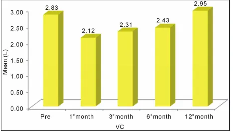
[image:2.595.307.541.526.658.2]The mean VC value significantly decreased post-oper- atively from 2.83 L preoperatively to 2.12 L at the first postoperative month. It progressively increased to 2.43 L six months postoperatively, but differences still remained statistically significant compared with preoperative val- ues (Figure 1).
3.3. Forced Vital Capacity
As with VC, the mean FVC value also decreased one month after resection to progressively increase thereafter. However, at the sixth postoperative month, it still was significantly lower than the preoperative value (Figure 2).
3.4. Forces Expiratory Volume in One Second
[image:3.595.58.288.312.444.2]The comparison of the mean values of FEV1 showed no statistically significant differences between the preopera- tive value and the value at six months postoperatively. However, there were statistically significant differences when the preoperative value was compared with those at the first and third postoperative months. In this case, a progressive recovery was observed during the first six months after lobectomy (Figure 3).
Figure 2. Mean forced vital capacity (FVC) values at the four timepoints: One preoperative and three postoperative. A statistically significant difference was found on compari- son of the four timepoints (p < 0.05).
Figure 3. Mean forced expiratory volume in one minute (FEV1) values of the four timepoints: one preoperative and three postoperative. No statistically significant difference (p > 0.05) was found between the preoperative value and that at sixth months postoperatively, but a significant difference (p < 0.05) was evident for the other two comparisons (pre- operative versus first month and preoperative versus third month). A progressive recovery of values was observed.
3.5. Peak Expiratory Flow
A significant decrease in PEF was observed across the postoperative period, with no significant recovery at six months postoperatively (Figure 4).
[image:3.595.306.540.338.477.2]3.6. Forces Expiratory Volume in One Second/Forced Vital Capacity
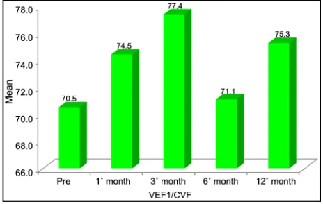
Figure 5 depicts the mean values of FEV1/FVC of four-
stages: pre-and postoperatively. There was nostatistically significant differences (p > 0.05) when compared with preoperative values. For this test, full recovery occurred six months after resection.
3.7. Forced Expiratory Flow
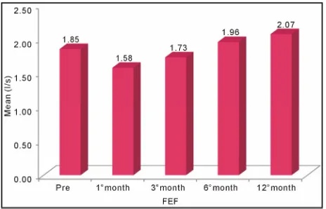
Figure 6 shows the mean values of FEF in four periods:
[image:3.595.58.289.502.648.2]pre- and postoperative. There was nostatistically signifi-cant difference (p > 0.05) among all comparisons with the initial FEF.
Figure 4. Mean PEF values of the four timepoints: one pre- operative and three postoperative. A statistically significant difference (p < 0.05) was evident on all comparisons with initial PEF, indicating that full recovery of the PEF had not occurred by the sixth month.
[image:3.595.306.538.545.692.2]Figure 6. Mean values of FEF four periods: pre- and post- operative. There was nostatistically significant difference (p > 0.05) among all comparisons with the initial FEF.
4. Discussion
Spirometry entails performing measurements of pulmo- nary flow and volumes obtained during the movements of forced inspiration and expiration which are interpreted by a device that transforms these into numbers and graphs.
The technique is indispensable in patients to be sub- mitted to surgical resection, particularly lung operations, because the measurements can help predict the risk of postoperative complications.
In the present study, spirometry was chosen because it is the method most commonly used in surgical practice, is available under our service, and also because the other known methods were difficult to obtain.
The election of tuberculosis as the focus for this study was due to the high incidence of TB patients in the state of Maranhão, the large number of symptomatic sequelae associated with the disease seen at the pulmonology and thoracic surgery outpatient units, and given the fact that these patients are uniform.
Lobectomy was selected as a variable of interest since this allowed comparison of the patient against themselves, after removal of the compromised lobe from the lung.
The curve of normality adopted in this study was that devised by Pereira in 2001 [21], the author responsible for the study of the Brazilian population.
FVC is considered the most important measurement of spirometry because it reflects the maximum limit of flow of expired air after a single maximal inspiration, per- formed in the shortest possible time.
Any process changing the pulmonary dynamic also affects the maximum flow, representing the most sensi- tive measurement to variations. In restrictive ventilatory defects, FVC values lie below reference values.
FEV1 corresponds to expirated volume within the first second of FVC, and constitutes the spirometry mea-
surement most representative of clinical alterations and restrictive ventilatory defects, and may be normal or re- duced.
In the study by Perin [19], he compared spirometric assessments of individuals submitted to abdominal der- molipectomy during the immediate pre-operative period and post-operative period of 19 to 36 months and these values showed no significant difference with correction of rectus musclesof the abdomen.
In the present study, a marked reduction in respiratory function was evident in the first month postoperatively, attributed to the great difficulty of the patient to execute the maneuvers as a result of trauma of the musculature of the chest wall and of pain associated with respiratory force.
Júnior [22], assessing respiratory function in individu- als submitted to abdominoplasty, at the preoperative stage and the fourth day postoperatively, observed a de- cline in respiratory function on the fourth day postopera- tively with normalization by the 30th postoperative day.
According to Tercan et al. [23], VC improves signifi- cantly up to the 30th postoperative day in abdomino- plasty, despite failing to return to pre-operative levels.
In the present study, the improvement in respiratory function was observed as of the 3rd postoperative month as a result of the absence of pain and improved lung com- pliance, with progressive recovery of the parameters at the 6th postoperative month.
The present study sought to assess whether full recov- ery of spirometric parameters occurs within a 180-day- postoperative period after lobectomies.
The results obtained showed no significant recovery in spirometric parameters, that did not reached the mean preoperative values 12 months after lung resection. This finding occurred only for FEV1, FVC and PEF meas- urements.
The authors suggest that studies of lung volumes be performed in order to verify whether the findings can be explained by the reduced lung volumes as a result of lobectomy.
5. Conclusion
Based on the results of the present study, it can be con- cluded that, at the 12th postoperative month, spirometric parameters of patients with tuberculosis sequelae who underwent lobectomy had returned to preoperative lev- els.
REFERENCES
Doença em Atividades Comprovada Bacteriologica- mente,” Jornal de Pneumologia, Vol. 28, No. 2, 2002, pp. 23-29. doi:10.1590/S0102-35862002000100006
[2] J. N. Muniz, A. Ruffino-Netto, M. Yamamura, T. C. S. Villa, R. Arcencio and R. I. Cardozo-Gonzales, “Aspectos Epidemiológicos da Co-Infecção Tuberculose e Vírus da Imunodeficiência Humana em Ribeirão Preto (SP), de 1998 a 2003,” Jornal Brasileiro de Pneumologia, Vol. 32, No. 6, 2006, pp. 529-534.
doi:10.1590/S1806-37132006000600010
[3] S. Bombarda, C. M. Figueiredo, M. B. G. Funari, J. Soares Jr., M. Seiscento and M. Terra, “Imagem em Tu- berculose pulmonar,” Jornal Brasileiro de Pneumologia, Vol. 27, No. 6, 2001, pp. 329-340.
[4] A. S. Ribeiro, “Tratamento Compulsório da Tuberculose: Avanço ou Retrocesso?” Jornal Brasileiro de Pneumo- logia, Vol. 29, No. 1, 2003, pp. 50-52.
[5] M. P. T. Silveira, R. F. R. Adorno and T. Fontana, “Perfil dos Pacientes com Tuberculose e Avaliação do Programa Nacional de Controle da Tuberculose em Bagé (RS),”
Jornal Brasileiro de Pneumologia, Vol. 33, No. 2, 2007, pp. 199-205.
doi:10.1590/S1806-37132007000200015
[6] A. Ruffino-Netto, “Ttuberculose: A Calamidade Negligen- ciada,” Revista da Sociedade Brasileira de Medicina Tro-
pical, Vol. 35, No. 1, 2002, pp. 51-58. doi:10.1590/S0037-86822002000100010
[7] Z. R. Cavalcanti, M. F. P. M. Albuquerque, A. R. L. Cam- pello, R. Ximenes, U. Montarroyos and M. K. A. Verçosa, “Característica da Tuberculose em Idosos no Recife (PE): Contribuição Para o Programa de Controle,” Jornal Bra- sileiro de Pneumologia, Vol. 32, No. 6, 2006, pp. 535- 543. doi:10.1590/S1806-37132006000600011
[8] Brasil, “Ministério da Saúde. Tuberculose no Brasil: Avan- ços e Perspectivas. Programa Nacional de Controle da Tuberculose,” 2010.
http://www.sam.pmrp.com.br/ssaude/programas/tubercul ose/tuberculose_no_brasil.pdf
[9] A. Castelo Filho, A. L. Kritski, A. W. Barreto, A. C. M. Lemos, A. Ruffino Netto, C. A. Guimarães, et al., “II Con- senso Brasileiro de Tuberculose. Diretrizes Brasileiras para Tuberculose 2004,” Jornal Brasileiro de Pneumo-
logia, Vol. 30, Suppl. 1, 2004, pp. S2-S56.
[10] T. M. Fernandes, “Sol e Trevas: Histórias Sociais da Tu- Berculose Brasileira,” História, Ciências, Saúde-Manguin-
hos, Vol. 11, No. 3, 2004, pp. 767-771. doi:10.1590/S0104-59702004000300012
[11] J. F. Murray, “A Century of Tuberculosis,” American Jour-
nal of Respiratory and Critical Care Medicine, Vol. 169, No. 11, 2004, pp. 1181-1186.
doi:10.1164/rccm.200402-140OE
[12] C. E. Gazetta, S. H. F. Vendramini, A. Ruffino-Netto, M. R. C. Oliveira and T. C. S. Villa, “Estudo Descritivo So- bre a Implantação da Estratégia de Tratamento de Curta Duração Diretamente Observado no Controle da Tuber-
culose em São José do Rio Preto e Seus Impactos (1998-2003),” Jornal de Pneumologia, Vol. 33, No. 2, 2007, pp. 192-198.
doi:10.1590/S1806-37132007000200014
[13] A. Gomes Neto, M. L. Medeiros and J. M. M. Gifoni, “Bronquiectasia Localizada e Multissegmentar: Perfil Clínico Epidemiológico e Resultado do Tratamento Cir- úrgico de 67 Casos,” Jornal de Pneumologia, Vol. 27, No. 1, 2001, pp. 1-6. doi:10.1590/S0102-35862001000100002
[14] M. S. Guerra, J. A. Miranda, F. Leal and L. Vouga, “Tra- tamento Cirúrgico Das Bronquiectasias,” Revista Portu- guesa de Pneumologia, Vol. 13, No. 5, 2007, pp. 691- 701.
[15] K. Balkanli, O. Genç, M. Dakak, S. Gurkök, A. Gözu- buyuk, H. Çaylakat, et al., “Surgical Management of Bronchiectasis: Analysis and Short-Term Results in 238 Patients,” European Journal Cardiothoracic Surgery, Vol. 24, No. 5, 2003, pp. 699-702.
doi:10.1016/S1010-7940(03)00497-4
[16] R. Saad Jr., T. Garrido, R. Stirbulov and F. Rafal, “Ava- liação da Função Respiratória de Doentes Submetidos à Operação Abdominal Alta,” Rev. Col. Bras. Cir., Vol. 21, No. 6, 1994, pp. 329-332.
[17] A. M. R. Pinto, “Estudo Comparativo da Função Pulmo- nar em Pacientes Pós-Revascularizados do Miocárdio, com CEC e sem CEC, com uso de Derivação Intralu- minal. Tese [Doutorado],” Faculdade de Ciências Mé- dicas da Santa Casa de Misericórdia de São Paulo, São Paulo, 1999.
[18] A. Helene Jr., “Avaliação da Função Respiratória em In- Divíduos Submetidos à Abdominoplastia. Tese [Dou- torado],” Faculdade de Ciências Médicas da Santa Casa de Misericórdia de São Paulo, São Paulo, 2005.
[19] L. F. Perin, “Avaliação Espirométrica de Indivíduos Sub- metidos à Dermolipectomia Abdominal. Tese [Mestra- do],” Faculdade de Ciências Médicas Santa Casa de Misericórdia de São Paulo, São Paulo, 2007.
[20] American Thoracic Society, “Standardization of Spiro- metry, 1994 Update. American Thoracic Society,” Ame- rican Journal of Respiratory and Critical Care Medicine, Vol. 152, No. 3, 1995, pp. 1107-1136.
doi:10.1164/ajrccm.152.3.7663792
[21] C. A. C. Pereira, “Testes de Função Pulmonar. Projeto Diretrizes. Associação Médica Brasileira e Conselho Fe- deral de Medicina,” 2001.
http://www.projetodiretrizes.org.br/projeto_diretrizes/090.pdf
[22] A. Helene Jr., “Avaliação da Função Respiratória em Indivíduos Submetidos à Abdominoplastia. Tese [Dou- torado],” Faculdade de Ciências Médicas da Santa Casa de Misericórdia de São Paulo, São Paulo, 2005.
[23] M. Tercan, M. Bekerecioglu, O. Dikensoy, H. Kocoglu, B. Atik, D. Isik, et al., “Effects of Abdominoplasty on Res- piratory Functions: A Prospective Study,” Annals of Plas-