Posthorax
®
Prevents Sternal Dehiscence and Instability:
Preliminary Results of a Prospective Randomized
Multicenter Trial
Santarpino Giuseppe, Fischlein Theodor, Steffen Pfeiffer
Department of Cardiac Surgery, Klinikum Nürnberg, Nuremberg, Germany. Email: g.santarpino@libero.it
Received August 21st, 2012; revised September 25th, 2012; accepted October 8th, 2013
Copyright © 2013 Santarpino Giuseppe et al. This is an open access article distributed under the Creative Commons Attribution Li-cense, which permits unrestricted use, distribution, and reproduction in any medium, provided the original work is properly cited.
ABSTRACT
Aim: A Prospective Randomized Multicenter Trial is ongoing to evaluate Posthoraxand prevention of sternal dehis- cence/instability: clinical percept is optimistic for Posthoraxuse. The aim of this mono-center analysis is to give a pre- liminary result of Posthoraxsupport vest after sternotomy. Methods: One hundred and eighty three cases elective pa- tients were consecutive operated and included in this study conducted in our department since June 2009. Patients were randomized as following:68 patients were treated with the Posthorax support vestand 115 received a standard bandage postoperatively. The primary endpoints were the Infective Events. Secondary endpoints included a composite of post- operative clinical variables and mortality. Results: The two groups are homogeneous for these characteristic except sex (more women in Control Group, p = 0.022). Operative data were comparable in both groups. Deep sternal infections occurred in four patients, all in Control Group (3.5% vs 0%, p = 0.153). At Follow up, we recorded 2 cases of superfi- cial infection in the control group versus 0 (1.7%, p = 0.394) and 1 case of wound dehiscence always in Control Group versus 0 (0.9%, p = 0.628). Cumulative Infective Events are statistically more in Control Group (7 cases 6.1% versus 0 cases, p = 0.036*). According to the secondary endpoints, there were also no differences between the two compared
groups except length of hospitalization (10.6 ± 4 days versus 13.4 ± 9.5, p = 0.019*). Conclusion: Preliminary results
show the Posthorax sternum support vest as a valuable adjunct to prevent sternum-related complications: We record a statistical reduction of length of hospital stay and infective events usingthe support vest in a 3-month follow-up.
Keywords: Chest Wall; Device; Wound Dehiscence; Wound Infection; Postoperative Care
1. Introduction
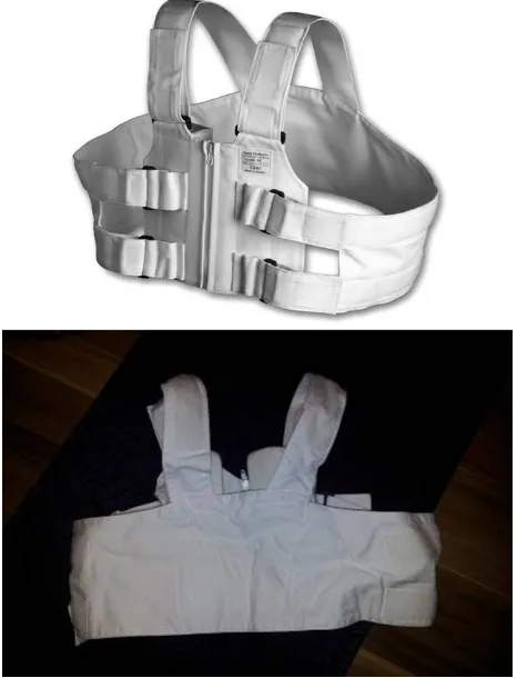
In cardiac surgery, sternal instability caused by dehis- cence or superficial/deep infection weigh on 1% to 10% [1-7]. Their consequences are an increase of morbidity and mortality (up to 10% and 40% respectively) and ad- ditional hospital costs [6,8-10]. Several factors (pre-, intra- and postoperatively) are associated with sternal dehiscence and the development of sternal wound infec- tion [11-19]. The Posthorax is a newly designed thorax support vest for prevention of sternum instability. The design of Posthorax® support vest (Epple Inc, Wien, Aus- tria) (Figure 1) ensures anterior-posterior stabilization of
the thorax: two pads placed on each side of the sternum, preventing intrinsic movement of the two sternum halves. Lateral flaps are designed for optimum fit and allow for stopping over-extension but guaranteed normal breathing. Posthorax vest is ergonomically made to fit, both male/
female as individual, for both thorax and abdominal re- gion by supporting it with upper straps [20]. The design of Posthorax® support vest could be usefull follow any kind of sternotomy both in cardiac surgery and in tho- racic surgery.
The literature shows a clinical efficacy for primary re- inforcement of the sternum [20] and is ongoing prospec- tive, randomized controlled multicenters study our Cen- ter as well [21]. The aim of this mono-center analysis is to give a preliminary result of Posthorax® support vest after sternotomy.
2. Material and Methods
Figure 1. Posthorax® support vest.
Langegasse 74, 1230 Vienna, Austria) and each patient providing informed consent.
All patients were stratified using a modified STS and LogEuroScore risk score analysis for cardiac surgery [22] and patients with age of under 20 years, congenital car- diac defect, mechanical reanimation, and irradiation of the thorax were excluded.
Patients were randomized immediately before opera- tion by automatic central system in Epple Posthorax Maurer Langegasse 74, 1230 Vienna, Austria.
Patients were randomized as follows:68 patients were treated with the Posthorax® support vest(Posthorax Group)
and 115 received a standard bandage postoperatively(Con- trol Group).
All patients received an identical perioperative pro- phylactic antibiotic treatment with 2 gr Cephazolin.
No patients underwent left and right internal mammary artery preparation at the same time. Internal mammary artery preparation was always non-skelletonized.
The technique of wound closure and disinfection pro- tocol was regulated: at the operating room, the skin is disinfected with alcohol solution preoperatively and with iodine solution postoperatively. We changed the gloves after the disinfection and after every suspect rupture.
Stainless steel wires (Assut Medical SrlW, Pully-Lau-
sanne, Switzerland) were used for sternum closure: four or more “figure-of-eight” sutures passed trans-sternal or peri-sternal. Trans-sternal sutures passed approxi-mately 1 cm on each side. The suture wires were crossed, pulled, and twisted. Care was taken that knot-ting only occurs at the point where desired, and is not tied under tension. No patients undergoing Robicsek sternum closure [23].
The method for wound suturing was the same for both groups: synthetic absorbable braided sutures 2 - 0 Vicryl were used subcutaneously. Going from deep up to the surface, the pre-sternal fascia was closed with 0 Vicryl sutures in a “U” stitch type. The wound closure was made intra-cutaneously with 4 - 0 Monocryl (Ethicon Inc., Somerville, NJ).
Postoperatively the pain score didn’t evaluated daily using the visual analog scale but Cough and Sneeze events were recorded with a 4 steps scale (1 = no event, 2 = few events with few pain, 3 = often enough with pain, 4 = many events with pain) [24].
Patients have Posthorax vest on in Intensive Care Unit as soon as they have a haemodynamic stability (usually 6 hours after extubation).
Patients who preoperatively refused the sternum vest were analyzed in the Control Group (n = 16) (Figure 2).
Patients who refused the sternum support were com- plaining about the close fit and slipping of the vest.
Patients’ data were collected during hospitalization using medical records and by telephone questionnaire after discharge during the 90-day follow-up period.
[image:2.595.56.289.100.405.2]The primary endpoints were the Infective Events: in- cidence hospital sternal dehiscence requiring reopera-tion as well as the rate of sternal dehiscence or wound infection for 90 days after cardiac surgery requiring re-hospitalization. Sternal wound infections included superficial infections, involving skin and subcutaneous tissue of the incision (SSWI), and deep infections (DSWI). The DSWI were defined according to the criteria proposed by the Centers for Disease Control as follows: 1) bacteria can be isolated from cultures of mediastinal tissue or fluid; 2) evidence of mediastinitis is seen during surgery; and 3) one of the following con-
[image:2.595.323.525.612.718.2]ditions: chest pain, sternal instability, or fever (≥38˚C) are present, and there is either purulent discharge from the mediastinum or bacteria can be isolated from a blood culture of drainage originating from the mediastinal area [25].
Secondary endpoints included a composite of postop- erative clinical variables and mortality. Baseline charac- teristics and perioperative variables were recorded. The SPSS statistical software package 15.0 for Windows (SPSS Inc., Chicago, IL) was used. The continuous and normally distributed data are presented as means ± SD. Categorical data were expressed as percentages.
Differences between the patient groups were carried out by using the χ2 and Fisher exact tests for categorical
variables, and unpaired t test or Mann-Whitney U test for continuous variables.
A p value less than 0.05 was considered to indicate statistical significance
3. Results
Preoperative patient characteristics including risk factors such as Age, Log EuroSCORE, NYHA class, STS score, BMI (Body mass Index), diabetes, chronic renal failure, peripheral artery disease, chronic obstructive pulmonary disease, myocardial infarction and Heart failure are sum- marized in Table 1. The two groups are homogeneous
for these characteristic except sex (more women in Con- trol Group, see Table 1). Operative data were compara-
ble in both groups (Table 2). Postoperative non-infective
details shown in Table 3 reached no statistical signifi-
cance including ventilation hours, mortality and non- infective morbidity, except of length of hospital stay. Three patients died (1 for Low Output Syndrome, 1 for Pulmonary Insufficence, 1 for MOF post-Sepsis) and 2 were in Posthorax Group but these never vest dressed. Follow up was 100% complete. Deep sternal infections occurred in four patients, all in Control Group (3.5% vs 0%, p = 0.153). These infections are related to S. Aureus in 2, to E. Faecalis in 1 and S. Epidermidis in 1 case. All these patients needed surgical revision and were treated by surgical debridement, VAC-system implantation. In hospital no cases of superficial infection or wound de- hiscence were recorded. At Follow up no cases of Deep sternal infections occurred but we recorded 2 cases of superficial infection in Control group versus 0 (1.7%, p = 0.394) and 1 case of wound dehiscence always in Control Group versus 0 (0.9%, p = 0.628). Of the 65 patients with sternum vest, sternal wound complications developed in 0 (0%). Cumulative Infective Events are statistically more in Control Group (7 cases 6.1% versus 0 cases, p = 0.036*) (
Table 4).
According to the secondary endpoints, there were also no differences between the two compared groups except
length of hospital stay (10.6 ± 4 days in Posthorax Group versus 13.4 ± 9.5 days in Control Group p = 0.019*).
4. Discussion
The aim of this mono-center analysis is to give a pre- liminary result of Posthorax support vest after ster- notomy. Prevention of sternal dehiscence/instability has a difficult solution in consideration of different variables causing sternum: to prevent these complications majority cardiac surgery centers use proper antibiotic prophylaxis, made attention to sterile technique and use antibiotic covered suture material. Thus the rate of infections in the most important study groups result in 3.6% deep sternum infection [3-7]. There was only 1 death in our entire co- hort caused by infection, which can be accounted to early recognition and aggressive treatment of sternal wound complications. Several study reports of a increased risk of sternal wound complications with obesity, diabetes, chronic obstructive pulmonary disease, NYHA score > 3, peripheral vascular disease, use of bilateral internal mam- mary arteries, duration of operation and ventilation [4, 8-12,16,17,20]. As well we record same pre-operative variable except for sex, Body Weigh and Body Height: no results show that women have more infective risk then men, and Body Weight/Height are higher in Posthorax Group (probably because more men) but BMI (indicative for obesity and more risk for infection) is not different between the two groups. The identification of reliable risk factors is important to carefully select patients’ need- ing special attention in perioperative and postoperative periods. We used a risk scoring system created by Fowler et al. [22] to identify patients undergoing cardiac surgery, who are at high risk of major infection (STS score). This risk score estimates that probability of infection at nine points is more than 3%. There was no difference between the vest and non-vest group in the identified risk score values.
Table 1. Preoperative variables.
Posthorax Control
68 patients 115 patients p
Log EUROscore (mean ± Standard deviation) 5.8 ± 8.5 7.5 ± 8.8 0.196 Age (mean ± Standard deviation) 64.9 ± 11.1 67.8 ± 11.6 0.101 Female [number (percentage)] 15 (22.1) 45 (39.1) 0.022*
NYHA class (mean ± Standard deviation) 2.6 ± 0.9 2.7 ± 0.8 0.240 STS score (mean ± Standard deviation) 11.9 ± 5.8 11.8 ± 6.3 0.919 Diabetes [number (percentage)] 16 (23.5) 29 (25.2) 0.472 Diabetes ID [number (percentage)] 7 (10.3) 10 (8.7) 0.454
Diabetes NID [number (percentage)] 9 (13.2) 19 (16.5) 0.355 Chronic pulmonary disease [number (percentage)] 5 (7.4) 18 (15.7) 0.077 Body weigh (Kg) (mean ± Standard deviation) 85 ± 16.6 80 ± 13.8 0.033*
Body height (cm) (mean ± Standard deviation) 172 ± 9.3 169 ± 8.5 0.006* BMI (Body mass index) (mean ± Standard deviation) 28.4 ± 4.8 28.1 ± 4.6 0.625
Creatinine ≥ 2 mg/dL [number (percentage)] 7 (10.3) 14 (12.2) 0.449
Heart failure [number (percentage)] 8 (11.8) 17 (14.8) 0.368
Myocardial infarct > 4 weeks [number (percentage)] 22 (32.4) 20 (17.4) 0.017*
Myocardial infarct ≤ 4 weeks [number (percentage)] 5 (7.4) 9 (7.8) 0.576
Extracardiac arteriopathy [number (percentage)] 11 (16.2) 21 (18.3) 0.442
* indicates values with statistical significance.
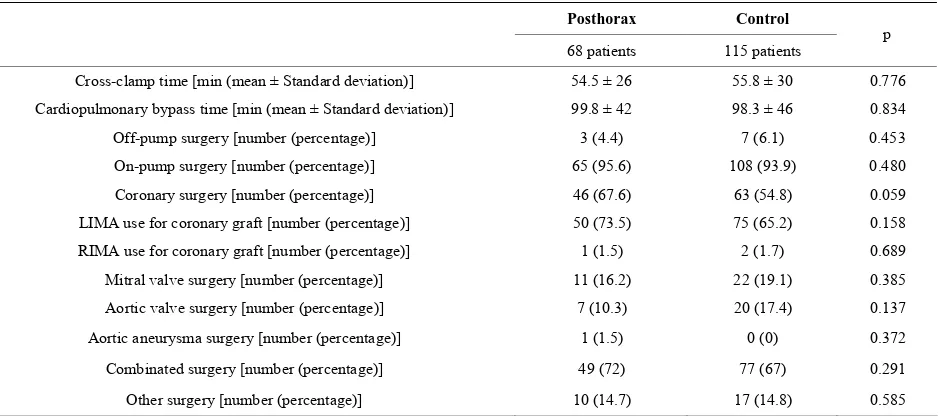
Table 2. Operative variables.
Posthorax Control
68 patients 115 patients p
Cross-clamp time [min (mean ± Standard deviation)] 54.5 ± 26 55.8 ± 30 0.776
Cardiopulmonary bypass time [min (mean ± Standard deviation)] 99.8 ± 42 98.3 ± 46 0.834 Off-pump surgery [number (percentage)] 3 (4.4) 7 (6.1) 0.453 On-pump surgery [number (percentage)] 65 (95.6) 108 (93.9) 0.480
Coronary surgery [number (percentage)] 46 (67.6) 63 (54.8) 0.059 LIMA use for coronary graft [number (percentage)] 50 (73.5) 75 (65.2) 0.158 RIMA use for coronary graft [number (percentage)] 1 (1.5) 2 (1.7) 0.689 Mitral valve surgery [number (percentage)] 11 (16.2) 22 (19.1) 0.385
Aortic valve surgery [number (percentage)] 7 (10.3) 20 (17.4) 0.137
Aortic aneurysma surgery [number (percentage)] 1 (1.5) 0 (0) 0.372
Combinated surgery [number (percentage)] 49 (72) 77 (67) 0.291
Other surgery [number (percentage)] 10 (14.7) 17 (14.8) 0.585
LIMA: Left Internal mammary artery; RIMA: Right Internal mammary artery.
the face of sternal separation and instability can then progress to DSWI [31]. On the other hand, the initial instability derives from the basic mechanism of an os- teomyelitis: this is the ground upon which the feared in- fection develops [32]. Experimental studies compared the mechanical stability of the sternum using a variety of
[image:4.595.70.539.433.641.2]Table 3. Postoperative variables.
Posthorax Control
68 patients 115 patients p Mortality (3 months) [number (percentage)] 2 (2.9) 1 (0.9) 0.311
Redo-surgery for Bleeding [number (percentage)] 2 (2.9) 4 (3.5) 0.604 Redo-surgery not for Bleeding or Infection [number (percentage)] 0 (0) 1 (0.9) 0.628 Blood unit transfusions (mean ± Standard deviation) 0.7 ± 1.3 9.9 ± 1.6 0.234
Plasma transfusion use [number (percentage)] 9 (13.2) 26 (22.6) 0.085 Platelets transfusion use [number (percentage)] 3 (4.4) 9 (7.8) 0.283 IABP use [number (percentage)] 0 (0) 6 (5.2) 0.059
Delirium/Mental confusion [number (percentage)] 7 (10.3) 19 (16.5) 0.172 Intubation time [hours (mean ± Standard deviation)] 13.7 ± 19 27.7 ± 66 0.092 Intensive care unit stay [days (mean ± Standard deviation)] 2.8 ± 2.6 3.4 ± 3.4 0.202 Hospital stay [days (mean ± Standard deviation)] 10.6 ± 4 13.4 ± 9.5 0.019*
Renal failure/Dialysis [number (percentage)] 3 (4.4)/1 (1.5) 10 (8.7)/3 (2.6) 0.217/0.524 Inotropic drugs use [number (percentage)] 65 (95.6) 112 (97.5) 0.396
Cough scale (mean ± Standard deviation) 2.2 ± 1.2 1.9 ± 0.9 0.096
[image:5.595.53.538.111.358.2]Sneeze scale (mean ± Standard deviation) 0.93 ± 0.4 0.99 ± 0.4 0.327 * indicates values with statistical significance.
Table 4. Infective outcome.
Posthorax Control
68 patients 115 patients p
Hospital SSWI [number (percentage)] 0 (0) 0 (0) /
Hospital DSWI [number (percentage)] 0 (0) 4 (3.5) 0.153
Follow up SSWI [number (percentage)] 0 (0) 2 (1.7) 0.394
Follow up DSWI [number (percentage)] 0 (0) 0 (0) /
Follow up wound dehiscence [number (percentage)] 0 (0) 1 (0.9) 0.628
Infective events [number (percentage)] 0 (0) 7 (6.1) 0.036*
SSWI: superficial sternal wound infections
DSWI: deep sternal wound infections
* indicates values with statistical significance.
tion of 2 mm at 46.8 mmHg of pressure: strong coughing during extubation periods or postoperative course in- creases the intrathoracic pressure to 300 mmHg [37]. Posthorax produces shearing forces in the anterior-pos- terior and lateral directions. We suppose that with a tra- ditional method of sternal closure the Posthorax ensures anterior-posterior stabilization of the thorax and prevent intrinsic movement of the two sternum halves. It was not our intent to establish multiple preoperative, intraopera- tive, and postoperative risk factors associated with an increased risk for sternal wound complications; these have been done by several other authors and could even be confirmed in this study [1,8,22,38-40]. This study contains the obvious limitation of missing documentation
use of Bone wax (our policy is a sporadic use, of course in <10% patients).
5. Conclusion
The Posthorax® design avoids tilting of the sternum and
and subsequent biomechanical studies are mandatory to evaluate geometrically forces provided by the thorax support vest. Nevertheless, in the 90 day follow-up pe- riod, a significantimpact on the reduction of infective events and of length of hospital stay usingthe “support vest was observed.
REFERENCES
[1] J. E. Losanoff, J. W. Jones and B. W. Richmann, “Pri- mary Closure of Median Sternotomy: Techniques and Principles,” Cardiovascular Surgery, Vol. 10, No. 2, 2002, pp. 102-110. doi:10.1016/S0967-2109(01)00128-4 [2] A. Negri, J. Manfredi, A. Terrini, et al., “Prospective
Evaluation of a New Sternal Closure Method with Ther- mo Reactive Clips,” European Journal Cardio-Thoracic Surgery, Vol. 22, No. 4, 2002, pp. 571-575.
doi:10.1016/S1010-7940(02)00411-6
[3] J. Zeitani, F. Bertoldo, C. Bassano, A. Penta de Peppo, A. Pellegrino, F. M. El Fakhri, et al., “Superficial Wound Dehiscence after Median Sternotomy: Surgical Treatment versus Secondary Wound Healing,” The Annals of Tho- racic Surgery, Vol. 77, No. 2, 2004, pp. 672-675.
doi:10.1016/S0003-4975(03)01594-7
[4] The Parisian Mediastinitis Study Group, “Risk Factors for Deep Sternal Wound Infection after Sternotomy: A Pro- spective Multicenter Study,” The Journal of Thoracic and Cardiovascular Surgery, Vol. 111, No. 6, 1996, pp. 1200- 1207. doi:10.1016/S0022-5223(96)70222-2
[5] M. G. Sarr, V. L. Gott and T. R. Townsend, “Mediastinal Infection after Sternotomy,” The Annals of Thoracic Sur- gery, Vol. 38, No. 4, 1984, pp. 415-423.
doi:10.1016/S0003-4975(10)62300-4
[6] F. D. Loop, B. W. Lytle, D. M. Cosgrove, S. Mahfood, M. C. McHenry, M. Goormastic, et al., “Sternal Wound Com- plications after Isolated Coronary Bypass Grafting: Early and Late Mortality, Morbidity and Cost of Care,” The Annals of Thoracic Surgery, Vol. 49, No. 2, 1990, pp. 179-187. doi:10.1016/0003-4975(90)90136-T
[7] E. A. Grossi, A. T. Culliford, K. H. Krieger, D. Kloth, R. Press, F. G. Baumann, et al., “A Survey of 77 Major In- fectious Complications of Median Sternotomy: A Review of 7949 Consecutive Operative Procedures,” The Annals of Thoracic Surgery, Vol. 40, No. 3, 1985, pp. 214-221. doi:10.1016/S0003-4975(10)60030-6
[8] J. C. Y. Lu, A. D. Grayson, P. Jha, A. K. Srinivasan and B. M. Fabri, “Risk Factors for Sternal Wound Infection and Mid-Term Survival Following Coronary Artery By-pass Surgery,” European Journal Cardiothoracic Surgery, Vol. 23, No. 6, 2003, pp. 943-949.
doi:10.1016/S1010-7940(03)00137-4
[9] M. A. Borger, V. Rao, R. D. Weisel, J. Ivanov, G. Cohen, H. E. Scully, et al., “Deep Sternal Wound Infection: Risk Factors and Outcomes,” The Annals of Thoracic Surgery, Vol. 65, No. 4, 1998, pp. 1050-1105.
doi:10.1016/S0003-4975(98)00063-0
[10] C. C. Shih, C. M. Shih, Y. Y. Su and S. J. Lin, “Potential Risk of Sternal Wires,” European Journal Cardiothoracic
Surgery, Vol. 25, No. 5, 2004, pp. 812-818. doi:10.1016/j.ejcts.2003.11.043
[11] M. A. Olsen, P. Lock-Buckley, D. Hopkins, L. B. Polish, T. M. Sundt and V. J. Fraser, “The Risk Factors for Deep and Superficial Chest Surgical-Site Infections after Coro- nary Artery Bypass Graft Surgery Are Different,” The Journal of Thoracic and Cardiovascular Surgery, Vol. 124, No. 1, 2002, pp. 136-145.
doi:10.1067/mtc.2002.122306
[12] W. E. Trick, W. E. Scheckler, J. L. Tokars, K. C. Jones, M. L. Reppen, E. M. Smith, et al., “Modifiable Risk Fac- tors Associated with Deep Sternal Site Infection after Coronary Artery Bypass Grafting,” The Journal of Tho- racic and Cardiovascular Surgery, Vol. 119, No. 1, 2000, pp. 108-114. doi:10.1016/S0022-5223(00)70224-8 [13] M. De Feo, A. Renzulli, G. Ismeno, R. Gregorio, A. Della
Corte, R. Utili, et al., “Variables Predicting Adverse Out- come in Patients with Deep Sternal Wound Infection,” The Annals of Thoracic Surgery, Vol. 71, No. 1, 2001, pp. 324-331. doi:10.1016/S0003-4975(00)02137-8
[14] J. H. Braxton, C. A. Marrin, P. D. McGrath, C. S. Ross, J. R. Morton, M. Norotsky, et al., “Mediastinitis and Long- Term Survival after Coronary Artery Bypass Graft Sur- gery,” The Annals of Thoracic Surgery, Vol. 70, No. 6, 2000, pp. 2004-2007.
doi:10.1016/S0003-4975(00)01814-2
[15] I. K. Toumpoulis, C. E. Anagnostopoulous, J. J. De Rose Jr. and D. G. Swistel, “The Impact of Deep Sternal Wound Infection on Long-Term Survival after Coronary Artery Bypass Grafting,” Chest, Vol. 127, No. 2, 2005, pp. 464-471. doi:10.1378/chest.127.2.464
[16] C. A. Milano, K. Kesler, N. Archibald, D. J. Sexton and R. H. Jones, “Mediastinitis after Coronary Artery Bypass Graft Surgery: Risk Factors and Long-Term Survival,” Cir- culation, Vol. 92, No. 8, 1995, pp. 2245-2251.
[17] L. Ridderstolpe, H. Gill, H. Granfeldt, H. Ahlfeldt and H. Rutberg, “Superficial and Deep Sternal Wound Compli- cations: Incidence, Risk Factors and Mortality,” Euro- pean Journal Cardiothoracic Surgery, Vol. 20, No. 6, 2001, pp. 1168-1175.
doi:10.1016/S1010-7940(01)00991-5
[18] A. Sachithanandan, P. Nanjaiah, P. Nightingale, I. C. Wil- son, T. R. Graham, S. J. Rooney, et al., “Deep sternal Wound Infection Requiring Revision Surgery: Impact on Mid-Term Survival Following Cardiac Surgery,” Euro- pean Journal Cardiothoracic Surgery, Vol. 33, No. 4, 2008, pp. 673-678. doi:10.1016/j.ejcts.2008.01.002 [19] F. Robicsek, A. Fokin, J. Cook and D. Bhatia, “Sternal
Instability after Midline Sternotomy,” The Journal of Thoracic and Cardiovascular Surgery, Vol. 48, No. 1, 2000, pp. 1-8. doi:10.1055/s-2000-9945
[20] M. Gorlitzer, S. Folkmann, J. Meinhart, P. Poslussny, M. Thalmann, G. Weiss, et al., “A Newly Designed Thorax Support Vest Prevents Sternum Instability after Median Sternotomy,” European Journal Cardiothoracic Surgery, Vol. 36, No. 2, 2009, pp. 335-339.
doi:10.1016/j.ejcts.2009.01.038
Meinhart, T. Fischlein, H. Reichenspurner and M. Gra- benwöger, “A Prospective Randomized Multicenter Trial Shows Improvement of Sternum Related Complications in Cardiac Surgery with the Posthorax® Support Vest,”
Interactive CardioVascular and Thoracic Surgery, Vol. 10, No. 5, 2010, pp. 714-718.
[22] V. G. Fowler Jr., S. M. O’Brien, L. H. Muhlbaier, G. R. Corey, T. B. Ferguson and E. D. Peterson, “Clinical Pre- dictors of Major Infections after Cardiac Surgery,” Cir- culation, Vol. 112, Suppl. 9, 2005, pp. I358-I365. [23] F. Robicsek, H. K. Daugherty and J. W. Cook, “The Pre-
Vention and Treatment of Sternum Separation Following Open-Heart Surgery,” The Journal of Thoracic and Car- diovascular Surgery, Vol. 73, No. 2, 1977, pp. 267-268. [24] S. L. Collins, R. A. Moore and H. J. McQuay, “The Vis-
ual Analogue Pain Intensity Scale: What Is Moderate Pain in Millimeters?” Pain, Vol. 72, No. 1-2, 1997, pp. 95-97. doi:10.1016/S0304-3959(97)00005-5
[25] A. J. Mangram, T. C. Horan, M. L. Pearson, L. C. Silver and W. R. Jarvis (for the Hospital Infection Control Prac- tices Advisory Committee), “Guideline for Prevention of Surgical Site Infection,” Infection Control and Hospital Epidemiology, Vol. 20, No. 4, 1999, pp. 424-429. doi:10.1086/501620
[26] C. Y. Bitkover and B. Gardlund, “Mediastinitis after Car- diovascular Operations: A Case-Control Study of Risk Factors,” The Annals of Thoracic Surgery, Vol. 65, No. 1, 1998, pp. 36-40. doi:10.1016/S0003-4975(97)01003-5 [27] J. Bellchambers, J. M. Harris, P. Cullinan, H. Gaya and J.
R. Pepper, “A Prospective Study of Wound Infection in Coronary Artery Surgery,” European Journal Cardiotho- racic Surgery, Vol. 15, No. 1, 1999, pp. 45-50.
doi:10.1016/S1010-7940(98)00255-3
[28] D. Jonkers, T. Elenbaas, P. Terporten, F. Nieman and E. Stobberingh, “Prevalence of 90-Days Postoperative Wound Infections after Cardiac Surgery,” European Journal Car- diothoracic Surgery, Vol. 23, No. 1, 2003, pp. 97-102. doi:10.1016/S1010-7940(02)00662-0
[29] A. T. Culliford, J. N. Cunningham Jr., R. H. Zeff, O. W. Isom, P. Teiko and F. C. Spencer, “Sternal and Costo-chondral Infections Following Open-Heart Surgery. A Review of 2594 Cases,” The Journal of Thoracic and Cardiovascular Surgery, Vol. 72, No. 5, 1976, pp. 714- 726.
[30] C. Schimmer, W. Reents, S. Berneder, P. Eigel, O. Sezer, H. Scheld, et al., “Prevention of Sternal Dehiscence and Infection in High-Risk Patients: A Prospective Rando- mized Multicenter Trial,” The Annals of Thoracic Sur- gery, Vol. 86, No. 6, 2008, pp. 1897-904.
doi:10.1016/j.athoracsur.2008.08.071
[31] D. H. Song, R. F. Lohmann, J. D. Renucci, V. Jeevanan- dam and J. Raman, “Primary Sternal Plating in High-Risk Patients Prevents Mediastinitis,” European Journal Car- diothoracic Surgery, Vol. 26, No. 2, 2004, pp. 367-372. doi:10.1016/j.ejcts.2004.04.038
[32] A. H. Kiessling, F. Isgro, U. Weisse, A. Möltner, W. Sag- gau and J. Boldt, “Advanced Sternal Closure to Prevent Dehiscence in Obese Patients,” The Annals of Thoracic Surgery, Vol. 80, No. 4, 2005, pp. 1537-1539.
doi:10.1016/j.athoracsur.2004.04.050
[33] W. E. McGregor, M. Payne, D. R. Trumble, K. M. Farkas and J. A. Magovern, “Improvement of Sternal Closure Stability with Reinforced Steel Wires,” The Annals of Thoracic Surgery, Vol. 76, No. 5, 2003, pp. 1631-1634. doi:10.1016/S0003-4975(03)00760-4
[34] J. E. Losanoff, A. D. Collier, B. W. Richman, H. Huff, F. Hsieh, et al., “Biomechanical Comparison of Median Stern- otomy Closures,” The Annals of Thoracic Surgery, Vol. 77, No. 1, 2004, pp. 203-209.
doi:10.1016/S0003-4975(03)01468-1
[35] F. C. Riess, N. Awwad, B. Hoffmann, R. Bader, H. Y. Helmold, C. Loewer, et al., “A Steel Band in Addition to 8 Wire Circlages Reduces the Risk of Sternal Dehiscence after Median Sternotomy,” Heart Surgery Forum, Vol. 7, No. 6, 2004, pp. 387-392. doi:10.1532/HSF98.200403114 [36] T. Bottio, G. Rizzoli and G. Gerosa, “Double Crisscross
Sternal Wiring and Chest Wound Infections: A Prospec- tive Randomized Study,” The Journal of Thoracic and Cardiovascular Surgery, Vol. 126, No. 5, 2003, pp. 1352- 1356. doi:10.1016/S0022-5223(03)00945-0
[37] R. S. Irwin, “Complications of Cough: ACCP Evidence- Based Clinical Practice Guidelines,” Chest, Vol. 129, Suppl. 1, 2006, pp. 54S-58S.
doi:10.1378/chest.129.1_suppl.54S
[38] R. Sharma, D. Puri, B. P. Panigrahi and I. S. Virdi, “A Modified Parasternal Wire Technique for Prevention and Treatment of Sternal Dehiscence,” The Annals of Tho- racic Surgery, Vol. 77, No. 1, 2004, pp. 210-213.
doi:10.1016/S0003-4975(03)01339-0
[39] J. E. Losanoff, B. W. Richman and J. W. Jones, “Disrup- tion and Infection of Median Sternotomy: A Comprehen- sive Review,” European Journal Cardiothoracic Surgery, Vol. 21, No. 5, 2002, pp. 831-839.
doi:10.1016/S1010-7940(02)00124-0