ESSENTIAL GUIDES FOR
ISOLATION/PURIFICATION OF CELLS
J. Bauer, Max-Planck-Institut fu(r Biochemie, Martinsried, Germany
Copyright^ 2000 Academic Press
In cell-separation technology the term ‘component’ of a mixture corresponds to a group of cells, which is usually called a cell population and shares a number of common features. How many common features a group of cells has to share in order to be called a cell population depends on the interest of the ‘separator’. For example, a T-cell population may be a group of mononuclear white blood cells bearing CD3 antigens, while a helper cell population usually comprises mononuclear white blood cells bearing CD3 and CD4 antigens.
Cells metabolize as long as they live independently, whether they remain in an actual state of activation or differentiation or they proceed to another one. This means that a whole cell must not change its appearance or functions, but some cell components are chemically modiRed either anabolically or catabolically. So for discussing cell separation the term ‘chemical modiRcation’ should be converted to ‘biological modiRcation’ and in this chapter the ex-pression ‘without biological modiRcation’ will be
de-Rned as puriRcation of cells without changes or sig-nals for changes of cellular states of activation and/or differentiation.
No technology has been developed so far which allows picking of cell populations directly out of pieces of plant or animal tissues. So a ‘mixture’ which will be separated is normally a suspension of single cells prepared from parts of plants, from organs or bodySuids of animals and humans or from two- or three-dimensional in vitro cell cultures. These cell sources already contain preselected groups of cells, the so-called organ orSuid (e.g. blood) speciRc cells. Still, a series of populations differing in important features are present in most plant or animal body compartments. In these instances, it may be of inter-est to separate cells for studying their biology or for using some of their capabilities in medicine or biotechnology.
Thus the following reSections on essential guides for separation/puriRcation of cells are based on separations deRned as processes of any scale by which cell populations of single-cell suspensions are separated from each other without biological modiRcation.
Methods for Cell Separation without
Biological Modi
\
cation
The above deRnition rules out some technologies, frequently used to prepare homogeneous cell popula-tions, because they include biological modiRcations of cells. For example, the enrichment of cell types of interest by establishing cell lines or cell clones will not be considered as a subject in this chapter. Cell lines or cell clones may be very useful sources of important genes and gene products. However, their cells are transformed in unnaturally fast growing states, in order to separate them from unwanted accompanying cells. Also cell separation/puriRcation techniques us-ing different capabilities of various kinds of cells to adhere to surfaces of, for example, culture dishes or
Rbres or to bind antibodies or macromolecules label-led bySuorescence dyes or magnetic beads, will not be described, because cell interactions with foreign components or antibody binding sites very often in-duce biological modiRcations. Of course, cell-puriR -cation methods like those mentioned above are very useful in research and biotechnology. The reader may
Rnd more informations regarding these techniques in the Further Reading.
This chapter focuses on application of counter-current centrifugal elutriation (CCE) and free-Sow electrophoresis (FFE). These methods use differences of physical cell parameters such as speciRc cell den-sity, cell size or negative surface charge density but do not include steps of cell labelling or cell transforma-tion. They have the advantage that cells can be
puri-Red within a short time while they are kept suspended in biocompatible Suids or even culture media. Cell contacts to foreign surfaces and/or biologically active substances are thereby minimized and signals of ac-tivation and differentiation are delivered to cells dur-ing the isolation procedure to a minimal extent. Both methods may help to obtain sufRcient numbers of identical cells with a high degree of purity and vitality for studying the biological role, which a deRned cell population may play within an organism or for transplantation of cells with states of activation and differentiation suitable toRt in the new organism of a recipient.
Single-Cell Suspensions
compartments such as peripheral blood, ascites, lymph or other body Suids already contain single cells. Cells of organs such as bone marrow, spleen or thymus can easily be removed, for example with the help of needles. The dissociation of single cells from two- or three-dimensional tissue cultures and from solid body tissues needs more rigorous methods. These cells not only adhere to each other, but are also more or less Rrmly attached to the extracelluar matrix, a complex network of collagen, proteog-lycans and cross-linking proteins such as laminin and
Rbronectin. Mechanical dissociation by scraping cell monolayers from their surfaces or by forcing tissue pieces or cell aggregates through oriRces or syringes or pipettes very often damages the cells and results in a poor yield. An enzymatic treatment or pretreatment of cell cultures or organs is thus often applied in order to digest the extracelluar matrix and/or to weaken the cell}cell attachment sites. The selection of the en-zymes, their concentration and their time and temper-ature of application depend on the type of organ and its originating organism. Enzymes frequently used for animal cell preparation are collagenase, trypsin, pronase, dispase, papain, chymotrypsin, hy-aluronidase, lysozyme and DNase, while cellulase is a typical enzyme for plant tissue dissociation. Some-times the action of the enzymes is supported by the presence of EDTA (ethylenediaminetetraacetic acid), which destroys binding sites mediated by Ca2#ions. Details regarding techniques of preparing single cells may be found in books quoted in the Further Read-ing. In general, the enzymatic treatment has to be optimized for each cell-separation process, because the enzymes may not only attack cell membrane com-ponents which keep the cells within the tissue but may also destroy important cell-membrane functions. If neither mechanical nor enzymatic methods lead to satisfactory results, an alternative way may be to incubate pieces of tissue on surfaces which challenge the cells to move out of the tissue and to form mono-layers surrounding the tissue. For example, cells of human prostate tumour sections, which cannot be dissociated in viable single cells by a number of mech-anical and enzymatic techniques, migrate out of the tissue and form a monolayer, when incubated in culture dishes for a few weeks. After removal of the tissue, the cells can easily be scraped off the plastic dish surface.
Pre-Separation
As soon as single cells are available, countercurrent centrifugal elutriation or cell electrophoresis may be applied. However, some cell-separation tasks need pre-enrichment of the cells of interest. Especially
where an investigator is interested in a peripheral blood leukocyte population such as a lymphocyte, granulocyte, monocyte or even reticulocyte popula-tion, the erythrocytes comprising more than 99% of the blood cells have to be removed before one of these white blood cell populations may be puriRed. In these instances, it has proved useful to perform aRrst step of density-gradient centrifugation, which does not need pre-labelling of cells. The method allows the separation of mononuclear leukocytes consisting mainly of lymphocytes and monocytes from granulocytes and erythrocytes. The separation prin-ciple is based on different speciRc densities of the various cell populations. In practice, a tube is Rlled with a biocompatible isotonic medium with a speciRc density adjusted between the speciRc densities of the cells to be separated and the cell mixture is layered on the top of this medium. Then the whole sample is exposed to a few hundred gby centrifugation. The forces cause mononuclear leukocytes with a density lower than the separation medium to remain at the top, while those with higher density sediment to the bottom. The speciRc density of the medium is ad-justed by silica colloids, which are coated with an inert material and have low osmolality. Although modern commercially available density-gradient sep-aration media are very inert and direct damage of the cells is seldom observed, the silica colloids are pinocytosed by some cells.
If this is a problem, prolonged centrifugation of whole blood may be an alternative route. During such a centrifugation procedure, a layer of white blood cells is formed above the erythrocytes. This layer, called a buffy coat, contains mononuclear as well as polymorphonucelated leukocytes and lies directly on the erythrocytes. The white cells may be collected. Although co-collection of a considerable number of red cells is usually unavoidable, a degree of white blood cell pre-enrichment can be achieved which allows reasonable further separation by, e.g. CCE.
Countercurrent Centrifugal Elutriation
(CCE)
Figure 1 Table-top elutriator (middle) together with an infusion pump driving the counterflow (left) and a volume analyser (right).
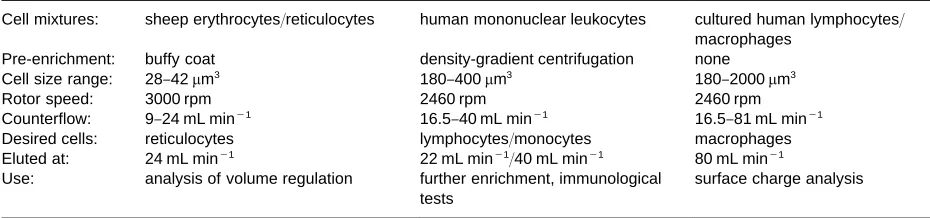
Table 1 Examples of counterflow and rotor speed adjustments using a Beckman elutriator equipped with JE-6 rotor (small separation chamber)
Cell mixtures: sheep erythrocytes/reticulocytes human mononuclear leukocytes cultured human lymphocytes/
macrophages
Pre-enrichment: buffy coat density-gradient centrifugation none
Cell size range: 28}42m3 180}400m3 180}2000m3
Rotor speed: 3000 rpm 2460 rpm 2460 rpm
Counterflow: 9}24 mL min\1 16.5}40 mL min\1 16.5}81 mL min\1
Desired cells: reticulocytes lymphocytes/monocytes macrophages
Eluted at: 24 mL min\1 22 mL min\1/40 mL min\1 80 mL min\1
Use: analysis of volume regulation further enrichment, immunological
tests
surface charge analysis
For details, see: Lauf PK and Bauer J (1987)Biochemical and Biophysical Research Communications 144: 849}855 and Bauer J and Hannig K (1984)Electrophoresis 5: 269}274.
and to a lesser extent on their speciRc densities. If the counterSow rate is increased by speeding up a pump and/or the sedimentation forces are decreased by re-ducing centrifugation velocity, the various cell popu-lations are washed out sequentially with increasing size ranges.
Commercially, small elutriation chambers with 5 mL separation volume and large ones with 40 mL are available together with suitable centrifuges and rotors from Beckman Instruments (Palo Alto, USA). They can accommodate up to 109 and 1010 cells,
respectively. A laboratory device has also been con-structed; it consists of a table-top centrifuge with a small rotor which has a separation chamber with a volume of 0.5 mL to which 106tissue cells or 2;107
mononuclear leukocytes may be loaded (Figure 1). In order to fractionate cells with different sizes into different fractions, counterSow rates and rotor speeds have to be adjusted depending on rotor size, chamber volume and the size of the cells to be separated. The result of each separation should be controlled by recording volume distributions of the cells of each fraction with the help of cell size analysers. Beckman
rotors are frequently operated at speeds ranging from 1000 to 4000 rpm. Dependent on the actual rotor speed, the counterSow through small chambers may be started at rates between 8 and 20 mL min\1and
increased for fractional elution stepwise up to 100 mL min\1 (Table 1); counterSow rates through
large separation chambers may start at 50 mL min\1.
The table-top centrifuge is operated at counterSow rates between 1 and 6 mL min\1, while the rotation
speeds are between 500 and 2200 rpm for tissue cell separation and between 1500 and 2800 rpm for leukocyte separation (Table 2). Using any of the in-struments, separation times are short and the cells may be kept suspended in culture medium. Thus unwanted exposure of the cells to stimulatory envi-ronments are minimized so that characteristics of separated populations will rather closely reSect the status of the original cells before fractionation.
Because of these advantages, CCE has proved most useful, if applied for the separation tasks listed below:
E For cell cycle analyses, the cellular DNA content is normally determined. However, cells have to be killed in order to make their DNA accessible for intercalatingSuorescence molecules. If living cells in different steps of the cell cycle need to be separ-ated, an increase of cell size during passage through the cell cycle may be used as a separation para-meter. With the help of CCE the small cells, which are in G1 phase can be separated from S-phase cells which have intermediary size and from the G2/ M-phase cells which have the largest size of the cell population. ManySow cytometric analyses of the DNA content of separated cells have already pro-ved that CCE enables the separation of cells of cell lines in fractions, which have up to 100% G1 phase cells, up to 80% S-phase cells and up to 80% G2/M phase cells, respectively.
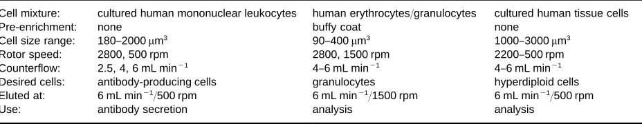
[image:3.568.50.515.578.687.2]Table 2 Examples of counterflow and rotor speed adjustments using the self-made table-top elutriator
Cell mixture: cultured human mononuclear leukocytes human erythrocytes/granulocytes cultured human tissue cells
Pre-enrichment: none buffy coat none
Cell size range: 180}2000m3 90}400m3 1000}3000m3
Rotor speed: 2800, 500 rpm 2800, 1500 rpm 2200}500 rpm
Counterflow: 2.5, 4, 6 mL min\1 4}6 mL min\1 4}6 mL min\1
Desired cells: antibody-producing cells granulocytes hyperdiploid cells
Eluted at: 6 mL min\1/500 rpm 6 mL min\1/1500 rpm 6 mL min\1/500 rpm
Use: antibody secretion analysis analysis
For details, see: Bauer J and Hannig K (1988)Journal of immunological Methods 112: 213}218 and Bauer J, Grimm D, Hofstaedter F and Wieland W (1992)Biotechnological Progress 8: 494}500.
foreign molecules and particles entering an organ-ism. So despite many alternative methods such as antibody-dependent sorting or panning, CCE, which does not involve cell adhesion to matrices or to antibodies, is often preferred, to separate mono-cytes from peripheral blood or bone marrow and to purify macrophages from alveolar tissues or Kup-ffer cells from liver and to enrich mast cells, if contacts to stimulatory surfaces and substances must be avoided.
E Problems still exist in the detailed study of the biological and physiological features of healthy and malignant animal tissue cells and plant proto-plasts. These cells have not yet been characterized, as well as, for example, lymphoid cells. Antibodies against the surface epitopes of such cells are not isolated in great abundance, so fractionation of single-cell populations, obtained from tissues of various organisms, by CCE, is a competitive way to provide important homogeneous cell populations for biological, toxicological and pharmacological studies.
E CD34-positive hematopoietic stem cells are very helpful to restore hematopoiesis of patients, who have to undergo whole-body radiation or rigorous chemotherapy. In the past, CD34-positive cells were separated either by panning, immunomag-netic sorting orSuorescence-activated cell sorting. All these techniques include expensive time-consuming steps of labelling cells by antibodies and generate problems of removing the antibodies/ ligands from the surface of the puriRed cells. CCE thus appears to be an alternative method for CD34-positive stem cell puriRcation as the stem cells have a similar volume as mononuclear leukocytes. However, resolution improvements still seem to be necessary.
Free-Flow Cell Electrophoresis (FFE)
Another method for purifying cell populations with-out antibody tagging or cell adherence is free-Sow
electrophoresis. Its basic principle has already been described and is repeated brieSy here. A laminar buffer stream Sows between two narrowly spaced parallel glass plates forming a separation chamber. Near one end of the chamber, a cell suspension is injected as a narrow band into theSuid Sow which carries the cells through an electricReld applied per-pendicularly to the carrierSuidSow. Cells exposed to the electricReld migrate laterally towards the posit-ively charged electrode with velocities depending on their negative surface charge densities. Thus cells with different negative surface charge densities mi-grate at different speeds, arrive at different points along the opposite edge line and can be collected for preparative isolation.
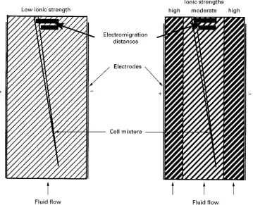
This principle is called ‘free-Sow zone electrophor-esis’ (FFZE) and is still the only electrophoresis mode applicable to cell separation, although it has poorer resolution than other electrophoresis modes such as isoelectric focusing (IEF) and isotachophoresis (ITP), because it is a non-focusing process. In addition, most whole cells do not tolerate aSuid pH below 6.9 and above 7.5 and need media which allow reasonable electrophoretic mobilities, but are simultaneously biocompatible. So for quite a long time, cell elec-trophoresis was rarely applied, particularly as re-solution was often not high enough to purify cell populations with different mean electrophoretic mobilities but overlapping distribution curves and this second drawback negatively inSuenced cell vital-ity. Cells had to be suspended in media lacking NaCl or other physiologically important ions, because too many ions in the chamber medium caused problems of performance such as overheating of the medium and short electromigration distances, as long as only conventional devices with homogeneous chamber media were available.
Figure 2 An Octopus free-flow electrophoresis apparatus with the electrophoresis chamber in vertical position (left) and imple-ments such as a power supply, a pump and the control unit (right). (A generous gift of the Dr. Weber GmbH, D-85551 Kirchheim, Germany. More information about the machine may be found at http://members.aol.com/ffeweber/default.htm.)
preparative cell electrophoresis but can easily be ad-justed to IEF and ITP of sub-cellular particles or molecular substances. Its electrophoresis chamber has a length of 500 mm and a width of 100 mm and can beRxed in a vertical or a horizontal position, as long as specimen sedimentation does not play a role. The thickness is variable, between 0.4 mm and 0.2 mm, so that heat-removal efRciency may be enhanced, if ions are required in cell suspension media and the applica-tion of high electric Relds is necessary. An optical particle detection system allows control of process stability.
The major advantage of the new system is that various media maySow through the chamber adjac-ent to each other and the sample may be introduced at the optimal site (Figure 3). This means for cell elec-trophoresis, that one central cell suspension medium, which may contain up to 50 mmol L\1 NaCl is
pumped between two margin media with elevated quantities of ions Sowing at both edges (Table 3). They cover the electrode membranes, protect the sep-aration medium from detrimental inSuences of the electrodes, prevent diminution of Na# and Cl\ion concentrations within the central chamber area and conduct the electric current to this area of cell trans-port with minimal voltage drop.
Like CCE, FFE is most advantageous if antibodies coupled toSuorescent dyes or magnetic beads are not
available or must not be applied. So the method is quite useful, when cells are separated by CCE because of the reasons explained above and the resulting frac-tions still contain cells which belong to different populations, but have equal size, while their elec-trophoretic mobilities are different. For example, cell fractions are routinely obtained, which contain more than 90% monocytes, if pre-enriched mononuclear leukocytes are elutriated. In such fractions, up to 0.2% antibody-producing cells with equal size as monocytes but different electrophoretic mobilities (EPM) are often co-collected. The antibody-produ-cing cells can be further enriched by FFE. Similarly, T-cell fractions obtained by CCE contaminated by accessory cells, of equal size have been submitted to a following step of FFE puriRcation. T-cells of indi-vidual blood donors were obtained, which did not respond to concanavalin A unless accessory cells were re-added.
A cell feature, which cannot be deRned by antibod-ies is the negative surface charge density. Its biolo-gical role is still very poorly understood. Observa-tions made during recent cell electrophoretic studies appear currently like very scattered mosaic stones which do not allow the whole picture to be revealed. For example, erythrocytes change their EPM in pa-tients suffering various kinds of diseases, monocytes change their EPM when maturing to non-activated macrophages, B-cells change their EPM when devel-oping to antibody-producing cellsin vivobut notin vitro, and mice with different erythrocyte EPM have different sensitivities to malaria infection (see Further Reading). These accumulating data suggest that fur-ther efforts in studying the biological relevance of the negative surface charge density by FFE will be worthwhile.
Since electrophoresis media with 20}50 mmol L\1
NaCl can be used for cell separation, tissue cells can be processed without clotting. Now it is possible to electrophorese cell suspensions obtained from tissues directly or indirectly after a few passages of culture. The separations performed so far have revealed quite interesting new tissue cell sub-populations. Hence, future application of FFE to fractionation of viable tissue cells appears promising.
Conclusions
Figure 3 Scheme of free flow electrophoresis chambers working with homogeneous (left) and segmented (right) carrier fluids.
Table 3 Examples of buffer systems for homogeneous and segmented FFE chamber fluids
Cell-suspension medium Margin buffers Electrode buffer(s)
Homogeneous
27 mmol L\1triethanolamine 342 mmol L\1triethanolamine
4 mmol L\1potassium acetate 40 mmol L\1potassium acetate
27 mmol L\1sucrose pH 7.2 adjusted by acetic acid
1 mmol L\1glucose 216 mmol L\1glycine pH 7.2 adjusted by acetic acid
Segmented
central: 10 mmol L\1triethanolamine anodal: 50 mmol L\1triethanolamine anodal: 200 mmol L\1sodium acetate
2 mmol L\1sodium acetate 250 mmol L\1Na
2SO3
50 mmol L\1NaCl pH 7.2 adjusted by acetic acid
2 mmol L\1glucose
180 mmol L\1sucrose cathodal: 50 mmol L\1triethanolamine cathodal: 100 mmol L\1HCl
pH 7.2 adjusted by acetic acid 250 mmol L\1NaCl 100 mmol L\1NaCl
75 mmol L\1sucrose 200 mmol L\1imidazole
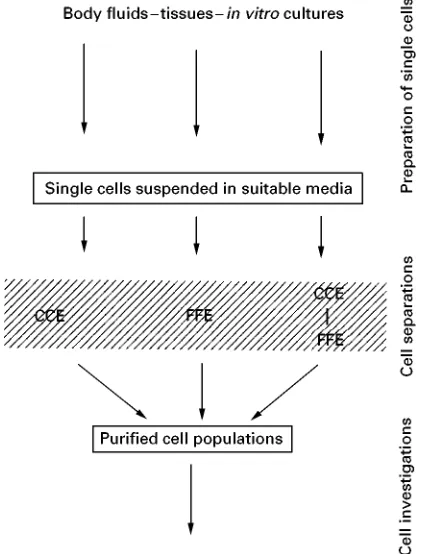
pH 7.2 adjusted by acetic acid suspended in suitable media after preparation from
human or animal bodySuids, from human, animal or plant tissues or fromin vitrocultures are prerequisites of such processes. If a cell suspension with a reason-able number of desired cells is availreason-able, methods such as countercurrent centrifugal elutriation and free-Sow electrophoresis may be applied, either each
[image:6.568.53.512.511.709.2]Figure 4 Flow diagram showing a survey of the processes of cell purification described in this article.
differentiation, even if appropriate antibodies are not available.
Cells puriRed without biological modiRcations may be especially useful if it is of interest to study their originalin vivostatus or to use them for transplanta-tion purposes and if size or surface charge-related phenomena are to be investigated. As knowledge of possible cellular characteristics and components is continuously accumulating, questions on their actual
expression under normal and pathological conditions will frequently arise. For studying such questions, homogeneous cell populations retaining their original
in vivo status may become so important that tech-niques and instruments required for their puriRcation will be further improved.
See also: Cells and Cell Organelles: Field Flow Frac-tionation.
Further Reading
Bauer J (1987) Electrophoretic separation of cells.Journal of Chromatography418: 359}383.
Bauer J (1994) Cell Electrophoresis. Boca Raton: CRC Press.
Bauer J (1998) Advances in cell separation: recent develop-ments in counterSow centrifugal elutriation and con-tinuousSow cell separation. Carrier free electrophor-esis.Electrophoresis19: Special issue.
Bauer J (1999)Journal of Chromatography722: 55}69. Coleman R, Wilton JC, Stone V and Chipman JK (1995)
General Pharmacology26: 1445}1453.
Dixon RA and Gonzales RA (1994) Plant Cell Culture: A Practical Approach. Oxford: Oxford University Press.
Merrill GF (1998) In: Mather JP and Barnes D (eds) Methods in Cell Biology, vol. 57, pp. 229}249. San Diego: Academic Press.
Pretlow TG and Pretlow TP (1982) Cell Separation: Methods and Applications. New York: Academic Press. Shapiro HM (1995) Practical Flow Cytometry, 3rd edn.
New York: Wiley-Liss.
Specto DL, Goldmann RD and Leinwand LA (1998)Cells. A Laboratory Manual, vol 1:Culture and Biochemical Analysis of Cells. New York: Cold Spring Harbor Laboratory Press.
ESSENTIAL GUIDES FOR ISOLATION/
PURIFICATION OF DRUG METABOLITES
I. P. Nnane and A. J. Hutt, Kings’ College London, UK L. A. Damani, Chinese University of Hong Kong, Hong Kong
This article is reproduced fromEncyclopedia of Analyti-cal Science, Copyright^ 1995 Academic Press
Metabolite Isolation and Identi
\
cation
Following the administration of drugs to either ani-mals or man, very few of the drugs are excreted
unchanged. The majority undergo biotransforma-tions by interaction with a complex series of enzymes. This process, known as drug metabolism, is not re-stricted to drugs but occurs with all chemicals that are taken in by living systems, including food additives, pesticides, carcinogens, etc. These chemicals are termed exogenous compounds, as opposed to endo-genous, or naturally present, compounds.