Circadian Clock Proteins and Immunity
Anne M. Curtis 1*, Marina M. Bellet 2, 4, Paolo Sassone-Corsi 2 & Luke A.J. O’Neill 1
1
School of Biochemistry and Immunology, Trinity Biomedical Sciences Institute, Trinity College Dublin, Dublin 2, Ireland
2
Center for Epigenetics and Metabolism, Department of Biological Chemistry, University of California, Irvine, Irvine, CA 92697, USA
4
Present address: Department of Experimental Medicine, University of Perugia, 06132 Perugia, Italy
*
Correspondence to Anne M. Curtis, Email: Acurtis@tcd.ie
Immune parameters change with time of day and disruption of circadian rhythms have been linked to inflammatory pathologies. A circadian clock-controlled immune system might allow an organism to anticipate daily changes in activity and feeding and the associated risk of infection or tissue damage to the host. Responses to bacteria have been shown to vary depending on time of infection, with mice being more at risk of sepsis when challenged ahead of their activity phase. Studies highlight the extent to which the molecular clock, most notably the core clock proteins BMAL1, CLOCK and REV-ERB control
fundamental aspects of the immune response. Examples include the BMAL1:CLOCK heterodimer regulating toll-like receptor (TLR9) expression and repressing expression of the inflammatory monocyte chemokine ligand (CCL2) as well as REV-ERBsuppressing the induction of interleukin-6.
Introduction
Organisms require the ability to tell the time of day in order to anticipate and respond to changes in the external environment imposed by solar time. This need is illustrated in the physiological changes required to accommodate the daily pattern of rest, activity and feeding. Circadian rhythms are these daily changes or oscillations in physiology and are due to numerous genes whose expression peaks and troughs approximately 12 hours apart to undergo a full cycle within 24 hours. The full breadth of control by the molecular clock on the genome was revealed by Zhang et al. who harvested multiple tissues at 2 hour intervals and found that more than a third of the mammalian genome is under clock control (Zhang R., 2013).
The master clock resides in the suprachiasmatic nucleus (SCN) of the brain and is oriented to the external environment via the retinohypothalamic tract receiving light information from the eye. The clock is cell autonomous and in its simplest form, consists as a transcription-translation oscillator loop. At the core of this oscillator lies the heterodimeric partnership of the basic helix loop helix PER-ARNT-SIM (PAS) domain proteins, BMAL1 (also known as ARNTL) and CLOCK, that bind E-box sites and induce the expression of the repressors Period (PER) and Cryptochrome (CRY) which in time translocate back into the nucleus and inhibit their own expression by interfering with the BMAL:CLOCK complex (Fig.1). As PER and CRY proteins are gradually degraded, the repression on BMAL1 and CLOCK is relieved and the cycle begins afresh with another 24h cycle. This machinery lies at the heart of the circadian clock in the SCN. However, an important finding was the existence of the same clockwork machinery in peripheral cells (Schibler and Sassone-Corsi, 2002) including fibroblasts cultured in vitro using what is now known as the serum shock model (Balsalobre et al., 1998). The serum shock model synchronizes cells so that circadian gene expression and outputs of the clock can be analyzed within an in vitro system. Since then, the molecular clock has been characterized in immune cells (Boivin et al., 2003) including macrophages (Keller et al., 2009), T-cells (Bollinger et al., 2011) dendritic T-cells and B-T-cells (Silver et al., 2012a).
BMAL1, CLOCKand REV-ERB can indeed impact on immune cell function, host defense and inflammation. The emerging picture is that time of day is critical to the nature of the immune response and that dysregulation may lead to inflammatory diseases or immunodeficiency. New therapeutic options are presenting themselves that might provide a new prospect for correcting pathologies associated with aberrant immunity and inflammation.
The Anatomy of the Molecular Clock
Molecularly, the core oscillator is comprised of at least three interlocking feedback loops. As stated above the central loop consists of the BMAL1:CLOCK heterodimer driving expression of the repressors, PER1-3 and CRY1-2, which feedback and limit their own expression (Fig. 1). This loop provides oscillations in Per and Cry
transcription and also oscillations in the activity of the BMAL:CLOCK heterodimer. The second loop consists of the nuclear receptors, RAR-related orphan receptor (ROR) () and REV-ERB ( (also known as NR1D1 and NR1D2) which through E-box activation by BMAL1:CLOCK can then translocate back into the nucleus to bind to receptor-related orphan receptor response elements (ROREs) in the promoter of Bmal1 (Fig. 1). RORs activate whereas REV-ERBs repress the expression of BMAL1 (Preitner et al., 2002; Sato et al., 2004). The third loop is comprised of the transcriptional activator albumin D-box binding protein (DBP), regulated through its E-box and the repressor nuclear factor interleukin 3 (NFIL3; also known as E4BP4), regulated via a RORE. These two factors then synergistically regulate the expression of D-box genes including that of Per. This tripartite system forms the foundation of the molecular oscillator and controls the circadian network of clock-controlled genes. In addition to this network of transcriptional control, posttranslational modifications provide a further layer of regulation onto this network with various kinases and phosphatases regulating the precision and function of the clock along with histone modifications and the epigenetic code. These areas have been reviewed extensively elsewhere (Aguilar-Arnal and Sassone-Corsi, 2013; Bellet and Sassone-Corsi, 2010; Feng and Lazar, 2012).
via the hypothalamus pituitary adrenal (HPA) axis and the autonomic nervous system (ANS), and their respective hormones glucocorticoids and the catecholamines (epinephrine and norepinephrine) which act as synchronizing messengers or “zeitgebers” to the peripheral clock (Curtis et al., 2007; Kalsbeek et al., 2012).
Circulating glucocorticoids, catecholamines and other hormones such as prolactin, melatonin and growth hormone, all of which affect the immune system, peak at certain times of the day. The control by the SCN on these autonomic and endocrine outputs keeps peripheral clocks, including that of immune cells, in phase with each other and allows for the coordination of a temporal program of physiology across many tissues at certain times of the day (Guo et al., 2006).
The Molecular Clock Controls Anti-bacterial Host Defense, Sepsis and Inflammation
(hpi) from the colon was greater in mice infected during the night (ZT16) than during the day (ZT4), however Clock mutant mice had lower numbers of bacteria at both time points. The authors found that the growth of Salmonella and its ability to compete with other microorganisms in the gut is dependent on the circadian regulation of antimicrobial peptides. They found that the antimicrobial peptide lipocalin-2, to which Salmonella is resistant, is higher during the day than at night. This suppresses the resident microbiota allowing for the higher outgrowth of
Salmonella during the day versus the night.
In addition, a circadian variation has been found in the ability of mouse macrophages to ingest particles with the increase observed ahead of the transition to activity (Hayashi et al., 2007). This may also enhance bacterial clearance at the beginning of the active phase. The induction of pro-inflammatory cytokines and chemokine ligands is far greater when mice are challenged with LPS at ZT12 versus ZT0. This includes a greater induction of interleukin-6 (IL6), IL-12(p40), chemokine (C-C motif) ligand 2 (CCL2) and CCL5 from macrophages challenged at ZT12 versus ZT0 (Gibbs et al., 2012). Again these differences go some way to explaining the boost in host defense as mice transition into activity, but equally the greater risk of sepsis due to an over active response. Keller et al. harvested peritoneal macrophages from mice every 4 hours for 24 hours and performed microarray analysis. They found that 8% of the macrophage
transcriptome was cycling with a circadian variation, and when they analyzed genes
involved in the toll-like receptor 4 (TLR4) pathway they found circadian control on
genes involved in all aspects of the TLR4 response. This indicates further that the
entire TLR4 pathway in macrophages is under tight circadian control and suggests
again that the circadian clock prepares the immune cell for an integrated response at
times of greatest risk.
Therefore the temporal increase in chemo-attractants, leukocyte trafficking, proinflammatory cytokines and phagocytic ability in the hours approaching the commencement of activity is interpreted as being indicative of clock-controlled enhanced sensitivity and immunosurveillance ahead of activity and feeding when the
risk of infection would be highest. Conversely, perturbations or desynchronization of
by experimental jet-lag (Castanon-Cervantes et al., 2010), a challenge of LPS in mice leads to an uncoordinated and more severe inflammatory response and increased mortality. One molecular feature of circadian desynchronization by jet lag is a sustained reduction in Bmal1 transcript (Castanon-Cervantes et al., 2010). This mirrors a ZT12 macrophage, in which the circadian phase of Bmal1 mRNA is at its lowest (Keller et al., 2009). Macrophages subjected to jetlag and ZT12 macrophages have a heightened inflammatory response once activated by LPS, as both have low expression of BMAL1 prior to activation. This suggests that BMAL1 may be one of the central gatekeepers governing the circadian inflammatory response (Fig. 3). BMAL1 will be discussed in more detail later.
The effects of the central clock in the SCN and its rhythmic control on autonomic and
endocrine regulators are important determinants of immune modulation (Logan and
Sarkar, 2012). The SCN produces diffusible signals as well as axonal projections to
both the paraventricular nucleus (PVN) and arcuate nucleus (ARC) in the
hypothalamus of the brain. For example the PVN governs noradrenergic input to the
spleen and releases norepinephrine to modulate the activity of natural killer (NK)
cells (Logan et al., 2011). Also, the enhanced recruitment of neutrophils to skeletal
tissues at ZT13 is dependent on circadian noradrenergic input to those skeletal tissues,
producing rhythmic expression of the adhesion molecule ICAM-1 (Scheiermann et
al., 2012). The glucocorticoids are under clock control. The SCN and adrenal clock
drive rhythms in glucocorticoid secretion from the adrenal glands. The SCN controls
the release of adrenocorticotropin (ACTH) from the pituitary gland and ACTH in turn
stimulates release of glucocorticoids from the adrenal gland (Dickmeis et al., 2013).
The adrenal clock then controls sensitivity of the adrenal gland to ACTH (Oster et al., 2006). Glucocorticoids exhibit broad anti-inflammatory properties and can control cytokine production and leukocyte trafficking (Coutinho and Chapman, 2011).
Glucocorticoids peak as the animals transition to the active phase, and may possibly
be required at that time due to the heightened risk of infection and injury. A
comprehensive analysis of the role and targets of glucocorticoids in the circadian
control of innate immunity has yet to be performed. Importantly, lack of endocrine
rhythms does not ablate the rhythms in immune cell function as mice lacking the
adrenal glands and splenocytes in culture still maintain rhythms of cytokine release
immune clock also plays a role in determining the circadian rhythm of immune
function.
In simple terms, the circadian system may partition the immune system into two states
over the circadian day (Fig. 2). One being a state of heightened alert as the animal
prepares to transition to activity and the risk of infection or injury is greatest. This
state would require an increase in leukocyte numbers along with increased sensitivity
of immune cells to infectious agents or danger signals. The second state would occur
when the animal rests and the risk of infection and injury is lessened. This state may
provide an opportunity for resolution of inflammation and repair of tissues.
It is no surprise that the immune system would be under circadian control given that a clock system would enable a precise, integrated and cohesive response across a number of immune cells. The molecular clock could act as a “ground controller” for
immune cells, orchestrating a response, dependent on the integration of a range of inputs, including neural, hormonal, and local factors along with the circadian phase of gene expression within the cell. There is a daily rhythm to life, and with that comes daily challenges for which an organism must prepare for and respond to. For example, the clock system prepares an organism to anticipate the onset of activity and feeding, but with that comes the increased threat of infection associated with foraging and feeding. Given the intense energy demands for mounting an immune response a clock might allow the body to utilise energy differently across tissues during periods of rest versus periods of activity. If the immune system needs to be “poised for attack” just as the animal enters the activity and feeding phase, a clock might then allow for the enhancement of immune function with resolution and repair of tissues occurring at alternative times (Fig. 2).
Specific Clock Proteins in Immune Cell Function
More recently, roles for the specific clock proteins in the immune response have been
studied. Mice with clock gene manipulations have unveiled the important contribution
core clock components, paying particular attention to BMAL1, CLOCK, REV-ERB
and RORand the emerging data surrounding their impact on the immune system.
BMAL1
BMAL1 is the central clock component and is the only single clock gene knockout in
which the mouse looses all rhythmic behavioral activity (Bunger et al., 2000). Global
deletion of Bmal1 causes a range of underlying pathologies under steady state
conditions related to accelerated aging (Kondratov et al., 2006) thus making them unsuitable for investigating immune function. Using a mouse model in which the
Bmal1 gene was selectively deleted from myeloid cells, Nguyen et al investigated the role of myeloid BMAL1 in the clearance of the gram-positive bacterium Listeria Monocytogenes (Nguyen et al., 2013). Ly6Chi monocytes provide the first line of defense against this bacterium and the authors had detected a diurnal variation in the absolute numbers of these specific monocytes in blood and in spleen under basal conditions, and enhanced recruitment of these monocytes to an inflamed peritoneum at ZT8 versus ZT0. Improved bacterial clearance was observed at ZT8 versus ZT0 and this was likely to be due to the higher recruitment of Ly6Chi monocytes at sites of infection and a higher production of the pro-inflammatory cytokines, IL-1, IL-6, tumor necrosis factor- (TNF-and interferon- (IFN- and the chemokine ligand
CCL2. The rhythmic oscillation in the numbers of Ly6Chi monocytes in circulation across 24 hours and enhanced recruitment of these cells into inflamed tissue was
entirely dependent on BMAL1 in myeloid cells as the circadian control over Ly6Chi
was abolished in the myeloid BMAL1-depleted mice. The authors found that BMAL1 binds to E-boxes in the promoters of Ccl2 (encoding chemokine ligand 2; Ccl2), Ccl8
and S100a8 (encoding S100 calcium binding protein A8) and recruits with it members of the polycomb repressor complex (PRC2) epigenetically marking histones for
repression. The authors went onto show that BMAL1 reduces Ccl2 transcription and attenuates Ly6Chi monocyte numbers and inflammation at an inflamed site.
Myeloid deletion of Bmal1 also exacerbates metabolic disease driven by a high fat diet. The authors speculated that this could be due to BMAL1 limiting Ly6Chi
monocytosis and recruitment into metabolically stressed tissues such as the fat pads.
resistance and hyperglycemia.
BMAL1 therefore functions as an anti-inflammatory molecule in monocytes in part
because of its repression on Ccl2. This anti-inflammatory effect of BMAL1 was confirmed previously in another study (Gibbs et al., 2012). Peritoneal macrophages
lacking BMAL1 produced higher amounts of IL-6 in response to LPS at ZT0 in
comparison to wild type peritoneal macrophages. In circulating monocytes and in
peritoneal macrophages, Bmal1 mRNA is high at ZT0 and low at ZT12 and this could
impact on the response to LPS or infection at these two times. However, it is not the
absolute amount of Bmal1 mRNA but the binding of BMAL1 to gene promoters that will determine the magnitude of response. ChIP-Seq analysis conducted on mouse
livers harvested every 4 hours across the 24 hours revealed that BMAL1 binding to
gene promoters rises from circadian time (CT, see glossary) CT0 to CT8 and this then
falls from CT12 to CT20 (Koike et al., 2012). When the animals transition to activity,
the reduced binding of BMAL1 to promoters may allow for a more robust
inflammatory response, with better clearance of a pathogen. However excessive
inflammation leading to sepsis can occur when BMAL1 is absent in myeloid cells.
Nguyen et al. demonstrated this as a non-lethal dose of Listeria in wild type mice caused massive lethality in mice lacking myeloid BMAL1 when challenged at both
ZT0 and ZT8.
adaptive immune response, suggesting that time of vaccination could have a significant impact on efficacy. If the phase of TLR9 expression is similar in humans, the optional time to vaccinate humans may also be during the middle of the night or early morning.
CLOCK
The BMAL1 binding partner CLOCK has also been shown to impact on immune
signals. Unlike transcription of Bmal1, Clock mRNA does not alter significantly
throughout the day in macrophages (Keller et al., 2009). However, CLOCK has
intrinsic histone acetyl transferase (HAT) activity capable of acetylating lysine
residues on histones and it is this HAT activity of CLOCK that is essential for
circadian gene expression (Doi et al., 2006). CLOCK can also acetylate the
glucocorticoid receptor and this suppresses binding of the glucocorticoid receptor to
its target genes (Nader et al., 2009). Although not yet investigated, this may have
strong implications regarding the effect of CLOCK on the immune system.
The daily variations observed in lethality from sepsis correlate with activation of the
NF- transcriptional complex and CLOCK converges directly on this key immune
transcription CLOCK is found in protein complexes with the NF-
p65 (RELA) and overexpression of CLOCK leads to enhanced phosphorylation and
acetylation of p65, leading to enhanced transcriptional activity of the NF-
(Spengler et al., 2012). BMAL1 attenuates the effect of CLOCK on NF-
likely by sequestering CLOCK. This is likely to be another mechanism by which
BMAL1 limits inflammation. Clock mutant mouse embryonic fibroblasts (MEFs) (Bellet et al., 2012) and bone marrow derived macrophages (BMDMs) (Bellet et al., 2013) are less responsive to LPS or TNF- in terms of NF- , and the
inflamed intestine of Clock mutant mice showed reduced expression of many pro-inflammatory, metabolic and circadian genes and reduced number of genes involved
in the immune response oscillating in a circadian manner, compared to wild type mice
compartments will provide a clearer picture on the function of CLOCK in the immune system.
The Nuclear Receptors REV-ERB and ROR
here is also compelling evidence for REV-ERBalso known as NR1D1)and
RORaffecting immunity.
A synthetic agonist for REV-ERB limits the release of IL-6 from macrophages
(Gibbs et al., 2012). This agonist was also shown to reduce Cxcl11, Ccl2, Cxcl6, Il19
mRNA but not Il8 mRNA. The mechanism behind this pharmacological effect has not
yet been described, however it may be due to the ability of REV-ERBto recruit the
repressor complex NCoR to selected pro-inflammatory genes including Il6.
REV-ERB also has effects on enhancer-derived RNAs (eRNAs). eRNAs are short RNA
strands produced from an enhancer site and play a role in the transcription of an
adjacent gene. Binding of REV-ERB proteins that bring in histone de-acetylases
repress the production of these eRNAs and thus lead to the inactivation of the adjacent gene (Lam et al., 2013). Target genes here include the metalloproteinase
Mmp9 and the chemokine receptor Cx3cr1, providing another mechanism for the anti-inflammatory effects of REV-ERB activation. In a macrophage, BMAL1 induces
the transcription of Nr1d1 (Fig.1), thus suggesting that some of the anti-inflammatory
effects of BMAL1 may be through its direct regulation on REV-ERB. In support of this, a recent study showed that REV-ERBalso represses the Ccl2 gene by binding
to an RORE sequence in the promoter of Ccl2 (Sato S, 2014). As stated above BMAL1 represses Ccl2 by binding to an E-box in the Ccl2 promoter (Nguyen et al., 2013). This indicates that BMAL1 and REV-ERBcooperate across the circadian cycle to repress Ccl2 transcription under inflammatory conditions. The effect of
REV-ERBis possibly cell type specific. REV-ERBhas been shown to drive TH17 cell
differentiation by repression of Nfil3 transcription which in turn will allow RORt to
induce IL17 production (Yu et al., 2013). In adaptive immunity REV-ERBmay
actually promote inflammation.
BMAL1 also controls the expression of ROR, and mice deficient in ROR (the
neurological mutant mouse Staggerer) are susceptible to LPS lethality (Stapleton et
of kappa light polypeptide gene enhancer in B-cells inhibitor, alpha) and limit NF- translocation to the nucleus. Therefore another mechanism whereby BMAL1 might
be anti-inflammatory could be via induction of RORwhich in turn will induce
Iand limit NF- (Delerive et al., 2001). Therefore both nuclear receptors REV-ERB and ROR that are components of the molecular clock, also have strong
anti-inflammatory capability.
PER and CRY
Two other clock proteins PER and CRY also modulate inflammation. The PER2
mutant leads to the loss of a daily rhythm in IFN- (Arjona and Sarkar, 2006). The
oscillation in mortality from LPS is abolished in PER2-deficient mice, with Per2
-/-mice being protected against LPS induced lethality at all time points analysed (Liu et
al., 2006). The serum concentrations of IFN- and IL-1 were lowered dramatically
in the Per2-/- mice with LPS injection, however amounts of TNF-, IL-6 and IL-10
were normal. The impaired IFN- was attributed to defective natural killer (NK) cell
function. It was also observed that Per2 mutants have reduced amounts of Tlr9
mRNA in macrophages and activation of TLR9 by CpG produced less TNF- and
IL-12 from these macrophages (Silver et al., 20IL-12b). Therefore PER2 may function to
upregulate pro-inflammatory cytokines at certain stages of the circadian cycle. Per2
mRNA rhythms are anti-phase to Bmal1 and Per2 mRNA peaks in macrophages just after the animals enter the active phase (Keller et al., 2009). PER2 is the negative
component of the feedback loop that inhibits BMAL1:CLOCK activity as the animal
enters the active phase (Fig.1). Therefore, PER2 may promote inflammation in part by
repressing the activity of BMAL1. PER2 abundance also inhibits the function of
REV-ERB(Preitner et al., 2002) and repressing REV-ERB could lead also to
greater inflammation from the mechanisms discussed above (Fig. 3). The other
components of the negative feedback loop are the cryptochromes (Fig. 1). Absence of
the cryptochrome CRY1 and CRY2 in fibroblasts and BMDMs leads to increase Il6,
Tnf, Cxcl1 and Inos mRNA at baseline. Upon LPS stimulation, IL-6 and TNF-
expression are significantly higher in the BMDMs lacking Cry1 and Cry2. In fibroblasts this was due to CRY1 binding adenylate cyclase and limiting cAMP
production. In the absence of CRY1, elevated cAMP increases PKA activity, leading
2012). Cry1-/-Cry2-/- mice have heightened inflammatory joint disease and enhanced
production of TNF-in a model of collagen induced arthritis (Hashiramoto et al.,
2010). Treatment of these mice with anti-TNF-antibody reduced the severity of the
disease in the Cry1-/-Cry2-/- mice and ectopic expression of Cry1 in mouse embryonic
fibroblasts from Cry1-/-Cry2-/- mice significantly reduced activation of the TNF- promoter luciferase construct. This may again be due to the effects of Cry1 to limit
PKA induced phosphorylation of p65 and NF- in fibroblasts
(Narasimamurthy et al., 2012).
Inflammation Disrupts the Molecular Clock
As discussed above, the clock can impact on the inflammatory process in a number of ways. In a reciprocal fashion inflammation induced by agents such as LPS, TNF- and IFN- (Cavadini et al., 2007a; Kwak et al., 2008; Lundkvist et al., 2002;
Marpegan et al., 2005; Okada et al., 2008) or acute bacterial infection (Bellet et al., 2013) can affect the circadian clock. The oscillations in the clock genes can be disrupted with inflammation and infection, with effects on the expression of core clock genes and clock-controlled genes, including clock controlled metabolic genes being reported (Bellet et al., 2013). Rodent studies indicate that LPS transiently suppresses clock gene expression and oscillation in the SCN (Okada et al., 2008) and phase-shift locomotor activity (Marpegan et al., 2005; Marpegan et al., 2009). One possible explanation for this is that TNF- and IL-1 can inhibit the ability of
BMAL1:CLOCK to induce activation of E-box dependent genes in the SCN and liver (Cavadini et al., 2007b). Also the NF-B subunit RelB interacts directly with BMAL1
to repress the circadian gene Dbp (Bellet et al., 2012). The mRNA expression of
Bmal1 was repressed in the spleens across the full circadian day in mice subjected to
collagen induced arthritis versus controls (Hashiramoto et al., 2010). The constitutively low expression of BMAL1 in this model of arthritis could promote further inflammation from immune cells by the mechanisms stated above (Fig. 3). The effect of cytokines on the circadian system is important given the increased use of biotherapeutics such as anti-TNF- for chronic inflammatory conditions such as arthritis. Anti-TNF-, by relieving the repression on the BMAL1:CLOCK
repress inflammatory genes (Fig. 3). This could be another mechanism of action for this biological therapy in chronic diseases such as rheumatoid arthritis (RA).
Molecular Clocks in Human Health and Disease
The recent work identifying the roles for clock proteins such as BMAL1 and REV-ERBin immunoregulation may provide new insights into the pathogenesis of
infectious and inflammatory diseases. It is well known that inflammatory diseases such as asthma (Barnes et al., 1980; Kraft et al., 1996) RA (Haus et al., 2012) and
atherosclerosis (Paschos and FitzGerald, 2010) have strong circadian components with exacerbations at night and into the early morning hours. There is also an intimate link between circadian rhythms and cancer, a disease associated with aberrant inflammation (Sahar and Sassone-Corsi, 2009). In healthy humans the pro-inflammatory cytokines, TNF- and IL-6, peak in serum at 3am and 6am respectively (Cutolo and Straub, 2008). In RA patients the peak of both cytokines shift forward to the early morning with peak amounts of IL-6 ten fold higher in the serum of patients with RA versus controls (Cutolo and Straub, 2008). In addition, oscillations in clock proteins is lost in synovial fibroblasts cultured from patients with RA (Kouri et al., 2013). These data suggest severe circadian disruption in RA. The irregularity in the amount and timing of these cytokines might be responsible for the symptoms of stiffness and pain for RA patients in the morning hours. Therefore, the underlying basis of many inflammatory conditions could depend on a perturbed clockwork system both systemically and locally. Chronic disruption of the external cues of light to the molecular clock, as imposed by shift work and airline travel, augments the inflammatory response (Castanon-Cervantes et al., 2010) and increases susceptibility to many metabolic diseases with inflammatory features, such as atherosclerosis, obesity and diabetes (Antunes et al., 2010). Our clock and output rhythms deteriorate
clock. It has also been concluded that ‘shift work that involves circadian disruption is probably carcinogenic to humans’ (Straif et al., 2007) and can lead to higher
incidence of cardiovascular disease and obesity (Karlsson et al., 2001; Stevens, 2009). Such studies on shift work might however be confounded by other variables such as gender, age and weight. Mice that undergo a model of jet lag however (akin to one
transatlantic flight every week for four weeks) followed by recovery for one week produce three times as much IL-6 upon challenge with LPS (Castanon-Cervantes et al., 2010). Conversely, recovery from sepsis induced in rats by caecal ligation and
puncture is impaired when the regular 12:12 light:dark cycle is replaced by constant conditions (either constant light or constant darkness) after surgery (Carlson and Chiu, 2008). These observations highlight the possibility that recovery of patients from sepsis could be improved by simple measures such as reducing nocturnal light and noise and improving quality of sleep in intensive care units (ICU) units (Hrushesky and Wood, 1997) (Herdegen, 2002).
Conclusions and Future Directions
The picture that is fast emerging is that time of day is critical in terms of the type of immune response generated by an organism. The type of immune response can be broadly partitioned into two states, one of heightened alert and the other of rest and repair. The last decade provided a number of descriptive insights regarding the rhythmic changes that occur in immune parameters, however this decade has provided some of the molecular mechanisms underpinning these descriptions. What is intriguing is that the key components of the molecular clock, whose expression and activity will change across the circadian day, have direct relationships with important components of the immune system. We have detailed the data that we know so far connecting the core clock components, most notably, BMAL1, CLOCK and REV-ERBand ROR, with important regulators of immune function and inflammation.
BMAL1 appears to be a central mediator connecting both the clock and immune system together, its role being to limit inflammation.
However we are only at the beginning of our journey to understand the tight coupling
lead to pathology and disease. There are still a number of questions to answer, amongst them:
1) What are the main signals that entrain or perturb the immune cell clock under different situations such as infection or inflammation? 2) What role do the clock proteins play in each type of immune cell? 3) What role does the clock play in the homeostatic functioning of the immune system? 4) Is the response of the human immune clock partitioned in the same way as the mouse immune clock? 5) Are inflammatory diseases in essence diseases of clock dysfunction? One question to investigate in this last regard is whether the anti-inflammatory effects of BMAL1 and REV-ERB are somehow disrupted in diseases such as RA giving rise to an inflammatory phenotype.
Given that the molecular clock may fundamentally regulate many if not all aspects of our immune system, an understanding of how the clock and immune function
intersect may reveal much needed therapeutic opportunities for some of our most common chronic diseases. These insights might also provide rationale for chronotherapy, the dosing of medications with reference to 24-hour rhythms of disease activity, for existing and upcoming treatments of immune diseases.
References
Aguilar-Arnal, L., and Sassone-Corsi, P. (2013). The circadian epigenome: how metabolism talks to chromatin remodeling. Current opinion in cell biology 25, 170-176.
Antunes, L.C., Levandovski, R., Dantas, G., Caumo, W., and Hidalgo, M.P. (2010). Obesity and shift work: chronobiological aspects. Nutrition research reviews 23, 155-168.
Arjona, A., and Sarkar, D.K. (2006). The circadian gene mPer2 regulates the daily rhythm of IFN-gamma. Journal of interferon & cytokine research : the official journal of the International Society for Interferon and Cytokine Research 26, 645-649.
Balsalobre, A., Damiola, F., and Schibler, U. (1998). A serum shock induces circadian gene expression in mammalian tissue culture cells. Cell 93, 929-937.
Bellet, M.M., Deriu, E., Liu, J.Z., Grimaldi, B., Blaschitz, C., Zeller, M., Edwards, R.A., Sahar, S., Dandekar, S., Baldi, P., et al. (2013). Circadian clock regulates the host response to Salmonella. Proc Natl Acad Sci U S A 110, 9897-9902.
Bellet, M.M., and Sassone-Corsi, P. (2010). Mammalian circadian clock and metabolism - the epigenetic link. Journal of cell science 123, 3837-3848.
Bellet, M.M., Zocchi, L., and Sassone-Corsi, P. (2012). The RelB subunit of NFkappaB acts as a negative regulator of circadian gene expression. Cell cycle 11, 3304-3311.
Boivin, D.B., James, F.O., Wu, A., Cho-Park, P.F., Xiong, H., and Sun, Z.S. (2003). Circadian clock genes oscillate in human peripheral blood mononuclear cells. Blood
102, 4143-4145.
Bollinger, T., Leutz, A., Leliavski, A., Skrum, L., Kovac, J., Bonacina, L., Benedict, C., Lange, T., Westermann, J., Oster, H., and Solbach, W. (2011). Circadian clocks in mouse and human CD4+ T cells. PloS one 6, e29801.
Bunger, M.K., Wilsbacher, L.D., Moran, S.M., Clendenin, C., Radcliffe, L.A., Hogenesch, J.B., Simon, M.C., Takahashi, J.S., and Bradfield, C.A. (2000). Mop3 is an essential component of the master circadian pacemaker in mammals. Cell 103, 1009-1017.
Carlson, D.E., and Chiu, W.C. (2008). The absence of circadian cues during recovery from sepsis modifies pituitary-adrenocortical function and impairs survival. Shock 29, 127-132.
Castanon-Cervantes, O., Wu, M., Ehlen, J.C., Paul, K., Gamble, K.L., Johnson, R.L., Besing, R.C., Menaker, M., Gewirtz, A.T., and Davidson, A.J. (2010). Dysregulation of inflammatory responses by chronic circadian disruption. J Immunol 185, 5796-5805.
Cavadini, G., Petrzilka, S., Kohler, P., Jud, C., Tobler, I., Birchler, T., and Fontana, A. (2007a). TNF-alpha suppresses the expression of clock genes by interfering with E-box-mediated transcription. Proc Natl Acad Sci U S A 104, 12843-12848.
Cavadini, G., Petrzilka, S., Kohler, P., Jud, C., Tobler, I., Birchler, T., and Fontana, A. (2007b). TNF-alpha suppresses the expression of clock genes by interfering with E-box-mediated transcription. Proc Natl Acad Sci U S A 104, 12843-12848.
Coutinho, A.E., and Chapman, K.E. (2011). The anti-inflammatory and immunosuppressive effects of glucocorticoids, recent developments and mechanistic insights. Molecular and cellular endocrinology 335, 2-13.
Curtis, A.M., Cheng, Y., Kapoor, S., Reilly, D., Price, T.S., and Fitzgerald, G.A. (2007). Circadian variation of blood pressure and the vascular response to asynchronous stress. Proc Natl Acad Sci U S A 104, 3450-3455.
Cutolo, M., and Straub, R.H. (2008). Circadian rhythms in arthritis: hormonal effects on the immune/inflammatory reaction. Autoimmunity reviews 7, 223-228.
Delerive, P., Monte, D., Dubois, G., Trottein, F., Fruchart-Najib, J., Mariani, J., Fruchart, J.C., and Staels, B. (2001). The orphan nuclear receptor ROR alpha is a negative regulator of the inflammatory response. EMBO reports 2, 42-48.
Dickmeis, T., Weger, B.D., and Weger, M. (2013). The circadian clock and glucocorticoids--interactions across many time scales. Molecular and cellular endocrinology 380, 2-15.
Doi, M., Hirayama, J., and Sassone-Corsi, P. (2006). Circadian regulator CLOCK is a histone acetyltransferase. Cell 125, 497-508.
Froy, O., and Chapnik, N. (2007). Circadian oscillation of innate immunity components in mouse small intestine. Molecular immunology 44, 1954-1960.
Gibbs, J.E., Blaikley, J., Beesley, S., Matthews, L., Simpson, K.D., Boyce, S.H., Farrow, S.N., Else, K.J., Singh, D., Ray, D.W., and Loudon, A.S. (2012). The nuclear receptor REV-ERBalpha mediates circadian regulation of innate immunity through selective regulation of inflammatory cytokines. Proc Natl Acad Sci U S A 109, 582-587.
Guo, H., Brewer, J.M., Lehman, M.N., and Bittman, E.L. (2006). Suprachiasmatic regulation of circadian rhythms of gene expression in hamster peripheral organs: effects of transplanting the pacemaker. The Journal of neuroscience : the official journal of the Society for Neuroscience 26, 6406-6412.
Halberg, F., Johnson, E.A., Brown, B.W., and Bittner, J.J. (1960). Susceptibility rhythm to E. coli endotoxin and bioassay. Proc Soc Exp Biol Med 103, 142-144. Hashiramoto, A., Yamane, T., Tsumiyama, K., Yoshida, K., Komai, K., Yamada, H., Yamazaki, F., Doi, M., Okamura, H., and Shiozawa, S. (2010). Mammalian clock gene Cryptochrome regulates arthritis via proinflammatory cytokine TNF-alpha. J Immunol 184, 1560-1565.
Haus, E., Sackett-Lundeen, L., and Smolensky, M.H. (2012). Rheumatoid arthritis: circadian rhythms in disease activity, signs and symptoms, and rationale for chronotherapy with corticosteroids and other medications. Bulletin of the NYU hospital for joint diseases 70 Suppl 1, 3-10.
Hayashi, M., Shimba, S., and Tezuka, M. (2007). Characterization of the molecular clock in mouse peritoneal macrophages. Biological & pharmaceutical bulletin 30, 621-626.
Herdegen, J.J. (2002). Intensive care unit sleep disruption: can the cycle be restored? Critical care medicine 30, 709-710.
Hrushesky, W.J., and Wood, P.A. (1997). Circadian time structure of septic shock: timing is everything. The Journal of infectious diseases 175, 1283-1284.
Kalsbeek, A., van der Spek, R., Lei, J., Endert, E., Buijs, R.M., and Fliers, E. (2012). Circadian rhythms in the hypothalamo-pituitary-adrenal (HPA) axis. Molecular and cellular endocrinology 349, 20-29.
Karlsson, B., Knutsson, A., and Lindahl, B. (2001). Is there an association between shift work and having a metabolic syndrome? Results from a population based study of 27 485 people. Occupational and Environmental Medicine 58, 747-752.
Keller, M., Mazuch, J., Abraham, U., Eom, G.D., Herzog, E.D., Volk, H.D., Kramer, A., and Maier, B. (2009). A circadian clock in macrophages controls inflammatory immune responses. Proc Natl Acad Sci U S A 106, 21407-21412.
Kohsaka, A., Laposky, A.D., Ramsey, K.M., Estrada, C., Joshu, C., Kobayashi, Y., Turek, F.W., and Bass, J. (2007). High-fat diet disrupts behavioral and molecular circadian rhythms in mice. Cell metabolism 6, 414-421.
Koike, N., Yoo, S.H., Huang, H.C., Kumar, V., Lee, C., Kim, T.K., and Takahashi, J.S. (2012). Transcriptional architecture and chromatin landscape of the core circadian clock in mammals. Science 338, 349-354.
Kondratov, R.V., Kondratova, A.A., Gorbacheva, V.Y., Vykhovanets, O.V., and Antoch, M.P. (2006). Early aging and age-related pathologies in mice deficient in BMAL1, the core componentof the circadian clock. Genes & development 20, 1868-1873.
Kraft, M., Djukanovic, R., Wilson, S., Holgate, S.T., and Martin, R.J. (1996). Alveolar tissue inflammation in asthma. American journal of respiratory and critical care medicine 154, 1505-1510.
Kwak, Y., Lundkvist, G.B., Brask, J., Davidson, A., Menaker, M., Kristensson, K., and Block, G.D. (2008). Interferon-gamma alters electrical activity and clock gene expression in suprachiasmatic nucleus neurons. Journal of biological rhythms 23, 150-159.
Lam, M.T., Cho, H., Lesch, H.P., Gosselin, D., Heinz, S., Tanaka-Oishi, Y., Benner, C., Kaikkonen, M.U., Kim, A.S., Kosaka, M., et al. (2013). Rev-Erbs repress macrophage gene expression by inhibiting enhancer-directed transcription. Nature
498, 511-515.
Liu, J., Malkani, G., Shi, X., Meyer, M., Cunningham-Runddles, S., Ma, X., and Sun, Z.S. (2006). The circadian clock Period 2 gene regulates gamma interferon production of NK cells in host response to lipopolysaccharide-induced endotoxic shock. Infect Immun 74, 4750-4756.
Logan, R.W., Arjona, A., and Sarkar, D.K. (2011). Role of sympathetic nervous system in the entrainment of circadian natural-killer cell function. Brain, behavior, and immunity 25, 101-109.
Logan, R.W., and Sarkar, D.K. (2012). Circadian nature of immune function. Molecular and cellular endocrinology 349, 82-90.
Lundkvist, G.B., Hill, R.H., and Kristensson, K. (2002). Disruption of circadian rhythms in synaptic activity of the suprachiasmatic nuclei by African trypanosomes and cytokines. Neurobiology of disease 11, 20-27.
Marpegan, L., Bekinschtein, T.A., Costas, M.A., and Golombek, D.A. (2005). Circadian responses to endotoxin treatment in mice. Journal of neuroimmunology
160, 102-109.
Marpegan, L., Leone, M.J., Katz, M.E., Sobrero, P.M., Bekinstein, T.A., and Golombek, D.A. (2009). Diurnal variation in endotoxin-induced mortality in mice: correlation with proinflammatory factors. Chronobiology international 26, 1430-1442. Nader, N., Chrousos, G.P., and Kino, T. (2009). Circadian rhythm transcription factor CLOCK regulates the transcriptional activity of the glucocorticoid receptor by acetylating its hinge region lysine cluster: potential physiological implications. FASEB journal : official publication of the Federation of American Societies for Experimental Biology 23, 1572-1583.
Narasimamurthy, R., Hatori, M., Nayak, S.K., Liu, F., Panda, S., and Verma, I.M. (2012). Circadian clock protein cryptochrome regulates the expression of proinflammatory cytokines. Proc Natl Acad Sci U S A 109, 12662-12667.
Nguyen, K.D., Fentress, S.J., Qiu, Y., Yun, K., Cox, J.S., and Chawla, A. (2013). Circadian gene Bmal1 regulates diurnal oscillations of Ly6C(hi) inflammatory monocytes. Science 341, 1483-1488.
Okada, K., Yano, M., Doki, Y., Azama, T., Iwanaga, H., Miki, H., Nakayama, M., Miyata, H., Takiguchi, S., Fujiwara, Y., et al. (2008). Injection of LPS causes transient suppression of biological clock genes in rats. J Surg Res 145, 5-12.
Oster, H., Damerow, S., Kiessling, S., Jakubcakova, V., Abraham, D., Tian, J., Hoffmann, M.W., and Eichele, G. (2006). The circadian rhythm of glucocorticoids is regulated by a gating mechanism residing in the adrenal cortical clock. Cell metabolism 4, 163-173.
Preitner, N., Damiola, F., Lopez-Molina, L., Zakany, J., Duboule, D., Albrecht, U., and Schibler, U. (2002). The orphan nuclear receptor REV-ERBalpha controls circadian transcription within the positive limb of the mammalian circadian oscillator. Cell 110, 251-260.
Raffatellu, M., George, M.D., Akiyama, Y., Hornsby, M.J., Nuccio, S.P., Paixao, T.A., Butler, B.P., Chu, H., Santos, R.L., Berger, T., et al. (2009). Lipocalin-2 resistance confers an advantage to Salmonella enterica serotype Typhimurium for growth and survival in the inflamed intestine. Cell host & microbe 5, 476-486.
Sahar, S., and Sassone-Corsi, P. (2009). Metabolism and cancer: the circadian clock connection. Nature reviews. Cancer 9, 886-896.
Sato S, S.T., Ogasawara J, Takahashi M, Izawa T, Imaizumi K, Taniguchi N, Ohno H, Kizaki T. (2014). A Circadian Clock Gene, Rev-erbα, Modulates the Inflammatory Function of Macrophages through the Negative Regulation of Ccl2 Expression. J Immunol. 192, 407-417.
Sato, T.K., Panda, S., Miraglia, L.J., Reyes, T.M., Rudic, R.D., McNamara, P., Naik, K.A., FitzGerald, G.A., Kay, S.A., and Hogenesch, J.B. (2004). A functional genomics strategy reveals Rora as a component of the mammalian circadian clock. Neuron 43, 527-537.
Scheiermann, C., Kunisaki, Y., Lucas, D., Chow, A., Jang, J.E., Zhang, D., Hashimoto, D., Merad, M., and Frenette, P.S. (2012). Adrenergic nerves govern circadian leukocyte recruitment to tissues. Immunity 37, 290-301.
Schibler, U., and Sassone-Corsi, P. (2002). A web of circadian pacemakers. Cell 111, 919-922.
Shackelford, P.G., and Feigin, R.D. (1973). Periodicity of susceptibility to pneumococcal infection: influence of light and adrenocortical secretions. Science 182, 285-287.
Silver, A.C., Arjona, A., Hughes, M.E., Nitabach, M.N., and Fikrig, E. (2012a). Circadian expression of clock genes in mouse macrophages, dendritic cells, and B cells. Brain, behavior, and immunity 26, 407-413.
Silver, A.C., Arjona, A., Walker, W.E., and Fikrig, E. (2012b). The circadian clock controls toll-like receptor 9-mediated innate and adaptive immunity. Immunity 36, 251-261.
Spengler, M.L., Kuropatwinski, K.K., Comas, M., Gasparian, A.V., Fedtsova, N., Gleiberman, A.S., Gitlin, II, Artemicheva, N.M., Deluca, K.A., Gudkov, A.V., and Antoch, M.P. (2012). Core circadian protein CLOCK is a positive regulator of NF-kappaB-mediated transcription. Proc Natl Acad Sci U S A 109, E2457-2465.
Stapleton, C.M., Jaradat, M., Dixon, D., Kang, H.S., Kim, S.C., Liao, G., Carey, M.A., Cristiano, J., Moorman, M.P., and Jetten, A.M. (2005). Enhanced susceptibility of staggerer (RORalphasg/sg) mice to lipopolysaccharide-induced lung inflammation. American journal of physiology. Lung cellular and molecular physiology 289, L144-152.
Stevens, R.G. (2009). Light-at-night, circadian disruption and breast cancer: assessment of existing evidence. International Journal of Epidemiology 38, 963-970. Straif, K., Baan, R., Grosse, Y., Secretan, B., El Ghissassi, F., Bouvard, V., Altieri, A., Benbrahim-Tallaa, L., and Cogliano, V. (2007). Carcinogenicity of shift-work, painting, and fire-fighting. The lancet oncology 8, 1065-1066.
Zhang R., L.N., Ballance H.I., Hughes M.E., Hogenesch J.B., (2013). A Transcriptional Map of Mouse Circadian Time and Space. Keystone Symposia on Molecular Clockworks and the Regulation of Cardio-Metabolic Function (C9) at Snowbird, UT 2013.
Figure Legends
Figure 1. The molecular clock consists of at least three interlocking feedback loops. Loop 1 consists of the core clock proteins BMAL1 and CLOCK binding to E-box elements within the genes encoding the repressor proteins PER and CRY and REV-ERB RORand DBP. After a period of time, PER and CRY can translocate back
into the nucleus and repress their own expression by interfering with the BMAL1:CLOCK complex on the gene promoters. The expression of these proteins is regulated by post-translational modifications such as phosphorylation of PER by CKI, which marks it for proteosomal degradation. Loop 2 consists of the alternate regulation by REV-ERB and RORon RORE promoter elements, which includes
Bmal1 and Nfil3. Loop 3 consists of the alternate regulation by NFIL3 and DBP on D
box promoter elements. The clock products from each of these loops can shuttle back to the nucleus and either repress or reactivate these loops. The transcription factors within each of these loops can also regulate clock-controlled genes (CCGs). These are genes that have a circadian profile of expression but do not feedback to affect the core molecular clock. If some of these CCGs are themselves transcription factors, they may confer a circadian profile of expression on their target genes. Therefore, the core clock components and CCGs have the capacity to regulate transcription of a wide variety of cellular programs that is independent of their function within the molecular clock.
at ZT8 and ZT10. This period also correlates with enhanced lethality from high doses of LPS observed at ZT10, greater induction of pro-inflammatory cytokines at ZT12, and enhanced numbers of leukocytes at ZT13. The opposing state of regeneration and repair is mainly speculative and requires further investigation. It does coincide however with the period of reduced induction of pro-inflammatory cytokines along with reduced clearance and lethality from bacteria. Whether similar processes occur in humans at the same states of transition have yet to be clearly determined.
Figure 3. BMAL1 is the central mediator on the circadian control of the immune system and promotes an anti-inflammatory state. BMAL1 directly represses Ccl2
gene expression leading to lower numbers of the Ly6Chi inflammatory monocytes in circulation and lower recruitment of these inflammatory monocytes into inflamed tissues. BMAL1 sequesters CLOCK and prevents it from acetylating and activating p65, this leads to lower amounts of transcriptionally active NF-B leading to less
induction of specific genes such as cytokines and regulators of survival and proliferation. BMAL1 drives the expression of Nr1d1 (Rev-Erbthat can inhibit Il6
and Ccl2 expression. BMAL1 drives the expression of Ror that can increase the
expression of IB, a major negative regulator of NF-B. This would have the effect of retarding the NF-B complex as a non-active form in the cytoplasm preventing it
from translocating into the nucleus and activating a range of genes including cytokines.
Glossary of Terms used in Circadian Biology
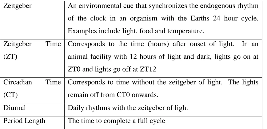
Zeitgeber An environmental cue that synchronizes the endogenous rhythm of the clock in an organism with the Earths 24 hour cycle. Examples include light, food and temperature.
Zeitgeber Time (ZT)
Corresponds to the time (hours) after onset of light. In an animal facility with 12 hours of light and dark, lights go on at ZT0 and lights go off at ZT12
Circadian Time (CT)
Corresponds to time without the zeitgeber of light. The lights remain off from CT0 onwards.
[image:22.595.85.519.554.765.2]E-box CLOCK BMAL1
CRY
PER
CRY
PER
CRY
REV-ERBa
RORE REV-ERBa
RORa
RORa
DBP
D-box
PER
DBP CCGs
Nucleus
NFIL3
CKIε
Proteasomal degradation
PER
CCG BMAL1
CCG
CCG PER
Cytoplasm Loop 1
Loop 2
Loop 3
NFIL3
ZT0 ZT12 ZT6 ZT18 Dawn Dusk
Ac ve
Inac ve ZT9 ZT15 ZT21 ZT3 Peak Leukocytes in Blood (ZT5) Increased Leukocytes in Tissue (ZT13) Increased lethality from LPS andD. Pneumoniae(ZT10) ,
Reduced clearance of
L. Monocytogenes(ZT0)
Enhanced clearance of
S. Thyphimurium(ZT16) Reduced
clearance of
S. Thyphimurium(ZT4)
Greater induc on of pro-inflammatory cytokines with LPS (ZT12) Smaller induc on
of pro-inflammatory cytokines with LPS (ZT0)
Increased TLR9 expression Increased sepsis from CLP (ZT19)
Reduced TLR9 expression Reduced sepsis from CLP (ZT7)
Immune System Poised for A ack
Immune System Undergoing Repair and Regenera on
Peak Ly6Chi monocytes in blood and enhanced clearance of
L. Monocytogenes(ZT8)
Reduced lethality from fromD. Pneumoniae
Nr1d1
(Rev-Erb
a)
Ccl2
IL6 Ccl2
BMAL1
CLOCK CLOCK
p65
BMAL1
REV-ERBa NCoR
Rora
RORa
IkB