THE
JOURNAL • RESEARCH • www.fasebj.org
Macrophage-derived insulin-like growth factor-1
is a key neurotrophic and nerve-sensitizing factor
in pain associated with endometriosis
Rachel Forster,* Alexandra Sarginson,* Atanaska Velichkova,* Chloe Hogg,* Ashley Dorning,* Andrew W. Horne,* Philippa T. K. Saunders,†and Erin Greaves‡,1
*Medical Research Council (MRC) Centre for Reproductive Health and†MRC Centre for Inflammation Research, The Queen’s Medical Research Institute, The University of Edinburgh, Edinburgh, United Kingdom; and‡Division of Biomedical Sciences, Warwick Medical School, University of Warwick, Coventry, United Kingdom
ABSTRACT:Endometriosis is a common incurable inflammatory disorder that is associated with debilitating pelvic pain in women. Macrophages are central to the pathophysiology of endometriosis: they dictate the growth and vascularization of endometriosis lesions and more recently have been shown to promote lesion innervation. The aim of this study was to determine the mechanistic role of macrophages in producing pain associated with endome-triosis. Herein, we show that macrophage depletion in a mouse model of endometriosis can reverse abnormal changes in pain behavior. We identified that disease-modified macrophages exhibit increased expression of IGF-1 in anin vitromodel of endometriosis-associated macrophages and confirmed expression by lesion-resident mac-rophages in mice and women. Concentrations of IGF-1 were elevated in peritoneal fluid from women with endo-metriosis and positively correlate with their pain scores. Mechanistically, we demonstrate that macrophage-derived IGF-1 promotes sprouting neurogenesis and nerve sensitizationin vitro. Finally, we show that the Igf-1 receptor inhibitor linsitinib reverses the pain behavior observed in mice with endometriosis. Our data support a role for macrophage-derived IGF-1 as a key neurotrophic and sensitizing factor in endometriosis, and we propose that therapies that modify macrophage phenotype may be attractive therapeutic options for the treatment of women with endometriosis-associated pain.—Forster, R., Sarginson, A., Velichkova, A., Hogg, C., Dorning, A., Horne, A. W., Saunders, P. T. K., Greaves, E. Macrophage-derived insulin-like growth factor-1 is a key neurotrophic and nerve-sensitizing factor in pain associated with endometriosis. FASEB J. 33, 000–000 (2019). www.fasebj.org
KEY WORDS:hyperalgesia • leukocytes • neurotrophin • nerve
Endometriosis is a chronic incurable estrogen-dependent inflammatory disorder affecting an estimated 176 million women worldwide (1). It is associated with debilitating
chronic pelvic pain and infertility and can significantly impair health-related quality of life (2–4). Endometriosis is defined by the attachment and growth of endometrial-like tissue outside the uterine cavity (endometriosis lesions), and current treatment options are limited to surgical abla-tion or excision of lesions or medical management to sup-press ovarian hormone production. However, symptoms recur within 5 yr in 40–50% of women following surgery (5), and medical management often has unwanted side-effects and is contraceptive (6). There is an unmet clinical need for new medical treatments for women with endometriosis.
Endometriosis lesions recruit sensory nerve fibers (7, 8) that innervate the ectopic endometrial tissue and can be activated by the local neuroinflammatory milieu (9). Re-peated interactions between peripheral sensory afferents (nociceptors) and cytokines and neurotrophins have been shown in other chronic pain conditions to cause nerve sen-sitization: an enhanced responsiveness of afferents (10), leading to a resultant increase in excitability of the ner-vous system and triggering pain hypersensitivity or allo-dynia (11, 12). In a mouse model of endometriosis (13) that
ABBREVIATIONS:Bdnf, brain-derived neurotrophic factor; Cy, cyanine; DRG, dorsal root ganglion; EAM, endometriosis-associated macrophage; EPHect, Endometriosis Phenome and Biobanking Harmonisation Project; hESC, human embryonic stem cell; IGF-1R, IGF-1 receptor; LpM, large peritoneal macrophage; Ly6C, lymphocyte antigen 6 complex, locus C; M0, unactivated macrophage; MDM, monocyte-derived macrophage; NGF, nerve growth factor; Nt-3, neurotrophin-3; PF, peritoneal fluid; PPP, picropodophyllin; qPCR, quantitative PCR; SCN, sodium voltage-gated channel; SpM, small peritoneal macrophage; TAC1, tachykinin precursor 1; TRP, transient receptor potential cation channel
1Correspondence: Division of Biomedical Sciences, Warwick Medical School, University of Warwick, CV2 2DX Coventry, United Kingdom. E-mail: erin.greaves@warwick.ac.uk
This is an Open Access article distributed under the terms of the Creative Commons Attribution 4.0 International (CC BY 4.0) (http://creativecommons. org/licenses/by/4.0/) which permits unrestricted use, distribution, and re-production in any medium, provided the original work is properly cited.
doi: 10.1096/fj.201900797R
recapitulates changes in sensory behavior and mirrors the range of painful manifestations observed in women with endometriosis, we have demonstrated molecular alterations along the pain axis resulting from the presence of endome-triosis lesions (14).
Monocytes and macrophages in tissues are known to play active roles in pain by producing a range of pronoci-ceptive molecules. These include cytokines, neurotrophins, and prostaglandins that can activate nerves by binding to their cognate receptors, triggering intracellular signaling cascades that induce sensitization by activation or up-regulation of nociceptive ion channels such as transient re-ceptor potential cation channel (TRP) A1, TRPV1, and the sodium ion channels Nav1.7–1.9 (10, 15, 16). The extent to which signals from macrophages generate changes in sen-sory behavior is dependent on the cause of pain; mechanical hypersensitivity caused by sterile incision as a model of tissue injury based inflammation is rescued by macrophage depletion, whereas monocytes seemingly have no effect (17). However, in a model of chemotherapy-induced neu-ropathic pain, monocytes migrate into peripheral nerves and produce reactive oxygen species that generate pain by activating TRPA1 (18). In disease models of pain, macro-phages play a key role. For example, in an osteoarthritis model, C-C motif chemokine receptor 2 (Ccr2) signaling, a key driver of macrophage recruitment, is required for movement-provoked pain behaviors (19), and in mice prone to lupus (systemic lupus erythematosus), blocking macrophage colony stimulating factor (m-csf; a factor crit-ical for macrophage recruitment and survival) can attenuate thermal hyperalgesia (20). In rats with diabetic neuropathy, macrophages have also been implicated in eliciting a pain response; depletion of macrophages after traumatic or metabolic nerve injury significantly reduces or prevents the progression of mechanical hyperalgesia and allodynia (21). Macrophages are considered central players in the pathophysiology of endometriosis, dictating both pro-liferation and vascularization of lesions (22, 23). They are observed clustered around nerve fibers in endometriosis lesions (24), and we have demonstrated a functional 2-way interaction between macrophages and nerves in endo-metriosis that is mediated by E2(25), a ligand that is
gen-erated in lesions by overexpression of steroidogenic enzymes, including aromatase (26). Specifically, an E2
-dependent increase in chemokine ligand 2 (Ccl-2) by nerve fibers recruits macrophages, which exhibit an increase in expression of brain-derived neurotrophic factor (Bdnf) and neurotrophin-3 (Nt-3), leading to concomitant neu-rotrophic effects on nerves (25). Although macrophages may promote nerve growth in endometriosis lesions, it is not known if they contribute to endometriosis-associated pain. Thus, we hypothesized that macrophages contribute to endometriosis-associated pain by secreting factors that encourage nerve growth and sensitization.
MATERIALS AND METHODS
Animals and reagents
C57BL/6 mice were purchased from Harlan (Indianapolis, IN, USA). To achieve macrophage depletion, liposomal clodronate
(Encapsula NanoSciences, Brentwood, TN, USA) or saline (con-trols) were injected intraperitoneally in 100ml volume. Liposomes were administered every 48 h. Linsitinib, an IGF-1 receptor (IGF-1R) inhibitor with modest activity of the insulin receptor (40 mg/kg; Selleckchem, Houston, TX, USA), or vehicle (30% polyethylene glycol 400, 0.5% Tween-80, and 5% propylene gly-col) was administered by oral gavage every 24 h. Mice had access to food and waterad libitum. Ambient temperature and humidity were 21°C and 50%, respectively.
Mouse model of endometriosis
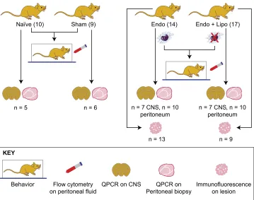
Endometriosis was induced in mice as previously described by Greaveset al. (13). In brief, donor mice were induced to undergo endometrial breakdown in a menses-like event (27). Donor mice were culled, and the endometrial tissue was collected by opening the decidualized uterine horn and scraping the endometrium away from the underlying myometrium. Approximately 40 mg of endometrial tissue was injected into the peritoneal cavity of E2-primed (500 ng E2valerate) recipient mice. Experiment 1 (mac-rophage depletion): 21 d post endometrial tissue injection, mice with induced endometriosis (n= 31) were randomly assigned to 1 of 2 groups: liposomal clodronate (n = 17) or saline (n = 14). Behavior assessments were performed to ascertain pretreatment recordings. Macrophage depletion was started on d 21 of the endometriosis protocol and maintained for an additional 7 d (21–28 d). At 28 d, mice were culled, and the following samples were recovered (seeFig. 1for flow diagram accounting for sample size for each endpoint): peritoneal lavage (n= 14 and 17; recovered by injecting 7 ml ice-cold DMEM into the peritoneal cavity followed by gentle massage and recovery), peritoneal bi-opsy (n= 10 each group), endometriosis lesions (n= 13 and 9), lumbar spinal cord (T13-L5;n= 7), and medial prefrontal cortex (brain;n= 7). Samples were also collected from naive (n= 10) and sham-treated (ovariectomy + E2 + intraperitoneal injection of PBS; n = 9) animals. Samples were collected into RNAlater (Thermo Fisher Scientific, Waltham, MA, USA) and frozen for quantitative PCR (qPCR) analysis (peritoneum, spinal cord, and brain), neutral-buffered formalin for paraffin embedding, and immunohistochemical analysis (endometriosis lesions). Sus-pected lesions were stained using hematoxylin and eosin and assessed for the presence of stroma+/2glands. Biopsies that did not include either epithelial or stromal compartments were not included in any further analysis. Experiment 2 (Igf-1r inhibition): In a separate experiment, mice with induced endometriosis (n= 24) were randomly assigned to 1 of 2 groups (vehicle,n= 12 or linsitinib,n= 12), and behavior assessments were performed.
Behavior assessments
Behavior assessments were performed as we have previously described in detail (14). In this study, behavioral assessments were performed in a blinded fashion, and all animals were acclima-tized to the apparatus and handling prior to the initiation of be-havior analysis. Spontaneous (grooming and activity) and evoked (mechanical hyperalgesia measured using von Frey filaments) behaviors were recorded. Experiment 1: Assessments were per-formed on 31 mice with induced endometriosis (saline,n= 14; liposomal, n= 17). Groups of naive (n = 10) and sham-treated (ovariectomy + E2+ intraperitoneal injection of PBS;n= 9) animals were included in all assessments. Experiment 2: Assessments were performed on 24 mice with endometriosis (vehicle,n= 12; linsitinib,n= 12), naive (n= 12), and sham-treated (n= 6) mice.
Patients and recovery of tissue and fluid samples
phase,n= 6; secretory phase,n= 7) were collected from women undergoing laparoscopic investigation for chronic pelvic pain with presence of endometriosis confirmed at the time of surgery. PF was also collected from women undergoing investigation for chronic pelvic pain with the absence of endometriosis (n = 9; proliferative phase,n= 5; secretory phase,n= 4). None of the women had taken exogenous hormones for$3 mo at the time of sampling. Samples were collected, stored, and processed in ac-cordance with Endometriosis Phenome and Biobanking Har-monisation Project (EPHect) guidelines (28–31). Endometriosis lesions were fixed in 4% neutral-buffered formalin and paraffin embedded. PF samples were centrifuged to pellet any cells and debris, divided into aliquots, and frozen at 280°. Pain scores were calculated using the Endometriosis Health Profile 30 questionnaire.
In vitrogeneration of endometriosis-associated macrophages
To isolate mononuclear leukocytes, blood was processed using dextran sedimentation and separated on a Percoll (GE Health-care, Waukesha, WI, USA) gradient with negative selection as previously described by Greaveset al. (25). Adherent monocytes were cultured on tissue culture plates in the presence of 4 ng/ml M-CSF 1 for 7 d to generate monocyte-derived macrophages (MDMs). Cells were cultured in DMEM (Thermo Fisher Scien-tific) containing 10% AB human serum (AMS Biotechnology, Milton, United Kingdom) in 12-well plates maintained at 37°C in 5% CO2.After differentiation to macrophages, MDMs were ac-tivated with PF from patients with (n= 7; secretory phase) or without (n = 8; secretory phase) endometriosis diluted 1:1 in medium for 24 h. Macrophages activated with PF from patients with endometriosis are referred to as in vitro generated endometriosis-associated macrophages (EAMs). We also in-cluded prototypically polarized macrophages as controls; from each blood preparation, some macrophages were activated with
20 ng/ml IFN-gand 50 ng/ml LPS to generate proinflammatory macrophages and others were activated with 20 ng/ml IL-4, IL-10, and TGF-b to generate prorepair macrophages; unac-tivated macrophages (M0s; medium only) were also included as controls. Conditioned medium was collected by removing PF or medium containing cytokines, washing once in fresh medium, and then incubating in medium for an additional 24 h. The con-ditioned medium was collected and frozen in aliquots at Oct4-80°C.
Human embryonic stem cell differentiation to sensory neurons
Human embryonic stem cells (hESCs), strain H9 (WiCell, Madi-son, WI, USA) were maintained and differentiated into sensory neurons using small molecule inhibitors as previously described in refs. 32 and 33. Differentiation was verified by confirming down-regulation of the pluripotency marker octamer-binding transcription factor 4 and up-regulation of the nociceptive genes tachykinin precursor 1 (TAC1), sodium voltage-gated channel (SCN) 9A, and SCN11A. Functionality of sensory neurons was confirmed by stimulating cells with 4 nM capsaiscin (Milli-poreSigma, Burlington, MA, USA) and recording intracellular calcium flux using a calcium indicator kit (BD Biosciences, San Jose, CA, USA), with calcium flux captured using a Novostar microplate fluorometer (BMG Labtech, Cary, NC, USA).
Neuronal outgrowth assay
Embryonic rat dorsal root ganglia (DRGs) were isolated as pre-viously described by Greaveset al. (34). Whole-ganglion explants were plated onto poly-D-lysine- and Matrigel-coated wells (BD Biosciences) in 48-well plates and incubated in DRG medium (high-glucose DMEM supplemented with 0.01% penicillin-streptomycin, 10% fetal calf serum). Positive controls were sup-plemented with nerve growth factor (NGF) (Bio-Rad, Hercules,
[image:3.594.51.410.49.332.2]CNS CNS
CA, USA) plus recombinant IGF-1 (R&D Systems, Minneapolis, MN, USA) (2–200 ng/ml)6125–500 nM Picropodophyllin (PPP; Tocris Bioscience, Bristol, United Kingdom). Some DRGs were incubated in PF or macrophage-conditioned medium diluted 1:1 in DRG medium6500 nM PPP. Images of explants were cap-tured using an Axiovert microscope (Carl Zeiss, Oberkochen, Germany), an Axiovision camera, and software.
Flow cytometry
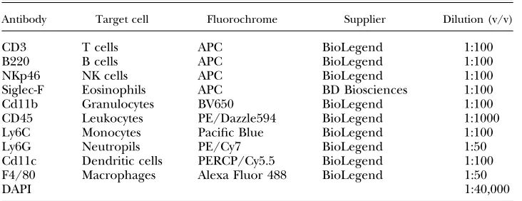
Red blood cells were lysed from peritoneal lavages, and equal numbers of cells were blocked with 0.025mg anti-CD16/32 (clone 93; BioLegend, San Diego, CA, USA) and then stained with a combination of antibodies shown inTable 1. Fluorescence minus 1 and negative controls were used to confirm gating strategies. Just prior to analysis, DAPI and 123count eBeads allowing ab-solute numbers of cells to be determined (Thermo Fisher Scien-tific) were added to samples. Samples were acquired using an LSRFortessa with FACSDiva software (BD Biosciences) and an-alyzed with FlowJo v.9 software (FlowJo, Ashland, OR, USA). Analysis was performed on single live cells determined using forward scatter heightvs.area and negativity for live or dead (DAPI).
Real-time qPCR
RNA was extracted from human and mouse tissues by ho-mogenization in Trizol reagent and chloroform phase sepa-ration prior to processing using an RNAeasy Kit (Qiagen, Hilden, Germany). RNA was extracted from cells using RLT (lysis) buffer and an RNAeasy Kit. Concentration and purity were assessed using a Nanodrop 1000 (Thermo Fisher Sci-entific). A standard curve was generated by pooling un-diluted RNA samples and performing four 10-fold dilutions. cDNA was synthesized using SuperScript Vilo Enzyme (Thermo Fisher Scientific) with 100 ng starting template in a 20-ml reaction. PCRs (10ml) were performed using the Roche Universal Library (Roche, Basel, Switzerland) and Express qPCR Supermix (Thermo Fisher Scientific). cDNA was added at 1ml per reaction, forward and reverse primers (Table 2) were added at 20mM, and thermal cycling conditions were performed on a 7900 Fast real-time PCR machine (Thermo Fisher Scientific) in 384-well plates with technical duplicates performed. 18S (Thermo Fisher Scientific) was selected as the reference gene. Data were analyzed using the relative stan-dard curve method, and samples were normalized to 1 con-sistent sample.
Immunofluorescence
Sections were antigen retrieved using citrate buffer, heat, and pressure (pH 6.0 for CD68 or pH 9.0 for IGF-1) or trypsin tablets dissolved in dH2O (for F4/80; MilliporeSigma) incubated with sections for 20 min at 37°C. Sections were blocked for endoge-nous peroxidase and nonspecific epitopes (species-specific serum diluted 1:5 in Tris-buffered saline and 5% bovine serum albumin) and incubated with primary antibody (Table 3) at 4°C overnight. Antibody detection was performed using a secondary pAb to IgG (horseradish peroxidase) and a tyramide signal amplification system kit with cyanine (Cy) 3 or fluorescein (1:50 dilution; Per-kinElmer, Waltham, MA, USA). For detection of the second an-tigen in dual immunofluorescence, sections were boiled in citrate buffer, and the second primary antibody was applied overnight and detected as before. Prior to mounting in Permafluor (Thermo Fisher Scientific), sections were counterstained with DAPI. Im-ages were captured using an LSM710 confocal microscope and AxioCam camera (Carl Zeiss). Human or mouse uterus was used as a positive control tissue, and negative controls had omission of the primary antibody.
IGF-1 ELISA
IGF-1 levels were detected in conditioned medium and PF using a Human IGF-1 DuoSet ELISA (R&D Systems) according to the manufacturer’s instructions.
Statistics
Initially, data were tested for normality using Shapiro-Wilk and Kolmogorov-Smirnov tests. Statistical analysis was performed using a Student’sttest or Mann-WhitneyUtest (nonnormal data) to compare 2 experimental groups or a 1-way ANOVA with a Tukey’s multiple comparison test to compare$3 experimental groups. For von Frey data, medians were plotted, and a Kruskal-Wallis test with a Dunn’s multiple comparison test was performed.
Study approval
[image:4.594.117.483.567.708.2]Mouse experiments were permitted under license by the United Kingdom Home Office and were approved by the University of Edinburgh Animal Welfare and Ethical Review Body (Edin-burgh, United Kingdom). Behavior assessments were performed in accordance with the Guidelines of the Committee for Research
TABLE 1. Flow cytometry antibodies
Antibody Target cell Fluorochrome Supplier Dilution (v/v)
CD3 T cells APC BioLegend 1:100
B220 B cells APC BioLegend 1:100
NKp46 NK cells APC BioLegend 1:100
Siglec-F Eosinophils APC BD Biosciences 1:100 Cd11b Granulocytes BV650 BioLegend 1:100 CD45 Leukocytes PE/Dazzle594 BioLegend 1:1000 Ly6C Monocytes Pacific Blue BioLegend 1:100 Ly6G Neutropils PE/Cy7 BioLegend 1:50 Cd11c Dendritic cells PERCP/Cy5.5 BioLegend 1:100 F4/80 Macrophages Alexa Fluor 488 BioLegend 1:50
DAPI 1:40,000
and Ethical Issues of the International Association for the Study of Pain. For the collection of patient biopsies, the study was ap-proved by the Lothian Research Ethics Committee (LREC 11/ AL/0376), and all samples were collected after informed consent was obtained in accordance with EPHect guidelines (29, 30). Human venous blood was collected from healthy female vol-unteers (n = 7) with informed consent and approval from the Local Lothian Research Ethics Committee (AMREC 15-HV-013).
RESULTS
Macrophages play a key role in
endometriosis-associated hyperalgesia in mice with induced endometriosis
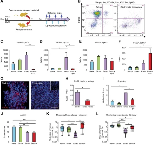
In C57BL/6 mice with induced endometriosis, macro-phages were depleted on d 21 post tissue injection using liposomal clodronate. Injections were repeated every 48 h (d 23, 25, and 27) to maintain depletion, and the mice were culled on d 28 (Fig. 2A). To confirm depletion of macro-phages, we performed flow cytometry on cells isolated from peritoneal lavage. Among CD45+, CD32, CD192, NKp462, Siglec-F2, lymphocyte antigen 6 complex, locus G2(Ly6G), and Cd11b+cells, 4 populations were sepa-rated based on expression of F4/80 and lymphocyte an-tigen 6 complex, locus C (Ly6C) (Fig. 2B). These were large peritoneal macrophages (LpMs; F4/80hi, Ly6C2), a pop-ulation of F4/80hiLy6C+cells that likely represent a subset of transient MDMs, and small peritoneal macrophages (SpMs; F4/80lo, Ly6C2) and monocytes (F4/802, Ly6C+). Administration of liposomal clodronate induced a signif-icant depletion of LpMs (Fig. 2C;P,0.001) and transient MDMs (Fig. 2D;P,0.05). There was no significant dif-ference in numbers of SpMs (Fig. 2E). Lipsomal depletion
[image:5.594.49.547.64.183.2]of macrophages in mice with endometriosis induced a significant increase in the number of monocytes compared with naive and sham-treated mice (Fig. 2F), but monocyte numbers were not significantly different to (nondepleted) mice with endometriosis. We observed a significant in-crease in the number of LpMs in mice with endometriosis compared with naive animals (Fig. 2C;P,0.01). We re-covered lesions from 13 (out of 14) mice treated with saline and 9 (out of 17) mice treated with liposomal clodronate. Using immunofluorescence, we identified a reduction in F4/80+ macrophages present in endometriosis lesions compared with control animals (Fig. 2G, H;P,0.05). Mice with endometriosis exhibited increased levels of sponta-neous abdominal grooming (Fig. 2I;P , 0.01) and de-creased levels of activity (Fig. 2J; P , 0.01) as well as decreased abdominal retraction (Fig. 2K;P,0.001) and paw withdrawal thresholds (Fig. 2L; P , 0.001) when stimulated with a punctate stimulus (von Frey filaments; an evoked measure of mechanical hyperalgesia) com-pared with naive or sham-treated animals [consistent with what we have observed previously (14)]. Following mac-rophage depletion with liposomal clodronate, the levels of grooming in the endometriosis mice declined such that they were no longer different to those observed in naive and sham-treated animals (Fig. 2I;P,0.05 compared with mice with induced endometriosis). Macrophage depletion did not rescue activity levels in endometriosis mice (Fig. 2J). However, there was a significant difference in ab-dominal retraction threshold between nondepleted and macrophage-depleted endometriosis mice (Fig. 2K; P, 0.001), with endometriosis mice withdrawing from a lighter stimulus than depleted animals. Depletion of macrophages also attenuated paw withdrawal thresholds in endometriosis mice (Fig. 2L;P,0.05 compared with
TABLE 3. Primary antibodies used in immunofluorescence
Antibody Host Species Supplier Dilution (v/v)
F4/80 Rat Mouse Thermo Fisher Scientific 1:600
CD68 Mouse Human Dako 1:800
[image:5.594.119.477.658.730.2]IGF-1 Rabbit Human Santa Cruz Biotechnology 1:150 Neurofilament Chicken Rat Covance 1:1000 TABLE 2. Primer sequences
Primer sequence, 59–39
Gene Forward Reverse UPL probe
Cox-2 GATGCTCTTCCGAGCTGTG GGATTGGAACAGCAAGGATTT 45
Tnf-a CTGTAGCCCACGTCGTAGC TTTGAGATCCATGCCGTTG 25
Igf-1 AGCAGCCTTCCAACTCAATTAT GAAGACGACATGATGTGTATCTTTATC 34
BDNF GTAACGGCGGCAGACAAA GACCTTTTCAAGGACTGTGACC 86
NT-3 CCCTTGTATCTCATGGAGGATT TTTCCGCCGTGATGTTCT 44
IGF-1 TGTGGAGACAGGGGCTTTTA ATCCACGATGCCTGTCTGA 67
SCN9A CAACTTTTAAGGGATGGACGA TCATATTTGGGCTGCTTGTCT 86
SCN11A ACCTGAGCCTGAACAACAGG TTTGAACTCTCTGGCTCGTG 2
TAC1 GCCTCAGCAGTTCTTTGGAT AGCCTTTAACAGGGCCACTT 89
nondepleted endometriosis mice). Thus, it appears that macrophages play a key role in altered sensory behavior in mice with endometriosis.
Depletion of macrophages attenuates markers of inflammatory pain
hypersensitivity in the CNS of mice with induced endometriosis
We have previously shown that the presence of endome-triosis lesions leads to increased expression of nociceptive and inflammatory markers (Trpv1, Scn11A, and Cox-2) in DRGs and spinal cords and the brains of mice with en-dometriosis (14). Cox-2 has previously been implicated as a marker of inflammatory pain hypersensitivity (35). In line with our previous findings, endometriosis mice exhibited increased mRNA expression ofCox-2(Fig. 3A; P,0.01) as well asTnf-a(Fig. 3B;P,0.05) in the spinal cord compared with naive and sham-treated animals, and these levels were attenuated following macrophage de-pletion using liposomal clodronate. The medial prefrontal cortices of the brains of mice with endometriosis also exhibited apparent increased mRNA expression of in-flammatory genes, withCox-2being significantly different to controls (Fig. 3C;P, 0.05). Expression ofCox-2 was reversed by macrophage depletion (P,0.05).Tnf-alevels in the brain were not significantly altered following mac-rophage depletion (Fig. 3D). We conclude that detectable molecular markers of inflammatory pain in the nervous system of mice with endometriosis can be attenuated by macrophage depletion.
Disease-modified macrophages in endometriosis exhibit elevated expression of IGF-1
To model EAMsin vitro, we activated human peripheral blood MDMs (see Supplemental Fig. S1 for characteriza-tion of monocyte-macrophage differentiacharacteriza-tion) collected from healthy female volunteers with PF from patients with endometriosis (Fig. 4A). M0s, proinflammatory macro-phages [M(LPS+IFN-g)], prorepair macrophages
[M(TGF-b + IL-10 + IL-4)], and macrophages activated with PF from women without endometriosis [M(No Endo)] were included for comparison. In order to investigate factors produced by macrophages that may contribute to pain in endometriosis, we analyzed mRNA expression of key neurotrophic genes. mRNA expression ofBDNFwas ele-vated in EAMs compared with M(No Endo) (Fig. 4B;P, 0.05). Concentrations ofNT-3also exhibit elevated levels in EAMs but this data did not reach statistical significance (Fig. 4C). mRNA concentrations of IGF-1 were signifi-cantly elevated in EAMs (P, 0.001) compared with all other macrophages, including macrophages activated with PF from women without endometriosis [M(No Endo);P,0.05, Fig. 4D)]. We aimed to further validate these data using patient biopsies. In endometriosis le-sions recovered at surgery from women during the se-cretory (progesterone-dominated) phase, we could detect macrophages (CD68) that coexpressed IGF-1 using dual
immunofluorescence (Fig. 4E). We also confirmed ex-pression of Igf-1 in F4/80+macrophages in mouse endo-metriosis lesions using immunofluorescence (Fig. 4F). In support of these findings, we also demonstrated thatIgf-1 mRNA concentrations were elevated in peritoneal bi-opsies of mice with endometriosis, and levels were sig-nificantly attenuated when macrophages were depleted (Fig. 4G;P,0.001).
IGF-1 is elevated in the PF of women with endometriosis and correlates with their pain scores
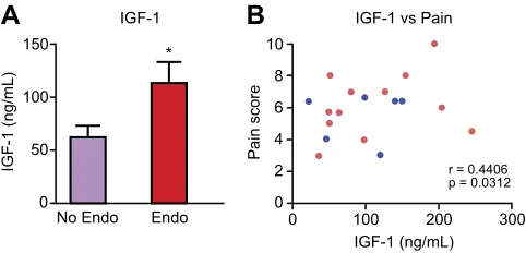
[image:7.594.302.545.48.302.2]We aimed to further explore the importance of IGF-1 in the mechanism behind endometriosis-associated pain in women using human samples of PF. IGF-1 protein con-centration was significantly elevated in the PF of women with endometriosis compared with those without disease (Fig. 5A;P,0.05) regardless of cycle phase (Supplemental Figure 3. Peripheral macrophages mediate endometriosis-associated inflammation in the CNS. qPCR analysis revealed evidence of apparent changes in the mRNA concentrations of key inflammatory genes in the spinal (T13-L5 segments) and medial prefrontal cortex area of the brains of mice with endometriosis. A) In the spinal cords of mice with endome-triosis (n = 7), Cox-2 mRNA was significantly elevated (P ,
0.01) compared with naive (n = 5) and sham-treated (n = 6) mice controls. This was attenuated following macrophage depletion (n= 7;P , 0.01). B)Tnf-a mRNA concentrations were increased in the spinal cords of mice with endometriosis (P , 0.05), and macrophage depletion attenuated levels.C)
Cox-2was also elevated in the brain (medial prefrontal cortex;
Fig. S2A). We also found that concentrations of IGF-1 in PF positively correlated with patient-reported pain scores (collected prior to surgery) in women with pelvic pain but no endometriosis and women with endometriosis and pelvic pain (Fig. 5B;P,0.05). Thus, we hypothesized that macrophage-derived IGF-1 might be a key factor involved in producing pain in endometriosis.
Macrophage-derived IGF-1 enhances sprouting neurogenesis and nociceptive gene expressionin vitro
IGF-1 is a known neurotrophic and sensitizing factor (36, 37). To determine potential mechanistic roles for macrophage-derived IGF-1 in endometriosis-associated pain, we explored the effects of EAM-conditioned medium on neuronal cell cultures. Recombinant IGF-1 (200 ng) stimulated sprouting neurogenesis in embryonic rat whole DRG explants (P , 0.001); this was specifically inhibited by 500 nM PPP (IGF-1R inhibitor;P,0.05;Fig. 6A, B). PF from women with endometriosis (PF Endo) and conditioned medium from EAMs also stimulated nerve
[image:8.594.97.540.40.446.2]growth compared with PF from women without disease (PF No Endo; Fig. 6A, B;P,0.01) or conditioned medium from macrophages activated with PF from women with-out disease [M(No Endo);P ,0.001]. Sprouting neuro-genesis was inhibited following addition of 500 nM PPP in each case (P,0.05 andP,0.01, respectively). Thus, the neurotrophic effects of PF and macrophages in endome-triosis are at least in part mediated by IGF-1. hESC-derived sensory neurons (Fig. 6C) express ion channels that are functionally active (32, 33). Incubation with EAM-conditioned medium enhanced mRNA expression of the nociceptive sodium voltage-gated ion channels SCN9A (Fig. 6D;P,0.01) and SCN11A (Fig. 6E;P,0.001) but not SCN3A, the vanilloid channel TRPV, or the purinergic channel purinergic receptor P2X 3 (Supplemental Fig. S3A–C). The neuropeptide Substance P (encoded by the gene TAC1) was significantly up-regulated (Fig. 6F;P, 0.001), but calcitonin gene-related peptide was not (Sup-plemental Fig. S3D). Changes in gene expression were attenuated by IGF-1R inhibitionviaPPP (P,0.01). Thus, we have shown a role for IGF-1 derived from EAMs in contributing to nerve growth and sensitizationin vitro. Figure 4.Disease-modified
mac-rophages in endometriosis ex-hibit elevated expression of IGF-1. A) Schematic showing generation of EAMs: peripheral blood monocytes from healthy female volunteers (n= 7) were differentiated into macro-phages for 7 d in the presence of recombinant M-CSF 1. Mac-rophages were then activated for 24 h with PF from patients with endometriosis (n = 7) or without endometriosis (n = 8) or activated with different cyto-kines to generate inflammatory (LPS + IFN-g) or repair
(TGF-b + IL-10 + IL-4; all n = 7) macrophages and compared with M0s. B–D) qPCR revealed that EAMs had significantly in-creased mRNA concentration of
BDNF (P , 0.05) (B), elevated levels of NT-3 (C), and signifi -cantly increased concentrations of IGF-1 (P , 0.001) (D). E) Dual immunofluorescence for CD68 (macrophages; red) and IGF-1 (green) revealed that macrophages in human endo-metriosis lesions express IGF-1.
F) In lesions recovered from mice with induced endometri-osis, we identified F4/80+ mac-rophages (red) that stained positive for Igf-1 (green). Scale bar, 50 mM. Top insets show negative controls where the primary antibody is omitted.G) In peritoneal biopsies recovered
from endometriosis mice (n= 10), qPCR analysis revealed that mRNA concentration ofIgf-1was elevated (P,0.05) compared with naive (n= 5) and sham-treated (n= 6) animals. Macrophage depletion in endometriosis mice (n= 10) significantly reducedIgf-1
Igf-1r inhibition attenuates hyperalgesia in mice with induced endometriosis
To further explore the role of Igf-1 in mice with endometriosis-associated pain, we inhibited Igf-1r using linsitinib, a selective IGF-1R inhibitor that prevents auto-phosphorylation and activation of downstream signaling. Treatment of endometriosis mice with 40 mg/kg linsitinib decreased grooming levels in mice with endometriosis (Fig. 7A;P,0.05). Although activity was reduced in mice with endometriosis, treatment with linsitinib did not res-cue this behavioral change (Fig. 7B). Abdominal retraction (P,0.01) and paw withdrawal thresholds (P,0.05) were higher in endometriosis mice treated with linsitinib com-pared with vehicle-treated mice (Fig. 7C, D) such that there was no significant difference between linsitinib-treated and control animals. These findings indicate that in-hibition of Igf-1 signaling in mice with endometriosis can attenuate endometriosis-associated-pain.
DISCUSSION
Endometriosis is a common incurable disease with dev-astating impacts on health-related quality of life. Despite its prevalence, there remains only limited treatment op-tions, and new therapeutic strategies are an area of unmet clinical need. Using a combination ofin vivo,ex-vivo, andin vitromodels, we show that macrophages play a key role in endometriosis-associated hyperalgesia, that disease-modified macrophages exhibit increased expression of IGF-1, and that inhibition of macrophage-derived IGF-1 can attenuate nerve growth and expression of nociceptive genes in vitro. Finally, we demonstrate that inhibition of Igf-1r in mice with endometriosis can attenuate hyper-algesia. Our study suggests that macrophages or signaling resulting from factors that they produce such as IGF-1 could be targeted to alleviate endometriosis-associated pain in women.
Pelvic pain is the most common presenting symptom of endometriosis (38). The pain is thought to be a manifes-tation of a 2-way dialogue between the nervous system and endometriosis lesions: the presence of endometrial-like tissue in the pelvic cavity of women with endometri-osis causes inflammation (39), and lesions are innervated by sensory nerve fibers that sense this peripheral in-flammatory environment and generate a nociceptive re-sponse resulting in pain perception. Macrophages are the most abundant immune cell present within the pelvic cavity (40), and in women with endometriosis, an increase in the number of macrophages both in the PF (41, 42) and in lesions (43) is evident; thus, these cells may con-tribute significantly to the inflammatory milieu present in the disease. In our study, we used an established mouse model of endometriosis to demonstrate that macro-phage depletion using liposomal clodronate can attenu-ate endometriosis-associattenu-ated spontaneous grooming and mechanical hyperalgesia (both locally and at a referred site). This finding complements and extends studies car-ried out in other rodent models of disease-related chronic pain that report a role for macrophages and monocytes in the generation of pain-related behaviors (19221).
We have previously established that the presence of endometriosis lesions in mice causes molecular changes in the CNS (14). In this study, we demonstrated that Cox-2 and Tnf-awere significantly elevated in the spinal cords and brains of mice with endometriosis and that deple-tion of peripheral macrophages attenuated expression of these inflammatory markers. The expression of proin-flammatory cytokines and Cox-2 in the CNS is a biomarker of spinal neuroinflammation and centralized inflamma-tory pain (12, 35, 44, 45). Our data suggest that macro-phages present in the pelvic cavity and in endometriosis lesions play a key role in establishing maladaptive changes in the CNS that are associated with enhanced pain per-ception. It was surprising that these changes could be re-versed, suggesting that in our model and at the time-points analyzed, CNS alterations are dynamic. The extent of spinal neuroinflammation in endometriosis remains un-known, although recently it was shown that in a mini-mally invasive model, mice with endometriosis exhibit astrocyte activation and subtle changes in immunoreac-tivity of microglia (46). Further characterizations of spinal maladaptation’s characteristic of neuroinflammation in en-dometriosis are warranted.
[image:9.594.50.291.34.150.2]Macrophages play diverse roles in health and disease in all tissues of the body and consequently exhibit tissue- and disease-specific transcriptional profiles (47). In line with this, peritoneal macrophages from women with endome-triosis exhibit an enhanced activation state characterized by enhanced expression of pro- and anti-inflammatory cytokines (48). Studies in mice have demonstrated that lesion-resident macrophages exhibit a phenotype that promotes lesion growth and enhances vascularization of lesions (22, 23), suggesting that EAMs exist as a disease-modified population that exhibits a wound-healing phe-notype. To investigate factors produced by macrophages that may be implicated in contributing to endometriosis-associated pain, we analyzed mRNA concentrations of neurotrophins inin vitrogenerated EAMs. We identified Figure 5.IGF-1 is elevated in the PF of women with
endome-triosis, and concentrations positively correlate with pain score.
that IGF-1 was significantly up-regulated in EAMs com-pared with control macrophages. Of note, expression of IGF-1 is a key characteristic of macrophages exhibiting a tissue-repair or wound-healing phenotype (49). This sup-ports the historical hypothesis that EAMs are prorepair (23). We confirmed expression of Igf-1 in lesion-resident macrophages in women with endometriosis and mice with induced endometriosis, and we hypothesized that macrophage-derived Igf-1 is likely to play a key role in the pathogenesis of the disease.
PF concentrations of IGF-1 have previously been shown to be elevated in patients with endometriosis compared with those without (50), and we confirmed this in the current study. IGF-1 present in the PF is thought to play a role in the pathophysiology of endometriosis by
[image:10.594.142.543.40.473.2]stimulating the growth of and preventing apoptosis of endometrial-like cells (51). In endometriotic stromal cells, IGF-1 up-regulates expression of estrogen receptorband aromatase, important drivers of endometriotic pathogen-esis (52). Although these limited studies have inferred a role for IGF-1 in the pathophysiology of endometriosis, the current study is the first to implicate a role for IGF-1 in endometriosis-associated pain. In support of our findings, we identified a positive correlation between PF IGF-1 concentrations and self-reported pain score in women with chronic pain with and without endometriosis, al-though there was no correlation with disease stage. There is increasing evidence that Igf-1 contributes to pain hy-persensitivity through binding to Igf-1r and activating Igf-1r–mediated PI3K, ERK, protein kinase B, or MAPK Figure 6. Macrophage-derived
IGF-1 enhances neuronal out-growth and nociceptive gene expressionin vitro.A) Quantifi -cation of neuronal outgrowth from rat DRG explants (be-tween 18 and 40 explants per group from embryos derived from 5 pregnant dams). Out-growth was maximal in the presence of recombinant NGF (positive control; P , 0.001 compared with negative con-trol, which was DRG me-dium in the absence of NGF). Recombinant IGF-1 (200 ng) also supported neuronal out-growth (P , 0.05 compared with negative control), and this was abrogated by coincubation with 500 nM PPP (specific IGF-1R inhibitor;P,0.05). PF from patients with endometriosis (n= 7) significantly enhanced neu-ronal outgrowth compared with PF from patients without endo-metriosis (n= 8;P,0.05). This outgrowth was reduced follow-ing addition of PPP. Condi-tioned medium from in vitro
generated EAMs (n= 7) signif-icantly enhanced neuronal out-growth (P , 0.05) compared with conditioned medium from macrophages activated with PF from patients without endome-triosis [M(No Endo)]; this was reduced following addition of PPP. B) Representative images of whole DRG explants follow-ing incubation under different conditions. +ve, DRG medium with recombinant NGF; 2ve, DRG medium without NGF;
CM, conditioned medium. Scale bar, 200 mM. C) Human sensory neurons were differentiated from H9 hESCs using small molecular inhibitors as previously described by Chamberset al. (32). Axons are visualized using with neurofilament (red), and nuclei are stained with DAPI.D,E) qPCR analysis revealed that incubation of sensory neurons with EAM-conditioned medium induced an increase in mRNA concentrations ofSCN9A (encodes Nav1.7;P ,0.01) (D) andSCN11A (encodes Nav1.9;P,
0.001) (E); this increase was abrogated following incubation with PPP (P,0.01).F) mRNA concentrations of the neuropeptide
intracellular signaling pathways (53). Igf-1r is widely expressed in small, medium, and large DRG neurons (37, 54), and in chronic inflammatory and tissue injury models, Igf-1 signaling enhances thermal and mechanical hyper-algesia (54, 55). Moreover, inhibition of Igf-1r can reverse mechanical allodynia and thermal hyperalgesia in a rat model of cancer bone pain (36). In the current study, Igf-1r inhibition had a profound effect on associated hyperalgesia by reversing endometriosis-associated spontaneous grooming as well as abdominal and hind-paw hyperalgesia, thus validating a strong as-sociation between IGF-1 signaling and endometriosis-associated pain.
Finally, we were able to infer a strong link between macrophage-derived IGF-1 and the observed endometriosis-associated changes in sensory behavior using mechanistic
in vitro models. We demonstrated that EAMs enhance sprouting neurogenesis in rat DRG explants as well as promote increased expression of nociceptive genes in hu-man stem cell–derived sensory neurons. IGF-1R inhibition attenuates these observed changes. Critically, enhanced expression of nociceptive genes is one of the first steps in generating primary afferent sensitization (10). We there-fore suggest that in endometriosis lesions macrophage-derived IGF-1 contributes to pain by promoting nerve growth in lesions and by sensitizing nerves by enhancing nociceptive gene expression.
Global inhibition of IGF-1 or IGF-1R is likely to have many off-target effects due to the pleiotropic roles of IGF-1. Thus, we suggest targeting disease-promoting macro-phages as a potential future treatment for endometri-osis. We now know that macrophages play a key role in growth, vascularization, and innervation of endometriosis lesions as well as generating endometriosis-associated pain, placing these cells at the center of the pathophysiol-ogy of a complex disorder. There are many pre- and early clinical trials in cancer that are successfully testing im-munotherapy, and key areas to target macrophages arevia inhibition of recruitment, direct killing, or re-education of disease-modified macrophages (56). However, before this can be a possibility, it is vital for us to know more about EAMs, their heterogeneity, and how they differ from healthy macrophages required for normal physiologic processes.
In summary, our study supports a previously un-recognized critical role for macrophages in endometriosis-associated hyperalgesia. The data suggest that macrophage-derived IGF-1 is a key driver of hyperalgesia in the disorder by promoting neurogenesis and nerve sensitization. By targeting specific populations of macro-phages that overexpress Igf-1, we may able to develop innovative new treatments for the debilitating pain asso-ciated with this common, incurable disease.
ACKNOWLEDGMENTS
The authors thank all the women who gave informed consent for the use of material in this study, research nurses Helen Dewart and Jennifer Devlin [National Health Service (NHS) Lothian, Edinburgh, United Kingdom] who organized tissue collection, and Halima Hassan and Kathryn Newton (University of Edinburgh) for technical support. The authors also thank Ronnie Grant (University of Edinburgh) for the figure preparation. This work was supported by a Medical Research Council Career Development Award (MR/ M009238/1; to E.G.), a Rosetree’s Trust research grant (A1759; to E.G.), a Medical Research Council Programme grant (MR/N024524/1; to P.T.K.S.), a Wellbeing of Women grant (R42533 to A.W.H.), and a Medical Research Council (MRC) Centre grant (MR/N022556/1). The authors declare no conflicts of interest.
AUTHOR CONTRIBUTIONS
[image:11.594.49.290.36.269.2]E. Greaves conceived and performed experiments, analyzed data and wrote manuscript; R. Forster and C. Hogg performed experiments and analyzed data; Figure 7.Igf-1r inhibition in mice with endometriosis rescues
A. Sarginson, A. Velichkova, and A. Dorning performed experiments; A. W. Horne and P. T. K. Saunders provided clinical samples; and A. W. Horne and P. T. K. Saunders provided feedback on experimental design and manu-script preparation.
REFERENCES
1. Johnson, N. P., and Hummelshoj, L.; World Endometriosis Society Montpellier Consortium. (2013) Consensus on current management of endometriosis.Hum. Reprod.28, 1552–1568
2. Gao, X., Yeh, Y. C., Outley, J., Simon, J., Botteman, M., and Spalding, J. (2006) Health-related quality of life burden of women with endo-metriosis: a literature review.Curr. Med. Res. Opin.22, 1787–1797 3. Mirkin, D., Murphy-Barron, C., and Iwasaki, K. (2007) Actuarial
analysis of private payer administrative claims data for women with endometriosis.J. Manag. Care Pharm.13, 262–272
4. Simoens, S., Dunselman, G., Dirksen, C., Hummelshoj, L., Bokor, A., Brandes, I., Brodszky, V., Canis, M., Colombo, G. L., DeLeire, T., Falcone, T., Graham, B., Halis, G., Horne, A., Kanj, O., Kjer, J. J., Kristensen, J., Lebovic, D., Mueller, M., Vigano, P., Wullschleger, M., and D’Hooghe, T. (2012) The burden of endometriosis: costs and quality of life of women with endometriosis and treated in referral centres.Hum. Reprod.27, 1292–1299
5. Guo, S. W. (2009) Recurrence of endometriosis and its control.Hum. Reprod. Update15, 441–461
6. Jacobson, T. Z., Duffy, J. M., Barlow, D., Koninckx, P. R., and Garry, R. (2009) Laparoscopic surgery for pelvic pain associated with endometriosis.Cochrane Database Syst. Rev.CD001300
7. Tokushige, N., Markham, R., Russell, P., and Fraser, I. S. (2006) Nerve fibres in peritoneal endometriosis.Hum. Reprod.21, 3001–3007 8. Berkley, K. J., Dmitrieva, N., Curtis, K. S., and Papka, R. E. (2004)
Innervation of ectopic endometrium in a rat model of endometriosis. Proc. Natl. Acad. Sci. USA101, 11094–11098
9. McKinnon, B. D., Bertschi, D., Bersinger, N. A., and Mueller, M. D. (2015) Inflammation and nervefiber interaction in endometriotic pain.Trends Endocrinol. Metab.26, 1–10
10. Basbaum, A. I., Bautista, D. M., Scherrer, G., and Julius, D. (2009) Cellular and molecular mechanisms of pain.Cell139, 267–284 11. Julius, D., and Basbaum, A. I. (2001) Molecular mechanisms of
nociception.Nature413, 203–210
12. Vardeh, D., Wang, D., Costigan, M., Lazarus, M., Saper, C. B., Woolf, C. J., Fitzgerald, G. A., and Samad, T. A. (2009) COX2 in CNS neural cells mediates mechanical inflammatory pain hypersensitivity in mice. J. Clin. Invest.119, 287–294
13. Greaves, E., Cousins, F. L., Murray, A., Esnal-Zufiaurre, A., Fassbender, A., Horne, A. W., and Saunders, P. T. (2014) A novel mouse model of endometriosis mimics human phenotype and reveals insights into the inflammatory contribution of shed endometrium. Am. J. Pathol.184, 1930–1939
14. Greaves, E., Horne, A. W., Jerina, H., Mikolajczak, M., Hilferty, L., Mitchell, R., Fleetwood-Walker, S. M., and Saunders, P.T. (2017) EP2 receptor antagonism reduces peripheral and central hyperalgesia in a preclinical mouse model of endometriosis.Sci. Rep.7, 44169 15. Ji, R. R., Chamessian, A., and Zhang, Y. Q. (2016) Pain regulation by
non-neuronal cells and inflammation.Science354, 572–577 16. Miller, R. J., Jung, H., Bhangoo, S. K., and White, F. A. (2009) Cytokine
and chemokine regulation of sensory neuron function.Handb. Exp.
Pharmacol.417–449
17. Ghasemlou, N., Chiu, I. M., Julien, J. P., and Woolf, C. J. (2015) CD11b+Ly6G- myeloid cells mediate mechanical inflammatory pain hypersensitivity.Proc. Natl. Acad. Sci. USA112, E6808–E6817 18. Old, E. A., Nadkarni, S., Grist, J., Gentry, C., Bevan, S., Kim, K. W.,
Mogg, A. J., Perretti, M., and Malcangio, M. (2014) Monocytes expressing CX3CR1 orchestrate the development of vincristine-induced pain.J. Clin. Invest.124, 2023–2036
19. Miller, R. E., Tran, P. B., Das, R., Ghoreishi-Haack, N., Ren, D., Miller, R. J., and Malfait, A. M. (2012) CCR2 chemokine receptor signaling mediates pain in experimental osteoarthritis.Proc. Natl. Acad. Sci. USA
109, 20602–20607
20. Yan, X., Maixner, D. W., Li, F., and Weng, H. R. (2017) Chronic pain and impaired glial glutamate transporter function in lupus-prone mice are ameliorated by blocking macrophage colony-stimulating factor-1 receptors.J. Neurochem.140, 963–976
21. Mert, T., Gunay, I., Ocal, I., Guzel, A. I., Inal, T. C., Sencar, L., and Polat, S. (2009) Macrophage depletion delays progression of neuropathic pain in diabetic animals. Naunyn Schmiedebergs Arch. Pharmacol.379, 445–452
22. Capobianco, A., Monno, A., Cottone, L., Venneri, M. A., Biziato, D., Di Puppo, F., Ferrari, S., De Palma, M., Manfredi, A. A., and Rovere-Querini, P. (2011) Proangiogenic Tie2(+) macrophages infiltrate human and murine endometriotic lesions and dictate their growth in a mouse model of the disease.Am. J. Pathol.179, 2651–2659
23. Bacci, M., Capobianco, A., Monno, A., Cottone, L., Di Puppo, F., Camisa, B., Mariani, M., Brignole, C., Ponzoni, M., Ferrari, S., Panina-Bordignon, P., Manfredi, A. A., and Rovere-Querini, P. (2009) Macrophages are alternatively activated in patients with endometriosis and required for growth and vascularization of lesions in a mouse model of disease.Am. J. Pathol.175, 547–556
24. Tran, L. V., Tokushige, N., Berbic, M., Markham, R., and Fraser, I. S. (2009) Macrophages and nervefibres in peritoneal endometriosis.
Hum. Reprod.24, 835–841
25. Greaves, E., Temp, J., Esnal-Zufiurre, A., Mechsner, S., Horne, A. W., and Saunders, P. T. (2015) Estradiol is a critical mediator of macrophage-nerve cross talk in peritoneal endometriosis. Am. J. Pathol.185, 2286–2297
26. Bulun, S. E., Gurates, B., Fang, Z., Tamura, M., Sebastian, S., Zhou, J., Amin, S., and Yang, S. (2002) Mechanisms of excessive estrogen formation in endometriosis.J. Reprod. Immunol.55, 21–33
27. Cousins, F. L., Murray, A., Esnal, A., Gibson, D. A., Critchley, H. O., and Saunders, P. T. (2014) Evidence from a mouse model that epithelial cell migration and mesenchymal-epithelial transition con-tribute to rapid restoration of uterine tissue integrity during men-struation.PLoS One9, e86378
28. Becker, C. M., Laufer, M. R., Stratton, P., Hummelshoj, L., Missmer, S. A., Zondervan, K. T., and Adamson, G. D.; WERF EPHect Working Group. (2014) World endometriosis research foundation endometriosis phenome and biobanking harmonisation project: I. Surgical phenotype data collection in endometriosis research.Fertil. Steril.102, 1213–1222; erratum: 104, 1047
29. Fassbender, A., Rahmioglu, N., Vitonis, A. F., Vigan`o, P., Giudice, L. C., D’Hooghe, T. M., Hummelshoj, L., Adamson, G. D., Becker, C. M., Missmer, S. A., and Zondervan, K. T.; WERF EPHect Working Group. (2014) World endometriosis research foundation endometriosis phenome and biobanking harmonisation project: IV. Tissue collection, processing, and storage in endometriosis research. Fertil. Steril.102, 1244–1253
30. Rahmioglu, N., Fassbender, A., Vitonis, A. F., Tworoger, S. S., Hummelshoj, L., D’Hooghe, T. M., Adamson, G. D., Giudice, L. C., Becker, C. M., Zondervan, K. T., and Missmer, S. A.; WERF EPHect Working Group. (2014) World endometriosis research foundation endometriosis phenome and biobanking harmonization project: III. Fluid biospecimen collection, processing, and storage in endometriosis research.Fertil. Steril.102, 1233–1243
31. Vitonis, A. F., Vincent, K., Rahmioglu, N., Fassbender, A., Buck Louis, G. M., Hummelshoj, L., Giudice, L. C., Stratton, P., Adamson, G. D., Becker, C. M., Zondervan, K. T., and Missmer, S. A.; WERF EPHect Working Group. (2014) World endometriosis research foundation endometriosis phenome and biobanking harmonization project: II. Clinical and covariate phenotype data collection in endometriosis research.Fertil. Steril.102, 1223–1232
32. Chambers, S. M., Qi, Y., Mica, Y., Lee, G., Zhang, X. J., Niu, L., Bilsland, J., Cao, L., Stevens, E., Whiting, P., Shi, S. H., and Studer, L. (2012) Combined small-molecule inhibition accelerates developmental timing and converts human pluripotent stem cells into nociceptors. Nat. Biotechnol.30, 715–720
33. Greaves, E., Grieve, K., Horne, A. W., and Saunders, P. T. (2014) Elevated peritoneal expression and estrogen regulation of nociceptive ion channels in endometriosis.J. Clin. Endocrinol. Metab.
99, E1738–E1743
34. Greaves, E., Collins, F., Esnal-Zufiaurre, A., Giakoumelou, S., Horne, A. W., and Saunders, P. T. (2014) Estrogen receptor (ER) agonists differentially regulate neuroangiogenesis in peritoneal endometriosis via the repellent factor SLIT3.Endocrinology155, 4015–4026 35. Samad, T. A., Moore, K. A., Sapirstein, A., Billet, S., Allchorne, A.,
Poole, S., Bonventre, J. V., and Woolf, C. J. (2001) Interleukin-1beta-mediated induction of Cox-2 in the CNS contributes to inflammatory pain hypersensitivity.Nature410, 471–475
up-regulation by insulin-like growth factor-1 in a rat model of bone cancer pain.Eur. J. Pain18, 774–784
37. Miura, M., Sasaki, M., Mizukoshi, K., Shibasaki, M., Izumi, Y., Shimosato, G., and Amaya, F. (2011) Peripheral sensitization caused by insulin-like growth factor 1 contributes to pain hypersen-sitivity after tissue injury.Pain152, 888–895
38. Coxon, L., Horne, A. W., and Vincent, K. (2018) Pathophysiology of endometriosis-associated pain: a review of pelvic and central nervous system mechanisms.Best Pract. Res. Clin. Obstet. Gynaecol.51, 53–67 39. Halme, J. (1991) Role of peritoneal inflammation in
endometriosis-associated infertility.Ann. N Y Acad. Sci.622, 266–274
40. Ho, H. N., Wu, M. Y., Chao, K. H., Chen, C. D., Chen, S. U., and Yang, Y. S. (1997) Peritoneal interleukin-10 increases with decrease in ac-tivated CD4+ T lymphocytes in women with endometriosis.Hum. Reprod.12, 2528–2533
41. Halme, J., Becker, S., and Haskill, S. (1987) Altered maturation and function of peritoneal macrophages: possible role in pathogenesis of endometriosis.Am. J. Obstet. Gynecol.156, 783–789
42. Gogacz, M., Galczynski, K., Wojtas, M., Winkler, I., Adamiak, A., Romanek-Piva, K., Rechberger, T., and Kotarski, J. (2017) Fas-related apoptosis of peritonealfluid macrophages in endometriosis patients: understanding the disease.J. Immunol. Res.2017, 3175394
43. Oosterlynck, D. J., Cornillie, F. J., Waer, M., and Koninckx, P. R. (1993) Immunohistochemical characterization of leucocyte subpopulations in endometriotic lesions.Arch. Gynecol. Obstet.253, 197–206
44. Ji, R. R., Xu, Z. Z., and Gao, Y. J. (2014) Emerging targets in neuroinflammation-driven chronic pain.Nat. Rev. Drug Discov.13, 533–548
45. Latremoliere, A., and Woolf, C. J. (2009) Central sensitization: a generator of pain hypersensitivity by central neural plasticity.J. Pain
10, 895–926
46. Dodds, K. N., Beckett, E. A. H., Evans, S. F., and Hutchinson, M. R. (2019) Spinal glial adaptations occur in a minimally invasive mouse model of endometriosis: potential implications for lesion etiology and persistent pelvic pain.Reprod. Sci.26, 357–369
47. Gautier, E. L., Shay, T., Miller, J., Greter, M., Jakubzick, C., Ivanov, S., Helft, J., Chow, A., Elpek, K. G., Gordonov, S., Mazloom, A. R., Ma’ayan, A., Chua, W. J., Hansen, T. H., Turley, S. J., Merad, M., and Randolph, G. J.; Immunological Genome Consortium. (2012) Gene-expression profiles and transcriptional regulatory pathways
that underlie the identity and diversity of mouse tissue macrophages.
Nat. Immunol.13, 1118–1128
48. Beste, M. T., Pf¨affle-Doyle, N., Prentice, E. A., Morris, S. N., Lauffenburger, D. A., Isaacson, K. B., and Griffith, L. G. (2014) Molecular network analysis of endometriosis reveals a role for c-Jun-regulated macrophage activation.Sci. Transl. Med.6, 222ra16 49. Wynn, T. A., and Vannella, K. M. (2016) Macrophages in tissue repair,
regeneration, andfibrosis.Immunity44, 450–462
50. Kim, J. G., Suh, C. S., Kim, S. H., Choi, Y. M., Moon, S. Y., and Lee, J. Y. (2000) Insulin-like growth factors (IGFs), IGF-binding proteins (IGFBPs), and IGFBP-3 protease activity in the peritonealfluid of patients with and without endometriosis.Fertil. Steril.73, 996–1000 51. Koutsilieris, M., Mastrogamvrakis, G., Lembessis, P., Sourla, A.,
Miligos, S., and Michalas, S. (2001) Increased insulin-like growth factor 1 activity can rescue KLE endometrial-like cells from apoptosis. Mol. Med.7, 20–26
52. Zhou, Y., Zeng, C., Li, X., Wu, P. L., Yin, L., Yu, X. L., Zhou, Y. F., and Xue, Q. (2016) IGF-I stimulates ERband aromatase expression via IGF1R/PI3K/AKT-mediated transcriptional activation in endome-triosis.J. Mol. Med. (Berl.)94, 887–897
53. Wang, H., Qin, J., Gong, S., Feng, B., Zhang, Y., and Tao, J. (2014) Insulin-like growth factor-1 receptor-mediated inhibition of A-type K(+) current induces sensory neuronal hyperexcitability through the phosphatidylinositol 3-kinase and extracellular signal-regulated ki-nase 1/2 pathways, independently of Akt.Endocrinology155, 168–179 54. Lin, S. F., Yu, X. L., Wang, B., Zhang, Y. J., Sun, Y. G., and Liu, X. J. (2016) Colocalization of insulin-like growth factor-1 receptor and T type Cav3.2 channel in dorsal root ganglia in chronic inflammatory pain mouse model.Neuroreport27, 737–743
55. Zhang, Y., Qin, W., Qian, Z., Liu, X., Wang, H., Gong, S., Sun, Y. G., Snutch, T. P., Jiang, X., and Tao, J. (2014) Peripheral pain is enhanced by insulin-like growth factor 1 through a G protein-mediated stimulation of T-type calcium channels.Sci. Sig-nal.7, ra94
56. Andon, F. T., Digifico, E., Maeda, A., Erreni, M., Mantovani, A., Alonso, M. J., and Allavena, P. (2017) Targeting tumor associated macrophages: the new challenge for nanomedicine.Semin. Immunol.
34, 103–113