Original citation:
Kakinen, Aleksandr, Javed, Ibrahim, Faridi, Ava, Davis, Thomas P. and Ke, Pu Chun. (2018) Serum albumin impedes the amyloid aggregation and hemolysis of human islet amyloid polypeptide and alpha synuclein. Biochimica et Biophysica Acta (BBA) - Biomembranes .
Permanent WRAP URL:
http://wrap.warwick.ac.uk/98815 Copyright and reuse:
The Warwick Research Archive Portal (WRAP) makes this work by researchers of the University of Warwick available open access under the following conditions. Copyright © and all moral rights to the version of the paper presented here belong to the individual author(s) and/or other copyright owners. To the extent reasonable and practicable the material made available in WRAP has been checked for eligibility before being made available.
Copies of full items can be used for personal research or study, educational, or not-for-profit purposes without prior permission or charge. Provided that the authors, title and full bibliographic details are credited, a hyperlink and/or URL is given for the original metadata page and the content is not changed in any way.
Publisher’s statement:
© 2018, Elsevier. Licensed under the Creative Commons Attribution-NonCommercial-NoDerivatives 4.0 International http://creativecommons.org/licenses/by-nc-nd/4.0/
A note on versions:
The version presented here may differ from the published version or, version of record, if you wish to cite this item you are advised to consult the publisher’s version. Please see the ‘permanent WRAP url’ above for details on accessing the published version and note that access may require a subscription.
Serum albumin impedes the amyloid aggregation and hemolysis
of human islet amyloid polypeptide and alpha synuclein
Aleksandr Kakinen,† Ibrahim Javed,† Ava Faridi,† Thomas P. Davis†§* and Pu Chun Ke†*
†ARC Centre of Excellence in Convergent Bio-Nano Science and Technology, Monash Institute of Pharmaceutical Sciences, Monash University, 381 Royal Parade, Parkville, VIC 3052, Australia §Department of Chemistry, University of Warwick, Gibbet Hill, Coventry, CV4 7AL, United Kingdom
Corresponding Author
ABSTRACT. Protein aggregation is a ubiquitous phenomenon underpinning the origins of a range
of human diseases. The amyloid aggregation of human islet amyloid polypeptide (IAPP) and alpha
synuclein (αS), specifically, is a hallmark of type 2 diabetes (T2D) and Parkinson’s disease
impacting millions of people worldwide. Although IAPP and αS are strongly associated with
pancreatic β-cell islets and presynaptic terminals, they have also been found in blood circulation
and the gut. While extensive biophysical and biochemical studies have been focused on IAPP and
αS interacting with cell membranes or model lipid vesicles, the roles of plasma proteins on the
amyloidosis and membrane association of these two major types of amyloid proteins have rarely
been examined. Using a thioflavin T kinetic assay, transmission electron microscopy and a
hemolysis assay here we show that human serum albumin, the most abundant protein in the plasma,
impeded the fibrillization and mitigated membrane damage of both IAPP and αS. This study offers
a new insight on the native inhibition of amyloidosis.
TOC GRAPHIC
Introduction
Human islet amyloid polypeptide (IAPP) and alpha synuclein (αS) are two major classes of
amyloid proteins serving both functional and pathogenic roles in biology. Specifically, monomeric
IAPP assists insulin for glycemic control, while monomeric αS is thought to be associated with
the modulation of neurotransmitter dopamine release, endoplasmic reticulum (ER)/Golgi
trafficking, and synaptic vesicles [1]. The endogenous stabilization of IAPP is provided by insulin,
physiological metal ions, low pH and zinc-mediated complexation of IAPP and C-peptide [2-8].
Likewise, transitions of αS from disordered monomers to partially folded intermediates and
amyloid fibrils may be promoted by changes in local pH, ionic strength and temperature, as well
as mutation [9, 10].
A large collection of literature has revealed that amphiphilic environments, such as cell membranes
and lipid vesicles, can initiate electrostatic interactions between the N-termini of amyloid proteins
and the anionic membranes [11-16], facilitated by hydrophobic interaction and hydrogen bonding
between the interactants. Such interactions have been shown to convert the proteins from
disordered monomers to α-helix and then β-sheet rich oligomers and protofibrils and, eventually,
cross-beta amyloid fibrils. The oligomers and amyloid fibrils show a high propensity for
partitioning into cell membranes, causing pore formation in a porin-like manner or extracting lipid
content to compromise membrane fluidity [17-20]. The endpoints of such interactions range from
generation of reactive oxygen species (ROS) to damage to ER, mitochondria, and DNA, followed
by cell degeneration. Furthermore, it has been found that the aromatic side chains of LANFLVH,
a fragment of IAPP, are nonessential in amyloid formation or membrane leakage [21], while
In addition, inhibition of IAPP amyloidosis and increased IAPP resistance to protease degradation
by copper (II) has also been reported [23].
IAPP amyloid aggregation has been implicated as causative to the onset of type 2 diabetes (T2D).
Likewise, the amyloid aggregation of αS into Lewy bodies or Lewy neurites is closely associated
with motor symptoms and Parkinson’s disease. While IAPP fibrils and plaques are generally found
in the extracellular space of pancreatic β-cell islets, Lewy bodies and Lewy neurites are located
intracellularly. Regardless, oligomeric IAPP and αS have been widely acknowledged as the toxic
species based on in vitro and animal studies [24, 25].
IAPP is stored at millimolar concentrations in pancreas beta islets and readily fibrillates at
micromolar concentrations [3]. Upon release into the bloodstream, IAPP is immediately exposed
to a milieu of plasma proteins and lipids [26, 27]. Consequently, we have recently applied the
concept of protein ‘corona’ [28, 29] originated from the field of nanomedicine for describing the
structural transformation as well as the toxicity of IAPP in circulation [27]. Here the protein corona
was rendered by model plasma proteins adsorbed on fibrillating IAPP or mature, hydrophobic
IAPP fibrils through electrostatic and hydrophobic interactions as well as H-bonding, similarly to
the formation of a protein corona adsorbed on nanoparticles [27, 29]. On the other hand, αS has
recently been found in the heart and the gut, with the latter being hypothesized as a possible origin
of the protein through the propagation of intestinal microbiota [30, 31]. Interestingly, human serum
albumin (HSA), which accounts for 55% of all plasma proteins, has recently been found to possess
a chaperone-like capacity against the amyloid aggregation of amyloid beta and transthyretin
[32-34], and this capacity of HSA was postulated to be realized through a ‘monomer-competitor’
We evaluated the effects of their binding on the conformation and toxicities of the amyloid proteins
using a thioflavin T (ThT, for fibrillization kinetics) assay and high-resolution transmission
electron microscopy (TEM, for mesoscopic morphology), as well as a red blood cell (RBC)
hemolysis assay (cell association and membrane leakage). Our study revealed a prolonged lag
phase of IAPP and suppressed fibrillization of αS with the increase of HSA concentration.
Additionally, the hemolysis assay demonstrated a protective effect of HSA on the toxicities of
IAPP and αS towards RBCs. Together, these results offered a new perspective on the natural
inhibition of IAPP and αS amyloidosis through the mediation of plasma proteins.
Results and discussion
Effects of HSA on IAPP and αS fibrillization
The fibrillization kinetics of IAPP and αS in the presence of different HSA concentrations was
examined using the amyloid specific dye ThT (Fig. 1). The lag phase of IAPP (50 µM)
fibrillization at 37 ºC was approximately 20 min, followed by an elongation phase and a saturation
phase after 2 h of incubation (Fig. 1A). In the presence of 10 µM HSA (5:1 molar ratio of IAPP to
HSA) the lag phase was increased considerably to 4.2 h and the elongation phase to 8 h. With the
HSA concentration increased to 50 µM (i.e., 1:1 IAPP to HSA molar ratio), the lag time was
extended to 5.4 h while the elongation phase up to 11 h. At the highest tested HSA concentration
of 100 µM (1:2 IAPP to HSA molar ratio), the lag time was further extended to 9 h while the
saturation phase was reached after 13 h. This result demonstrates the deceleration effect of HSA
on the fibrillization kinetics of IAPP, which could be due to the attraction between cationic IAPP
and negatively charged HSA at physiological pH, or through hydrophobic interaction between the
amyloidogenic region of IAPP (i.e. residues 20-29) and the hydrophobic domains of HSA to
aggregation inhibition of amyloid beta in the presence of HSA at comparable HSA/amyloid protein
ratios [35].
Our electron microscopy imaging confirmed the formation of IAPP amyloid fibrils after 17 h for
the control (Figs. 2A,B) and for IAPP incubated with HSA (Figs. 2C-H). However, the presence
of HSA altered the IAPP fibril morphology. Namely, at IAPP to HSA molar ratio of 5:1 the IAPP
amyloid fibrils were stacked together into multi-fibrillar bundles (Figs. 2C,D). At a higher HSA
concentration of 50 µM, IAPP fibrillated into large aggregates displaying significant intertwining
(Figs. 2E,F). At excess HSA concentration of 100 µM we observed clear co-aggregation of fibrillar
IAPP with HSA (Figs. 2G,H).
In addition, our statistical analysis of the TEM images revealed that HSA exerted a
concentration-dependent influence on IAPP fibril length and rigidity (Figs. 4A-D). The persistence length (λ), a
major indicator of rigidity [36], is 2,885 nm for (control) IAPP fibrils (Fig. 4A), which is
comparable to that of beta lactoglobulin and amyloid-beta fibrils [37, 38]. The lowest HSA
concentration did not significantly affect the persistence length or contour length of IAPP (Fig.
4B). However, at 1:1 IAPP to HSA molar ratio we noted a three-fold softening of the amyloid
fibrils (λ = 846 nm) and a narrower distribution of the fibril contour length (Fig. 4C). It should be
acknowledged that the analysis of amyloid aggregates is not straightforward and may result in
underestimated fibril contour length. Interestingly, the highest HSA concentration yielded more
rigid fibrils (λ = 3,684 nm, or 27% increase from that of the control) (Fig. 4D).
Understandably, the kinetics of αS fibril formation was markedly slower compared to IAPP, partly
due to the much larger molecular weight of the neuronal protein (140 residues vs. 37 residues, or
the amyloid proteins (sequence, charge, and hydrophobicity etc.). The lag phase of αS at 50 µM,
37 ºC and with 500 rpm of shaking was up to 60 h, followed by a steep elongation phase up to 125
h (Fig. 1B; Figs. 3A,B). In the presence of the lowest HSA concentration (10 µM, 5:1 IAPP to
HSA molar ratio), αS fibrillization was markedly supressed according to the ThT fluorescence
intensity, but was not entirely halted according to TEM imaging (Figs. 3C,D). At lower αS to HSA
molar ratios (i.e. 1:1 and 1:2), no increase of ThT fluorescence (Fig. 1B) and no fibrils – according
to TEM (Figs. 3E-H) – were observed up to 125 h of incubation. Due to the shaking applied to the
samples to speed up fibril formation, the contour length analysis yielded relatively short fibrils
only (Fig. 4E). The persistence length of αS fibrils was found to be 1,141 ± 9 nm, or 40% that of
IAPP fibrils, whereas the presence of HSA increased the fibril persistence length up to 2,771 ± 3
nm (Fig. 4E), comparable to that of IAPP control in the absence of the plasma protein.
It has been proposed that the effect of (external) ligands on protein amyloid aggregation depends
on the relative strength between protein-protein binding and protein-ligand binding [39]. In the
case of IAPP, electrostatic attraction between IAPP (isoelectric point or pI: 8.9) [40] and HSA (pI:
4.7) at equal molar concentration likely compromised the assembly of β sheets in amyloid fibril
formation, resulting in both softer and shorter fibrils (Figs. 2E,F). This electrostatic interaction
mediated the bundling of IAPP fibrils at the highest IAPP:HSA molar ratio (Figs. 2C,D), while
caused IAPP fibrils to stiffen through the adsorption of an HSA corona at the highest plasma
protein concentration (Figs. 2G,H). In the case of αS, HSA adsorption yielded a corona layer at
the 5:1 αS:HSA molar ratio (Figs. 3C,D), while at lower αS:HSA molar ratios the repulsion
between the like-charged neuronal protein (pI: 4.67) [41] and plasma protein as well as their
hydrophobic interactions could interfere with the self-assembly of αS, thereby inhibiting αS fibril
Ex vivo hemolysis of red blood cells
One facile system for screening the toxicity of exogenous substances is the ex vivo hemolysis assay
[42]. In this assay, the erythrocyte membranes serve as a model system for probing interactions
between toxic compounds and the lipid bilayer. This model has been previously adopted to
evaluate the endosomolytic behavior of cell-penetrating peptides, pH-responsive endosomolytic
agents for cytosolic delivery [43], as well as other polymeric gene delivery systems [42, 44, 45].
Here we applied the hemolysis assay to examine the interactions of red blood cells (RBCs) with
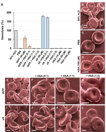
IAPP and αS w/o HSA (Fig. 5A). Evidently, both IAPP and αS demonstrated hemolytic activities
against RBCs confirming their toxicity. Consistently with the ThT assay and TEM imaging, the
hemolysis assay revealed a minor effect induced by the amyloid proteins mixed with HSA of 10
µM (5:1 molar ratio). However, no hemolysis of RBCs was detected at both 1:1 and 1:2 amyloid
proteins to HSA molar ratios alluding to the protective effect of the plasma protein. Using helium
ion microscopy we observed noticeable damage to RBCs in DI water (Fig. 5B), or when the cells
were exposed to IAPP (Fig. 5E), αS (Fig. 5I), or the amyloid proteins mixed with HSA of 10 µM
(Figs. 5F,J). In those cases, the RBC cells appeared deformed from their usual biconcave-discoid
shape, and at times were fully ruptured. No membrane damage was visible at higher HSA
concentrations for either IAPP (Figs. 5G,H) or αS (Figs. 5K,L), while the RBC cell membranes
displayed more rugged morphologies than the control likely resulting from HSA adsorption.
Amyloid formation is a complex, multistep process, comprising the lag, elongation and saturation
phases. Oligomeric species, formed during the lag process as an intermediate state, are postulated
to be the most toxic of amyloidogenic proteins [3], including IAPP and αS. Over the past decade,
a number of molecular mechanisms have been proposed for amyloid oligomer toxicity, indicating
oligomeric toxicity is excessive formation of ROS during amyloid fibrillization. Despite free
radicals are a normal component of cellular oxygen metabolism in mammals, free
radical-associated damage is an important factor in many pathological processes, including amyloid
diseases. Albumin, on the other hand, has many known physiological functions, including the
binding and transport of various endogenous and exogenous substances [47, 48]. Moreover, HSA
has been found to be an important circulating antioxidant [49]. Thus, HSA can protect RBCs from
hemolysis through its physical interaction with toxic oligomeric species, or its antioxidative
property.
Conclusion
This study has generated several key findings concerning the interactions between plasma protein
HSA and amyloid proteins IAPP and αS, an important aspect relevant to amyloidosis but has not
been examined extensively in the literature. It has been shown that the major plasma protein HSA
impeded the fibrillization of IAPP and αS, likely through competitive protein and
protein-ligand interactions [39], consistent with the ‘monomer-competitor’ mechanism proposed for the
chaperone-like activity of HSA against the aggregation of amyloid beta [35]. Specifically, HSA
displayed a dose-dependent suppression of IAPP fibrillization kinetics, leading to delayed but not
diminished amyloid fibril formation. In comparison, HSA completely inhibited the fibrillization
of αS up to 125 h, also coupled with the fact that the neuronal protein, much larger in molecular
weight but opposite in charge, fibrillates significantly slower than IAPP. In addition, we have
found that HSA can protect RBCs from hemolysis incurred by the toxic IAPP and αS, which can
be attributed to the protective mechanism of the plasma protein through its physical interaction
Materials and Methods
Materials
Human islet amyloid polypeptide (IAPP; 37 residues, 2-7 disulfide bridge, 3,904 Da, >95% pure
by HPLC) and α-synuclein (αS; 140 residues, 14,460 Da, >95% pure by SDS-PAGE and mass
spectrometry) were obtained in lyophilized monomeric form from AnaSpec, and prepared in
Milli-Q water (Millipore) at a stock concentration of 100 µM at room temperature immediately prior to
use. Thioflavin T (ThT) dye and human serum albumin (HSA, 66,478 Da, ≥ 97% pure by gel
electrophoresis) were both acquired from Sigma-Aldrich.
Thioflavin T (ThT) assay
IAPP and αS fibrillization in the presence or absence of HSA was analyzed by a ThT assay. For
this 50 µM ThT dye, 50 μM of IAPP or αS monomers and 10 µM, 50 µM or 100 µM HSA were
mixed, and the ThT fluorescence (read from bottom) was measured at 37 °C over 17 h for IAPP
and 125 h (500 rpm linear shaking) for αS, using an EnSpire Multimode Plate Reader (Perkin
Elmer; excitation/emission: 440 nm/485 nm) and a 96-well plate (Costar black/clear bottom). The
ThT kinetic assay was performed in technical triplicate, and average spectra of 3 measurements
were analyzed and presented by blank subtraction and normalized by IAPP/αS fluorescence at
saturation.
Transmission electron microscopy (TEM) and analysis
IAPP and αS amyloids fibrillated (17 h for IAPP and 125 h for αS) alone or with different ratios
solution was pipetted onto glow discharged (15 s) copper grids (400 mesh; ProSciTech), followed
by 1 min of adsorption. Excess samples were then drawn off using filter paper and the grids were
washed by Milli-Q water with the excess drawn off. Then the grids were stained with a drop of
1% uranyl acetate for 30 s. The excess stain was drawn off and the grids were air dried. Imaging
was performed by a Tecnai G2 F20 transmission electron microscope (FEI) operated at a voltage
of 200 kV. Images were recorded using a Gatan UltraScan 1000 (2k×2k) CCD camera (Gatan,
California, USA) and Gatan Microscopy Suite control software.
Statistical analysis of amyloid fibrils
The persistence length (λ) and contour length of IAPP and αS fibrils were analyzed using open
source tracking and analysis software – FiberApp by a semi-automated fibril tracking procedure
based on the A* pathfinding algorithm (minimal-cost path calculation or Dijkstra’s method) (Fig.
4H) [36]. The persistence length reflects an intrinsic property of a polymer, denoting its rigidity
and is mathematically defined by the bond correlation function (BCF) in 3D or 2D as the length
over which angular correlations in the tangential direction decrease by a factor of e (Fig. 4G). Here
the λ values of IAPP and αS fibrils were estimated using the average values derived from three
methods: BCF, mean-squared end-to-end distance (MSED) and mean-squared midpoint
displacement (MSMD). The contour length of a polymer corresponds to the end-to-end length
along its contour/backbone (Fig. 4G). Statistical analysis was performed by tracking 97~336 fibrils
in multiple TEM images per sample condition (Figs. 4A-F, n value).
Ex vivo hemolysis assay
IAPP, αS and their mixtures with HSA at different molar ratios were subjected to a hemolysis
assay to assess the toxicities of the amyloid proteins to RBCs. Blood was collected from a healthy
Human ethics approval 1443420 and the Australian National Health and Medical Research
Council Statement on Ethical Conduct in Human Research. The freshly drawn blood was
centrifuged at 2 g for 5 min and RBCs were collected as pellets. The RBCs were washed three
times with Dulbecco phosphate buffer saline (PBS) by centrifugal washings and diluted up to 5×
with PBS. 100 µL of RBCs were incubated with 100 µL of each sample. The final sample
concentration after mixing with RBCs was 50 μM for IAPP and αS, and 10 µM, 50 µM and 100
µM for HSA. Samples containing αS, IAPP and their mixtures with HSA were pre-incubated in
water for 2 days (500 rpm linear shaking) and 1 h at 37 °C, respectively, and mixed with PBS of
1× in final concentration. After mixing with RBCs, the samples were incubated for 2 h at 37 ºC,
centrifuged at 2 g for 5 min to pellet down the non-lysed RBCs, and hemolysis was measured by
analyzing the absorbance of free hemoglobin leaked out of compromised RBCs in the supernatants
at 541 nm (EnSpire Multimode Plate Reader, PerkinElmer). The RBCs incubated with PBS and
50% water (1:1 cells in PBS with Milli-Q water) were used as 0% and 100% hemolysis controls.
The percentage hemolysis was determined as:
btAb Ab Ab Ab Hemolysis t t 100 0
% , (1)
where Abt, Ab0 and Ab100 are absorbance values of the sample, the negative and the positive control,
respectively. The hemolysis assay was performed in technical triplicate and average values of 3
measurements were analyzed and presented.
Helium ion microscopy
RBCs treated by IAPP, αS and their combinations with HSA at different molar ratios were
concentrations of ethanol, i.e., 20%, 40%, 60%, 80% and 100%, with 2 h incubation at each
gradient. A drop of RBCs in 100% ethanol was placed on a carbon tape and the sample
morphologies were visualized by helium ion microscopy (Orion NanoFab, Zeiss, USA).
ACKNOWLEDGMENT
This work was supported by ARC Project CE140100036 (Davis). Davis is thankful for the award
of an ARC Australian Laureate Fellowship.
Author contributions
P.C.K. and T.P.D. conceived the project. A.K. performed TEM imaging and statistical analysis.
A.K. and A.F. carried out the ThT assay. I.J., A.K. and A.F. performed the hemolysis assay and
helium ion microscopy. A.K. and P.C.K. wrote the manuscript.
Conflicts of interest
REFERENCES
[1] M. Vilar, H.T. Chou, T. Lührs, S.K. Maji, D. Riek-Loher, R. Verel, G. Manning, H.
Stahlberg, R. Riek, The fold of α-synuclein fibrils, Proc. Natl. Acad. Sci. USA, 105 (2008)
8637-8642.
[2] L. Haataja, T. Gurlo, C.J. Huang, P.C. Butler, Islet amyloid in type 2 diabetes, and the toxic
oligomer hypothesis, Endocr. Rev., 29 (2008) 303-316.
[3] P.C. Ke, M.A. Sani, F. Ding, A. Kakinen, I. Javed, F. Separovic, T.P. Davis, R. Mezzenga,
Implications of peptide assemblies in amyloid diseases, Chem. Soc. Rev., 46 (2017)
6492-6531.
[4] J.R. Brender, K. Hartman, R.P.R. Nanga, N. Popovych, R. de la Salud Bea, S. Vivekanandan,
E.N.G. Marsh, A. Ramamoorthy, Role of zinc in human islet amyloid polypeptide
aggregation, J. Am. Chem. Soc., 132 (2010) 8973-8983.
[5] R.P.R. Nanga, J.R. Brender, S. Vivekanandan, A. Ramamoorthy, Structure and membrane
orientation of IAPP in its natively amidated form at physiological pH in a membrane
environment, Biochim. Biophys. Acta, 1808 (2011) 2337-2342.
[6] J.L. Larson, A.D. Miranker, The mechanism of insulin action on islet amyloid polypeptide
fiber formation, J. Mol. Biol., 335 (2004) 221-231.
[7] M.F. Sciacca, D. Milardi, G.M. Messina, G. Marletta, J.R. Brender, A. Ramamoorthy, C.
La Rosa, Cations as switches of amyloid-mediated membrane disruption mechanisms:
calcium and IAPP, Biophys. J., 104 (2013) 173-184.
[8] X. Ge, A. Kakinen, E.N. Gurzov, W. Yang, L. Pang, E.H. Pilkington, P.
Govindan-Nedumpully, P. Chen, F. Separovic, T.P. Davis, P.C. Ke, F. Ding, Zinc-coordination and
C-peptide complexation: a potential mechanism for the endogenous inhibition of IAPP
aggregation, Chem. Commun., 53 (2017) 9394-9397.
[9] R. Khurana, C. Ionescu-Zanetti, M. Pope, J. Li, L. Nielson, M. Ramírez-Alvarado, L. Regan,
A.L. Fink, S.A. Carter, A general model for amyloid fibril assembly based on morphological
[10] L. Xu, B. Ma, R. Nussinov, D. Thompson, Familial mutations may switch conformational
preferences in α-synuclein fibrils, ACS Chem. Neurosci., 8 (2017) 837-849.
[11] P. Cao, A. Abedini, H. Wang, L.-H. Tu, X. Zhang, A.M. Schmidt, D.P. Raleigh, Islet
amyloid polypeptide toxicity and membrane interactions, Proc. Natl. Acad. Sci. USA, 110
(2013) 19279-19284.
[12] Y. Porat, S. Kolusheva, R. Jelinek, E. Gazit, The human islet amyloid polypeptide forms
transient membrane-active prefibrillar assemblies, Biochemistry, 42 (2003) 10971-10977.
[13] M. Duan, J. Fan, S. Huo, Conformations of islet amyloid polypeptide monomers in a
membrane environment: implications for fibril formation, PLOS ONE, 7 (2012) e47150.
[14] C. Guo, S. Côté, N. Mousseau, G. Wei, Distinct helix propensities and membrane
interactions of human and rat IAPP 1–19 monomers in anionic lipid bilayers, J. Phys. Chem.
B, 119 (2015) 3366-3376.
[15] D.H.J. Lopes, A. Meister, A. Gohlke, A. Hauser, A. Blume, R. Winter, Mechanism of islet
amyloid polypeptide fibrillation at lipid interfaces studied by infrared reflection absorption
spectroscopy, Biophys. J., 93 (2007) 3132-3141.
[16] S.M. Patil, S. Xu, S.R. Sheftic, A.T. Alexandrescu, Dynamic α-helix structure of
micelle-bound human amylin, J. Biol. Chem., 284 (2009) 11982-11991.
[17] R.A. Ritzel, J.J. Meier, C.-Y. Lin, J.D. Veldhuis, P.C. Butler, Human islet amyloid
polypeptide oligomers disrupt cell coupling, induce apoptosis, and impair insulin secretion
in isolated human islets, Diabetes, 56 (2007) 65-71.
[18] M. Gao, R. Winter, The effects of lipid membranes, crowding and osmolytes on the
aggregation, and fibrillation propensity of human IAPP, J. Diabetes. Res., 2015 (2015) 21.
[19] Y. Hirakura, W.W. Yiu, A. Yamamoto, B.L. Kagan, Amyloid peptide channels: blockade
by zinc and inhibition by Congo red (amyloid channel block), Amyloid, 7 (2000) 194-199.
[20] R. Kayed, Y. Sokolov, B. Edmonds, T.M. McIntire, S.C. Milton, J.E. Hall, C.G. Glabe,
Permeabilization of lipid bilayers is a common conformation-dependent activity of soluble
[21] D. Milardi, M.F. Sciacca, M. Pappalardo, D.M. Grasso, C. La Rosa, The role of aromatic
side-chains in amyloid growth and membrane interaction of the islet amyloid polypeptide
fragment LANFLVH, Eur. Biophys. J., 40 (2011) 1-12.
[22] M.F. Sciacca, F. Lolicato, G. Di Mauro, D. Milardi, L. D’Urso, C. Satriano, A.
Ramamoorthy, C. La Rosa, The role of cholesterol in driving IAPP-membrane interactions,
Biophys. J., 111 (2016) 140-151.
[23] A. Sinopoli, A. Magri, D. Milardi, M. Pappalardo, P. Pucci, A. Flagiello, J.J. Titman, V.G.
Nicoletti, G. Caruso, G. Pappalardo, G. Grasso, The role of copper(II) in the aggregation of
human amylin, Metallomics, 6 (2014) 1841-1852.
[24] S. Zraika, R.L. Hull, C.B. Verchere, A. Clark, K.J. Potter, P.E. Fraser, D.P. Raleigh, S.E.
Kahn, Toxic oligomers and islet beta cell death: guilty by association or convicted by
circumstantial evidence? Diabetologia, 53 (2010) 1046-1056.
[25] T.P.J. Knowles, M. Vendruscolo, C.M. Dobson, The amyloid state and its association with
protein misfolding diseases, Nat. Rev. Mol. Cell. Biol., 15 (2014) 384-396.
[26] Y. Xing, E.H. Pilkington, M. Wang, C.J. Nowell, A. Kakinen, Y. Sun, B. Wang, T.P. Davis,
F. Ding, P.C. Ke, Lysophosphatidylcholine modulates the aggregation of human islet
amyloid polypeptide, Phys. Chem. Chem. Phys., 19 (2017) 30627-30635.
[27] E.H. Pilkington, Y. Xing, B. Wang, A. Kakinen, M. Wang, T.P. Davis, F. Ding, P.C. Ke,
Effects of protein corona on IAPP amyloid aggregation, fibril remodelling, and cytotoxicity,
Sci. Rep., 7 (2017) 2455.
[28] T. Cedervall, I. Lynch, S. Lindman, T. Berggård, E. Thulin, H. Nilsson, K.A. Dawson, S.
Linse, Understanding the nanoparticle–protein corona using methods to quantify exchange
rates and affinities of proteins for nanoparticles, Proc. Natl. Acad. Sci. USA, 104 (2007)
2050-2055.
[29] P. Ke, S. Lin, W. Parak, T. Davis, F. Caruso, A decade of the protein corona, ACS Nano, 11
(2017) 11773–11776.
[30] T.R. Sampson, J.W. Debelius, T. Thron, S. Janssen, G.G. Shastri, Z.E. Ilhan, C. Challis, C.E.
regulate motor deficits and neuroinflammation in a model of Parkinson's disease, Cell, 167
(2016) 1469-1480.
[31] H. Malkki, Could gut microbiota influence severity of Parkinson disease?, Nat. Rev. Neurol.,
13 (2016) 66.
[32] M. Algamal, J. Milojevic, N. Jafari, W. Zhang, G. Melacini, Mapping the interactions
between the Alzheimer's Ab-peptide and human serum albumin beyond domain resolution,
Biophys. J., 105 (2013) 1700-1709.
[33] T.E. Finn, A.C. Nunez, M. Sunde, S.B. Easterbrook-Smith, Serum albumin prevents protein
aggregation and amyloid formation and retains chaperone-like activity in the presence of
physiological ligands, J. Biol. Chem., 287 (2012) 21530-21540.
[34] M. Algamal, R. Ahmed, N. Jafari, B. Ahsan, J. Ortega, G. Melacini, Atomic-resolution map
of the interactions between an amyloid inhibitor protein and amyloid beta (Aβ) peptides in
the monomer and protofibril states, J. Biol. Chem., DOI: 10.1074/jbc.M117.792853.
[35] J. Milojevic, A. Raditsis, G. Melacini, Human serum albumin inhibits Aβ fibrillization
through a “monomer-competitor” mechanism, Biophys. J., 97 (2009) 2585-2594.
[36] I. Usov, R. Mezzenga, FiberApp: an open-source software for tracking and analyzing
polymers, filaments, biomacromolecules, and fibrous objects, Macromolecules, 48 (2015)
1269-1280.
[37] A. Kakinen, J. Adamcik, B. Wang, X. Ge, R. Mezzenga, T. Davis, F. Ding, P. Ke, Nanoscale
inhibition of polymorphic and ambidextrous IAPP amyloid aggregation with small
molecules, Nano Res., DOI: 10.1007/s12274-017-1930-7.
[38] J. Adamcik, J.-M. Jung, J. Flakowski, P. De Los Rios, G. Dietler, R. Mezzenga,
Understanding amyloid aggregation by statistical analysis of atomic force microscopy
images, Nat. Nanotech., 5 (2010) 423.
[39] A. Gladytz, B. Abel, H.J. Risselada, Gold‐induced fibril growth: the mechanism of
[40] S. Li, M. Micic, J. Orbulescu, J.D. Whyte, R.M. Leblanc, Human islet amyloid polypeptide
at the air–aqueous interface: a Langmuir monolayer approach, J. Royal Soc. Interface., 9
(2012) 3118-3128.
[41] N. Gould, D.E. Mor, R. Lightfoot, K. Malkus, B. Giasson, H. Ischiropoulos, Evidence of
native α-synuclein conformers in the human brain, J. Biol. Chem., 289 (2014) 7929-7934.
[42] K. Saar, M. Lindgren, M. Hansen, E. Eiríksdóttir, Y. Jiang, K. Rosenthal-Aizman, M.
Sassian, Ü. Langel, Cell-penetrating peptides: a comparative membrane toxicity study, Anal.
Biochem., 345 (2005) 55-65.
[43] B.C. Evans, C.E. Nelson, S.S. Yu, K.R. Beavers, A.J. Kim, H. Li, H.M. Nelson, T.D.
Giorgio, C.L. Duvall, Ex Vivo red blood cell hemolysis assay for the evaluation of
pH-responsive endosomolytic agents for cytosolic delivery of biomacromolecular drugs, J. Vis.
Exp., (2013) 50166.
[44] A. Kichler, C. Leborgne, E. Coeytaux, O. Danos, Polyethylenimine-mediated gene delivery:
a mechanistic study, J. Gene. Med., 3 (2001) 135-144.
[45] M.F. Sohail, H.S. Sarwar, I. Javed, A. Nadhman, S.Z. Hussain, H. Saeed, A. Raza, N. Irfan
Bukhari, I. Hussain, G. Shahnaz, Cell to rodent: toxicological profiling of folate grafted
thiomer enveloped nanoliposomes, Toxicol. Res., 6 (2017) 814-821.
[46] R. Kayed, C. Lasagna-Reeves, Molecular mechanisms of amyloid oligomers toxicity, J.
Alzheimers Dis., 33 (2013) S67-S78.
[47] U. Kragh-Hansen, V.T.G. Chuang, M. Otagiri, Practical aspects of the ligand-binding and
enzymatic properties of human serum albumin, Biol. Pharm. Bull., 25 (2002) 695-704.
[48] B. Carlo, D. Enrico, Reversible and covalent binding of drugs to human serum albumin:
methodological approaches and physiological relevance, Curr. Med. Chem., 9 (2002)
1463-1481.
[49] M. Roche, P. Rondeau, N.R. Singh, E. Tarnus, E. Bourdon, The antioxidant properties of
FIGURES AND CAPTIONS
Figure 3. Transmission electron microscopy imaging of αS amyloid fibril formation w/o and with
Figure 4. Contour length and persistence length (λ) analyses of IAPP (50 µM) (A-D) and αS (50
µM) (E-F) fibrils incubated with 10 µM (5:1 ratio), 50 µM (1:1 ratio) and 100 µM (1:2 ratio) HSA.
The contour length of αS with HSA at 50 µM and 100 µM was not analyzed due to the lack of
Figure 5. (A) Determination of the in vitro toxicities of IAPP, αS and their combinations with
HSA against red blood cells (RBCs) with an absorbance-based hemolysis assay. (B-L)
[image:24.612.123.492.73.534.2]