Original Article
DSTYK kinase domain ablation impaired the mice
capabilities of learning and memory in water maze test
Kui Li1,2, Ji-Wei Liu1, Zhi-Chuan Zhu1, Hong-Tao Wang1, Yong Zu1, Yong-Jie Liu1, Yan-Hong Yang1, Zhi-Qi Xiong1,
Xu Shen2, Rui Chen3, Jing Zheng1, Ze-Lan Hu1
1Shanghai Key Laboratory of New Drug Design, School of Pharmacy, East China University of Science and
Technology, 130 Meilong Road, Shanghai, China; 2Shanghai Institute of Materia Medica, Chinese Academy of
Sciences, Shanghai, China; 3Department of Molecular and Human Genetics, Baylor College of Medicine, Houston,
Texas, United States of America
Received July 23, 2014; Accepted August 23, 2014; Epub September 15, 2014; Published October 1, 2014
Abstract: DSTYK (Dual serine/threonine and tyrosine protein kinase) is a putative dual Ser/Thr and Tyr protein kinase with unique structural features. It is proposed that DSTYK may play important roles in brain because of its high expression in most brain areas. In the present study, a DSTYK knockout (KO) mouse line with the ablation of C-terminal of DSTYK including the kinase domain was generated to study the physiological function of DSTYK. The DSTYK KO mice are fertile and have no significant morphological defects revealed by Nissl staining compared with wildtype mice. Open field test and rotarod test showed there is no obvious difference in basic motor and balance capacity between the DSTYK homozygous KO mice and DSTYK heterozygous KO mice. In water maze test, however, the DSTYK homozygous KO mice show impaired capabilities of learning and memory compared with the DSTYK heterozygous KO mice.
Keywords: DSTYK, knockout mice, brain development, learning and memory
Introduction
DSTYK (dual serine/threonine and tyrosine pro-tein kinase) is originally identified as a novel receptor interacting protein (RIP)-homologous protein designated as receptor interacting pro-tein kinase 5 (RIPK5) [1], because its kinase domain shares homology with kinase domain of RIP protein family. However, evidences from evolution analysis showed DSTYK probably does not belong to RIP protein family, but a divergent protein kinase and is distant from PSK (protein serine/threonine kinase) and PTK (protein tyrosine kinase) [2]. Finally, HUGO (human genome organization) Gene Nomen- clature committee (HGNC) changed the official name “RIPK5” to “DSTYK”.
DSTYK widely exists in multicellular organisms. The absence of DSTYK from unicellular organ-ism and some invertebrates and ubiquitous existence in vertebrates suggest that DSTYK is a result of evolution, which may play important roles in higher animals [2]. The kinase domain
of DSTYK is strictly conserved, so it has been proposed to have kinase activity to play some potentially important functional roles in the brain and other tissues [2].
com-pared with DSTYK heterozygous KO mice. But the homozygous KO mouse showed impaired capabilities of learning and memory in water maze test, which suggested DSTYK may play roles in the process of learning and memory.
Materials and methods
Animals
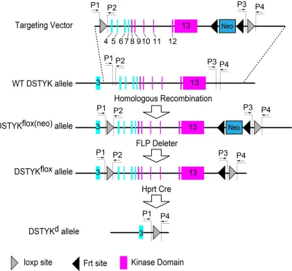
The DSTYK heterozygous knockout mice was generated in Baylor College of Medicine using a strategy similar as previously reported [3]. Briefly, a DSTYK-targeting vector containing two loxp sites flanking the exons 4-13 of DSTYK gene was constructed, with an Frt-Neo-Frt cas-sette for selecting (Figure 1). The targeting
vec-tor was linearized, purified, and electroporated into murine embryonic stem cells to obtain high quality male chimeras. Then the chimeras were crossed with C57BL/6 to generate DSTYKflox(neo)
mice. DSTYKflox(neo) mice were crossed with Frt
deleter mice to generate DSTYKflox mice. To
obtain a DSTYK knockout mouse line, the Hprt Cre mice were crossed with DSTYKflox mice to
generate DSTYK null allele (DSTYKd) as the
[image:2.612.98.519.73.463.2]DSTYK heterozygous mouse to generate off-springs to perform behavioral test.
Genotyping
Polymerase chain reaction (PCR) was used to confirm the genotype of the offsprings. PCR Primer sequences are: P1, 5’-GATGCAATGTT- TGGCTTAAATAGATA-3’; P2, 5’-GCATAAAAAGTT- TTGACATGTAACTCC-3’; P3, 5’-CAGCACTGTCTT- CCTGTTCCTAC-3’; P4, 5’-CTGCCTCTGAGACTG- TGAGAAAC-3’. Since the binding sites of P2 and P3 were deleted in DSTYK knockout mouse, using primer sets P1/P2 or P3/P4 can’t get specific products in DSTYK knockout mouse. While it can get specific products in wildtype mouse using these primer sets. As there is more than 16 kb distance between primer bind-ing sites of P1 and P4 in wildtype mouse
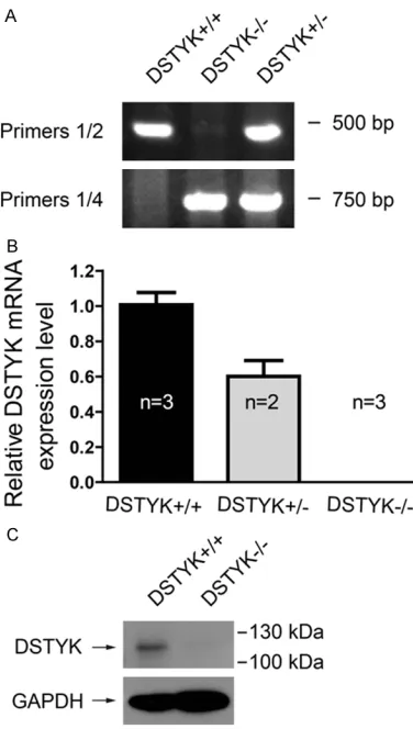
genome, no specific product can be amplified in wildtype mouse with limited extension time. While in DSTYK knockout mouse, it can gener-ate an about 700 bp product with primer set P1/P4 since the DSTYK kinase domain ablation shortened the distance between P1 and P4. Thus P1/P2 or P3/P4 can be used to identify wildtype DSTYK genome, and P1/P4 can be used to identify DSTYK mutant genome. An example of genotyping was provided (Figure
2A).
Quantitative PCR
Adult male mice were decapitated, and brain tissues were rapidly dissected to homogenate in Trizol (Invitrogen, Carlsbad, USA) reagent. Total RNA was extracted, reverse transcribed (RT M-MLV, Promega, Beijing, China) and ana-lyzed by quantitative real-time PCR. Quantitative PCR kit SYBR Premix Ex Taq (Takara, Dalian, China) was used following the instructions of the manufacturer. The primer sets for quantita-tive PCR were: DSTYK, 5’-TGCCGCTTACCAT- GTTGAGG-3’, 5’-CACCCATGTGATACGCTGGAT-3’; β-Actin, 5’-GGCTGTATTCCCCTCCATCG-3’, 5’-CC- AGTTGGTAACAATGCCATGT-3’.
Western blot
Mice were decapitated, and the brain cerebral cortex tissues were rapidly dissected. Tissues were then immersed in liquid nitrogen until homogenization using Dounce homogenizer. Tissues were lysed in RIPA lysis buffer (Beyotime, Nantong, China) supplemented with 1 x PMSF (Beyotime, Nantong, China) and 1 x protease inhibitor cocktail (Sangon, Shanghai, China). 8% SDS-PAGE was used to separate the proteins. We used antibodies specific for DSTYK (Santa Cruz: sc-162109, Shanghai, China), horseradish peroxidase-conjugated donkey anti-goat secondary antibody (Santa Cruz: sc-2020, Shanghai, China) and GAPDH (Kangchen, Shanghai, China). The antibody dilution rates were: anti-DSTYK, 1:200, donkey goat secondary antibody, 1:4000, anti-GAPDH, 1:8000.
Nissl staining
[image:3.612.93.281.70.402.2]Adult male mice were anesthetized and per-fused with saline and 4% paraformaldehyde, and their brains were removed for cryosections at 40 μm per slice for Nissl staining. Tissue sec-tions were mounted on gelatin-coated slides Figure 2. Genotyping, mRNA expression and protein
expression of mice with kinase domain ablation of DSTYK. A. Representative Genotyping PCR results us-ing different primer sets. B. The mRNA level of DSTYK in DSTYK+/+, DSTYK+/- and DSTYK-/- mice analyzed by
quantitative PCR. Sample size was marked in the col-umns. C. DSTYK protein expression in DSTYK+/+ and
and baked for more than two hours in 50°C oven. The sections were defatted in a solution containing 50% chloroform and 50% alcohol for 30 min. Then the sections were hydrated in 100% alcohol (for 5 min), 95% alcohol (for 5 min), 70% alcohol (for 5 min), 50% alcohol (for 5 min) and distilled water (for 5 min). The hydrat-ed sections were stainhydrat-ed in Nissl staining solu-tion for 30 min. To make Nissl staining solusolu-tion, 60 ml 0.24% (w/v) cresyl violet in distilled water, 30 ml 1.36% (w/v) sodium acetate in distilled water and 470 ml 6 mM acetic acid were mixed and the pH was adjusted to 3.5. Then the sec-tions were rinsed with distilled water and dehy-drated in 50% alcohol (for 5 min), 70% alcohol (for 5 min), 95% alcohol (for 5 min) and 100% alcohol (for 5 min). Finally, slides were cleared in xylene for 2 × 5 min and covered with glass coverslips. Images were captured using micro-scope Nikon Ti-s.
Open field test
The open field test box was a plastic box with 49 cm long, 49 cm wide and 49 cm high. Its walls and floor were all white. Two 8W fluores-cent tube were placed on two top sides of the box. Mice were allowed to explore for 30 min. Their movements were automatically recorded and analyzed by AniLab Software (Ningbo, China).
Rotarod test
Rota rod test was performed as previously reported [4]. The rota rod test device and asso-ciated control software was bought from
tion of each quadrant. Both visible platform water maze test and hidden platform water maze test were performed. In visible platform water maze test, the platform was set just above the water with a black triangle paper over it. Every mouse was tested for four trials per day. The location of the platform and the drop location of mice always changed but kept a constant distance between them in the four trials. In hidden platform water maze test, the platform was put just under the water and its position was fixed at northwest. Every mouse was also trained for four trials per day. The order of drop location (east, south, west and north) changed among 7 training days. After 7 days of training, the hidden platform was removed and mice were giving one probe trial test. AniLab Software (Ningbo, China) was used to record and analyze the data.
Data analysis
Data were presented as means ± standard error of mean (SEM). Data were analyzed by t test using Graphpad Prism 5 software. The lev-els of significance were measured based upon respective graph values. Symbols * and *** represent significant values P < 0.05 and P < 0.001, respectively.
Results
The expression of DSTYK mRNA and protein is deleted in DSTYK homozygous KO mice
[image:4.612.90.322.73.239.2]To compare DSTYK mRNA expression level in DSTYK+/+, DSTYK+/- and DSTYK-/- mouse, 2-3
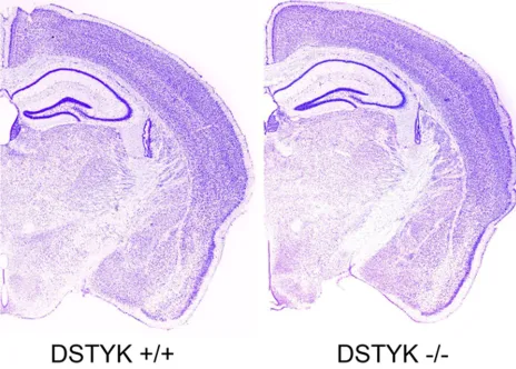
Figure 3. Representative Nissl staining of brain sections showed no significant morphological defects of mouse brain.
Taimeng Software (Chengdu, China). When training, the speed was fixed at 24 rpm. Mice were trained 4 trials per day, less than 1 minute in a trial. Intervals between two training trails were about 10 minutes. After 3 consecutive days of training, the test tri-als were performed on the fourth day. When testing, the speed of the rod acceler-ated from 0 to 35 rpm in 180 seconds. Mice were tested for 3 trials per mouse and the average latency to fall from the rod was calculated.
Morris water maze test
loca-mice for each genotype were sacrificed to obtain mRNA from cerebral cortex. Quantitative PCR analysis showed that the DSTYK mRNA level of DSTYK+/- mouse were reduced to about
60% of DSTYK+/+ mouse and no DSTYK mRNA
was detected in DSTYK-/- mice (Figure 2B). To
confirm the deletion of full-length DSTYK pro-tein in DSTYK-/- mice, adult male mice were
sac-rificed to obtain protein from cerebral cortex. Results from western blot showed that a band about 119 kDa disappeared in DSTYK-/- mice,
which confirmed the knockout of DSTYK. The observed protein size of DSTYK was larger than the predicted size 105 kDa, which may be due
to unknown posttranslational modification. The antibody manufacturer also observes the change in molecular weight of DSTYK protein in mice.
DSTYK-/- mice showed no detectable
devel-opmental defects in brain revealed by Nissl staining
The knockout of DSTYK is not lethal to mouse. The DSTYK-/- mice can normally develop and is
fertile. To check the effect of DSTYK knockout on the brain development, adult DSTYK+/+ and
[image:5.612.94.516.71.261.2]DSTYK-/- mice were sacrificed to conduct Nissl
Figure 4. DSTYK knockout mice showed no significant defects in basic motor and balance capacity. A. Total distance moved in open field test. B. Rota rod test showed no significant difference between DSTYK heterozygous and homo-zygous knockout mice. N.S. No Significance.
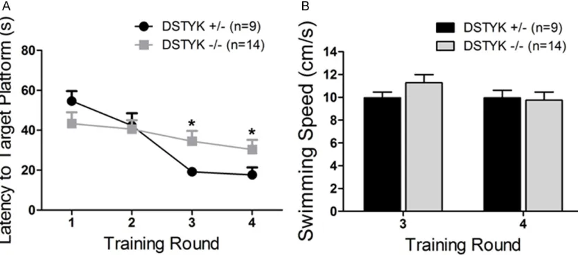
Figure 5. Visible task results of water maze test. A. DSTYK-/- mice showed a longer Latency to target platform than
DSTYK+/- mice in training round 3 and 4. B. There is no significant difference in average speed between DSTYK
[image:5.612.97.514.321.505.2]staining. The results showed there are no detectable defects both in DSTYK+/+ and
DSTYK-/- mice (Figure 3).
DSTYK-/- mice showed no basic locomotion
defects
To identify the effect of DSTYK knockout on the basic locomotion activity of mouse, open field test and rotarod test were performed. The total distance moved in open field test (Figure 4A) and the latency to fall from rod in rota rod test
(Figure 4B) were not significantly different
between DSTYK+/- and DSTYK-/- mice. These
results indicated that DSTYK knockout did not cause severe defects in basic movement and balance capacity.
DSTYK-/- mice showed poor performance in
morris water maze test
To test if DSTYK knockout has any effects on spacial learning and memory, 9 DSTYK+/- mice
and 14 DSTYK-/- mice were given morris water
showed no significant difference between DSTYK+/- and DSTYK-/- mice. Taken together, our
findings suggest that DSTYK may be involved in the process of spacial learning and memory.
Discussion
DSTYK is reported to be widely distributed in multiple tissues of vertebrates from the devel-opmental stage to the adult [2]. Human DSTYK full-length (7.9 kb) mRNA is presented in many organs and tissues such as heart, brain, pla-centa, skeletal muscle, kidney, lung and pan-creas assessed by northern blot analysis. Another short DSTYK mRNA (about 3.6 kb) was detected in testes [2]. Like human DSTYK, mouse DSTYK also located on Chromosome 1, containing 13 exons and encoding a protein with 927 amino acid residues. Northern Blot experiments found that full length mRNA (7.6 kb) of DSTYK existed in E9.5 mouse embryo and in adult mouse brain, heart, lungs, kidneys, and skin [2]. A smaller mRNA fragment (3.6 kb) Figure 6. Probe trial results of water maze test. A. Target platform latency. B.
Number of target platform crossing. C. Time spent in target quadrant. D. Rela-tive total distance moved. DSTYK+/-, n=9; DSTYK-/-, n=14. “***”, P < 0.001.
maze test. In trial 1-2 of visi-ble platform water maze test, DSTYK-/- mice did not show
great difference compared with DSTYK+/- mice (Figure
5A). But In trial 3-4 of visible platform water maze test, DSTYK-/- mice cost
significant-ly more time to swim to the target platform than DSTYK
+/-mice (Figure 5A). The poor performance of DSTYK-/- mice
may not be caused by the motor defects but by the defects in learning and mem-ory because there was no sig-nificant difference in the aver-age swimming speed of trial 3-4 between DSTYK+/- and
DSTYK-/- mice (Figure 5B). In
the probe trial of hidden plat-form water maze test, the latency to target platform of DSTYK-/- mice were
signifi-cantly higher than that of DSTYK+/- mice (Figure 6A). But
the other index in probe trial such as number of target plat-form crossing (Figure 6B), time spent in target quadrant
(Figure 6C) and relative total
was also detected in mouse testes [2]. Results from fluorescence in situ hybridization showed that DSTYK mRNA exists in E14.5 mouse embryo and in adult mouse brain, heart, kidney and other organs, which is consistent with results of northern blot [2]. Western Blot test showed that DSTYK protein exists in the mouse brain, heart, kidney, lung and skeletal muscle [2]. Northern Blot has also detected the expres-sion of DSTYK in multiple tissues of rats and chickens [2]. However, few studies are focusing on the physiological function of DSTYK until now.
In vitro study revealed that DSTYK could induce cell death in a dose dependent manner and also cause DNA fragmentation [1]. Pan-caspase inhibitors did not rescue the cell death induced by DSTYK, suggesting that DSTYK induce cell death through caspase-dependent and cas-pase-independent pathways [1]. DSTYK knock-down in zebrafish caused severe developmen-tal defects. Many organs such as heart, fins, tail, and cloaca were not normally developed. Cloaca of zebra fish is considered as the homol-ogous organ of the urogenital system of higher animals [5]. Genome analysis of a family suffer-ing from a congenital kidney disease pointed out a linkage between DSTYK and kidney devel-opment [5]. In this family, a mutation in DSTYK genomic DNA was detected between exon 2 and exon 3, which was believed to cause mRNA splicing errors and the formation of mRNA miss-ing 27 bp in exon 2. The protein product of this abnormal mRNA lacked amino acid residues 210-218 (VTMHHALLQ), which was considered as the cause of congenital kidney disease in this family. DSTYK mutations were also detect-ed in 2.3% of 311 other unrelatdetect-ed patients suf-fering from congenital kidney disease. Thus mutations in DSTYK were considered as one of the causes of congenital kidney disease [5]. In summary, previous studies showed that DSTYK plays multiple roles in cell death and the development of many organs. To our sur-prise, our results showed that kinase domain ablation of DSTYK did not cause severe devel-opmental defects especially in brain develop-ment. It is noted that the identified DSTYK mutant in the family suffering from congenital kidney disease occurs at exon 2-3. But exon 1-3 is not ablated in the knockout mice we used. The truncated DSTYK protein may be translated from exons 1-3 in this DSTYK
knock-out mice line, which may still play some roles as wildtype DSTYK in development. In addition, we can’t exclude the possibility that other genes may compensate the function of DSTYK. Thus the ablation of full-length DSTYK in mouse is needed to further clarify the role of DSTYK in the development of mouse.
Disclosure of conflict of interest
None.
Address correspondence to: Dr. Ze-Lan Hu or Dr. Jing Zheng, Shanghai Key Laboratory of New Drug Design, School of Pharmacy, East China University of Science and Technology, 130 Meilong Road, Shanghai, China. Tel: 0086-21-64250608; Fax: 0086-21-64250608; E-mail: [email protected] (ZLH); Tel: 64250608; Fax: 0086-21-64250608; E-mail: [email protected] (JZ)
References
[1] Zha J, Zhou Q, Xu LG, Chen D, Li L, Zhai Z and Shu HB. RIP5 is a RIP-homologous inducer of cell death. Biochem Biophys Res Commun 2004; 319: 298-303.
[2] Peng J, Dong W, Chen Y, Mo R, Cheng JF, Hui CC, Mohandas N and Huang CH. Dusty protein kinases: primary structure, gene evolution, tis-sue specific expression and unique features of the catalytic domain. Biochim Biophys Acta 2006; 1759: 562-572.
[3] Fernandez-Valdivia R, Jeong J, Mukherjee A, Soyal SM, Li J, Ying Y, Demayo FJ and Lydon JP. A mouse model to dissect progesterone signal-ing in the female reproductive tract and mam-mary gland. Genesis 2010; 48: 106-113. [4] Hu ZL, Huang Y, Tao XR, Qi ZH, Chen JY and
Ding YQ. Inducible Prrxl1-CreER(T2) recombi-nation activity in the somatosensory afferent pathway. Genesis 2012; 50: 552-560.