Original Article
Relationship between changes in the cochlear blood
flow and disorder of hearing function induced
by blast injury in guinea pigs
Wei Chen1, Jianmin Wang1, Jing Chen1, Jichuan Chen2, Zhiqiang Chen1
1Research Institute of Surgery, State Key Laboratory of Trauma, Burns and Combined Injury, Daping Hospital,
Third Military Medical University, Chongqing 400042, China; 2Department of Otorhinolaryngology, Daping
Hospi-tal, Third Military Medical University, Chongqing 400042, China
Received November 20, 2012; Accepted January 11, 2013; Epub February 15, 2013; Published March 1, 2013
Abstract: The auditory system is the most susceptible to damages from blast waves. Blast injuries always lead to
varying degrees of hearing impairment. Although a disorder of the cochlear blood flow (CoBF) has been considered
to be related to many pathological processes of the auditory system and to contribute to various types of hearing loss, changes in the CoBF induced by blast waves and the relationship between such changes and hearing
impair-ment are undefined. To observe the changes in the cochlear microcirculation after exposure to an explosion blast, investigate the relationship between changes in the CoBF and hearing impairment and subsequently explore the mechanism responsible for the changes in the CoBF, we detected the perfusion of the cochlear microcirculation and hearing threshold shift after exposure to an explosion blast. Then, an N-nitro-L-arginine-methyl ester (L-NAME, NO
synthase inhibitor) solution and artificial perilymph were applied to the round window (RW) of the cochlea before the
blast exposure, followed by an evaluation of the CoBF and hearing function. The results indicated that the changes
in the CoBF were correlated to the strength of the blast wave. The cochlear blood flow significantly increased when the peak value of the blast overpressure was greater than approximately 45 kPa, and there was no significant change in the cochlear blood flow when the peak value of the blast overpressure was less than approximately 35
kPa. Following local administration of the NO synthase inhibitor L-NAME, the increase in the CoBF induced by the
blast was inhibited, and this reduction was significantly associated with the hearing threshold.
Keywords: Cochlea, blood flow, hearing function, blast injury, guinea pigs
Introduction
Blast injuries can occur in normal life, during military acts and in the workplace. They can cause injuries to various organ systems [1]. These injuries differ, ranging from relatively minor to lethal, in accordance with the magnitude of the blast wave [2, 3]. As an air-containing organ, the auditory system is considered to be most susceptible to blast wave-associated damage. Such damages can be full range, affecting the region from the tympanic membrane to the inner ear, and lead to temporary and permanent losses of hearing sensitivity [4]. These structural damages cause conductive hearing loss, sensorineural hearing loss or both [1-3].
experimental conditions, criteria and research methods, the results have been varied [10-14]. A better understanding of the CoBF changes will be helpful for the prevention and management of hearing disorders resulting from blast damage.
Materials and methods
Subjects
A total of 45 guinea pigs of both sexes were used, weighing 250-350 g and with normal Preyer’s reflexes. The animals were housed under standard laboratory conditions with free access to food and water. Examinations of the tympanic membrane revealed no evidence of pathology. All operative procedures were per-formed in a state of deep surgical anaesthesia with pentobarbital sodium (30 mg/kg) adminis-tered intraperitoneally.
Experimental protocols were approved by the Management Committee of Experimental Animals of Third Military Medical University, China.
Surgery and preparation
The animals were initially anaesthetised with pentobarbital sodium (3 mg/kg intraperitoneal injection). This injection was repeated every 60 min using half of the initial dose. The left bulla was exposed and opened via a retroauricular approach. A round hole (2 mm diameter) was drilled in the upper portion of the bulla to observe the position of the laser Doppler probe, and a small hole (1 mm diameter) was drilled under the observation hole for the insertion of a stainless steel tube (1 mm outer diameter and 0.5 mm inner diameter) that was used to bolster the probe. The tip of the tube was placed on the bony surface of the basal turn of the cochlea. The probe was fixed, and the holes were sealed with dental self-curing resin. The incision was sutured, and the outer tip of the tube protruded beyond the skin. The systemic blood pressure (BP) of all animals was recorded by a pressure transducer (MLT0380/D Reusable BP transducer, AD Instruments Pty Ltd, Australia) through a catheter that was inserted into the left femoral artery before sur-gery, and the data were collected using a quad bridge (ML118, AD Instruments Pty Ltd). The BP data were recorded and analysed using
Chart Software (v5.5.6, Copyright: 1994-2008, AD Instruments Pty Ltd, Austrilia), and after being divided into 5 s segments, each individu-al segment was averaged and converted to the percentage change.
Histopathology of cochlea
At the end of each experiment, the animals were killed under pentobarbital anaesthesia (30 mg/kg, intraperitoneal) followed by cervical dislocation. The temporal bone was removed, and the bulla was opened immediately. A small hole was created in the apex of the bone shell by a needle, and the round window was opened by the same method. The fixative solution (4% paraformaldehyde, buffered at pH 7.3) was perfused three times into the cochlea through the apical hole using a pipette. Then, the cochleae were immersed in the same fixative solution at 4°C overnight. After fixation, the excess bone around the cochlea was removed, and the cochlea was decalcified in 10% EDTA solution (buffered at pH 7.3) for 5 days. Next, the specimens were embedded in paraffin and sectioned in the horizontal plane at a thickness of 5 μm. The sections were stained with hae-matoxylin-eosin and studied by light microscopy.
Detonation and measurement of blast wave
To generate the blast wave, a detonator (paper shell, 600 mg cyclotrimethylenetrinitramine, RDX inside) was fixed in an open space at a height of 0.4 meter and detonated by electrodes. The electric pressure transducers were fixed at the same level and distance to record the pressure of the blast waves. A dynamic data acquisition meter (TESTELECTRONIC Inc, Chengdu, China) was used to record the signal from the transducers. The signal was recorded and analysed using DAP Software (v3.01, copyright: 2005.6.6). Measurement of CoBF by laser doppler
an optic fibre. The blood flow meter was connected to a Power Lab System (AD Instruments), and the percentage of back scatter (BSC) and the laser Doppler flowmetry output signal were recorded using Chart Software (v5.5.6, Copyright: 1994-2008, AD Instruments). Both the LDF and BSC outputs were factory calibrated to produce a certain voltage output per calibrated unit. The BSC output represents the relative strength of the returned signal.
Evaluation of hearing function
To assess the hearing function, the auditory brain stem response (ABR) was measured. An evoked potential instrument (MK-NMD, MINGKAN Technologies, Chongqing, China) was used to generate acoustic stimuli and subsequently record the evoked potentials. An alternating broadband clicking sound (repetition rate of 11/s) was given to test the ear and used as the stimulus sound, and white noise sound was used as a contralateral masking noise. To record the bioelectrical potentials, subdermal stainless-steel needle electrodes were inserted at the vertex (active), dorsolateral to the measured ear (reference) and the nasal root (ground). The thresholds were determined from a set of responses at varying intensities with 5 dB SPL intervals, and the electrical signals were averaged from 512 to 1024 repetitions. The thresholds at each frequency were verified twice.
Blast exposure and evaluation of CoBF and hearing function
To ensure that the animals were exposed to a suitable intensity of blast overpressure, the
animals were placed at distances from 0.5 meter to 0.9 meter at 0.1 meter intervals (3 animals for each distance). The blast overpressures were measured at the same distances and times. The normal LDF was recorded for 5 minutes a half hour before the exposure to the explosion blast, and the same recording process was performed immediately and 1 hour, 3 hours, 1 day and 2 days after the exposure. The ABR was recorded immediately and 1 hours, 3 hours, 1 day and 1 week after the exposure to the explosion blast. The CoBF and ABR of an additional 3 animals were measured at same time points, serving as the blank group.
Measurement of CoBF and evaluation of hear-ing function after L-NAME infusion and blast exposure
For this experiment, 27 guinea pigs were divided into 3 groups. L-NAME (Beyotime Institute of Biotechnology, China)was dissolved in a 1% artificial perilymph solution (137 mM NaCl, 5 mM KCl, 2 mM CaCl2, 1 mM MgCl2, 1 mM NaH2PO4, 12 mM NaHCO3, and 10 mM glu-cose; pH adjusted to 7.4 at 37°C) [15]. Group A consisted of 9 animals that received a 2 μl infu-sion of a 1% L-NAME solution into the RW by a micropipette (75N 5 μl SYR, HAMILTON CO). Group B consisted of the same number of ani-mals that received the same infusion. Group B consisted of 9 animals that received the same dose of artificial perilymph infused into the RW. Both groups A and C were exposed to a blast generated by the same detonator at the dis-tances of 0.5 meter, 0.6 meter and 0.7 meter (3 animals for each distance) 15 minutes after the RW infusion. The CoBF and ABR were mea-sured in all animals of the 3 groups 10 minutes before the blast exposure; the CoBF was recorded immediately, 1 hour, 2 hours, and 3 hours after the blast exposure, and the ABR was measured 2.5 hours after the blast expo-sure in groups A, B and C.
Statistical analysis
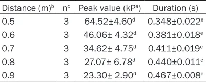
[image:3.612.91.292.97.177.2]Peak values and durations of the overpressure, BP, data of CoBF, and TS of all groups in each distance were averaged and analyzed using a 1-way analysis of variance (ANOVA). Data of CoBF and TS between group A and group C were averaged and analyzed using independent samples T-test. Correlation between the
Table 1. Peak value and duration of overpres-surea
Distance (m)b nc Peak value (kPa) Duration (s)
0.5 3 64.52±4.60d 0.348±0.022e
0.6 3 46.06± 4.32d 0.381±0.018e 0.7 3 34.62± 4.75d 0.411±0.019e 0.8 3 27.07± 6.78d 0.440±0.011e 0.9 3 23.30± 2.90d 0.467±0.008e aResults are expressed as mean ± S.E.M. bThe heads of
the transducers were placed at the same distances as the external auditory canal. cThe tests of the blast wave were
repeated three times for each distance. dSignificantly dif
-ferent between the peak values of the blast waves at each distance (P<0.05). eSignificantly different between the dura
maximum values of the CoBF baseline and peak values of the blast overpressure was analyzed using bivariate correlation analysis.
All analysis processes were conducted using SPSS statistics 17.0. The distribution are reported as mean±S.E.M.
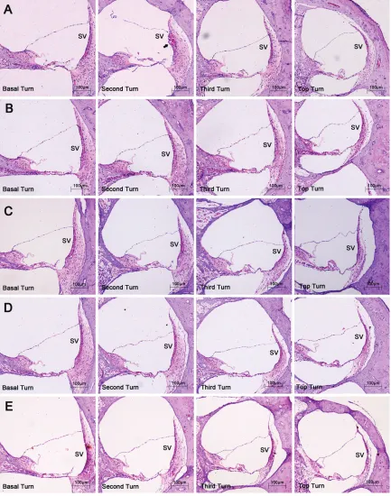
Figure 1. The pathological changes of the inner ear. Haematoxylin-eosin, original magnification ×100. SV, stria
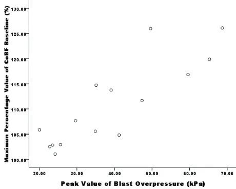
[image:4.612.91.524.72.622.2]Figure 3. Correlation between the maximum values of the CoBF baseline and peak values of the blast overpressure. The
correlation is significant at the 0.01 level (2-tailed) (analysed
by SPSS 17.0.0).
Results
The systemic blood pressure, which was record-ed until the end of each experiment, showrecord-ed values within the normal range, and the per-centage changes were minimal in all animals. Pressure changes of the blast waves at differ-ent distance
As an open field explosion, free-field blast waves were produced with a characteristic
[image:5.612.94.325.71.239.2]pressure-time history. The intensity of the blast wave decreased with the increase in the distance from the epicentre, and the positive phase duration had the opposite relationship.
Table 1 shows the peak value and duration of the overpressure at each distance. The nearest point had the highest peak over-pressure value of 63.64±5.78 kPa and the shortest duration time of 0.348±0.022 s. The farthest distance had the weakest overpressure intensity of 23.34± 1.52 kPa and the longest duration of 0.467±0.008 s. Histopathological changes of the cochlea The pathological changes of the cochlea were not significant under the light micro-scope. Disorder of the organ of Corti was common for all distances. Although the irregular arrangement of sensory cells could be observed for the close distances (0.5 meter) and was relatively distinct in the basal and second turns, the complete detachment of the sensory epithelium was not observed. Structural damage to the later wall of the cochlea was rare, with only one case of rupture of the stria vascularis found in the second turn at the distance of 0.5 meter. Figure 1 shows the pathological changes of the inner ear for each distance.
Blood flow measured by LDF
[image:5.612.90.320.328.512.2]After exposure to a blast wave, the cochle-ar blood flow (CoBF) showed appropriate distance and time-dependent patterns. At close distances (0.5 meter and 0.6 meter), the baseline CoBF increased immediately after the blast exposure, reached a peak value (0.5 meter, 227.76% of the initial level; 0.6 m, 142.79% of the initial level) at 2 to 3 hour (0.5 meter, 3 hours; 0.6 meter, Figure 2. Time course of the cochlear blood flow (CoBF) mea
-sured by LDF at different distances. The y axis depicts varia-tions in the percentage of CoBF compared with the initial baseline levels, and the x axis depicts the time. The values are means ± S.E.M.
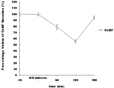
Figure 5. Percentage changes in the CoBF following the admin-istration of 2 μl of 1% L-NAME in the RW. The CoBF decreased
after the RW infusion, reached its lowest point at 2 hour, and then recovered after 3 hour. The data points are means±S.E.
Hearing threshold shift after blast
Hearing threshold shifts (TS) were significant with 87.5 dB (0.5 meter) to 47.5 dB (0.9 meter) (P<0.01), immediately after the blast wave exposure for all distances, followed by a gradu-al recovery course. Although the hearing thresh-old shift also showed a time-dependent pat-tern, the recovery courses were not so quick compared with the CoBF changes. The TS remained at 55 dB (0.5 meter) to 31.25 dB (0.9 meter) even 1 week after the blast exposure. At every time point of the test, the TS values were
significantly different between each dis-tance (Figure 4).
Observation of CoBF and hearing thresh-old shift after L-NAME infusions and blast exposure
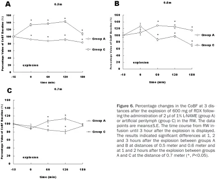
The BP was simultaneously recorded using the same Chart Software after anaestheti-sation until the end of the experiment. The results showed values within the normal range, and the percentage changes were minimal in all animals from the 3 groups. The changes in the CoBF presented differ-ent patterns in the 3 groups during the experiment. Following the L-NAME RW administration, the CoBF decreased and reached its lowest baseline value (percent-age value of change: 55.3±2.8%) (Figure 5) at 2 hours in the animals that were admin-istered L-NAME (group B). This decrease lasted for 3 hours, followed by recovery to the normal level. Varying degrees of the decrease in the CoBF were also detected in the animals that were exposed to the blast at 3 distances after the L-NAME RW admin-istration (group A). Contrasting changes were observed in the animals that were administered artificial perilymph (group C), with the CoBF increasing after the blast exposure (P<0.05) at all 3 distances; these changes lasted for 3 hours. At most of the time points, the differences between groups A and C were significant (Figure 6). The hearing threshold shifts (TS) were sig-nificant for all distances between group A (75.0±7.1 dB at 0.5 meter, 70.0±9.1 dB at 0.6 meter, 56.3±4.8 dB at 0.7 meter) and group C (57.5±6.5 dB at 0.5 meter, 58.8±4.8 dB at 0.6 meter, 42.5±8.7 dB at 0.7 meter). Meanwhile, the TS values were min-imal (1.3±2.3 dB) between before and 3 hours after the L-NAME solution RW infusion in group B (P>0.05).
Discussion
[image:6.612.89.325.307.489.2]Blast pressure can produce a wide range of traumas, especially in air-containing structures. The auditory system has been considered the most susceptible organ and can endure seri-ous injuries after a blast explosion [16]. The severity of the resulting hearing dysfunction is Figure 4. Time course of the hearing threshold shift. The y axis
depicts the hearing threshold shift in dB SPL, and the x axis de-picts the time. The values are means ± S.E.M. The difference
related to the distance from the epicentre in a simple open-space explosion. However, the trauma can be fatal if the injuries occur near a violent explosion, and the management of such trauma would focus on life support measures and non-auditory organ systems. Therefore, a moderate intensity of blast is necessary to evaluate injuries of the auditory system indi-vidually. In this study, we generated a relatively mild blast with a small power explosive (600 mg RDX) and placed the animals at moderate distances (0.5 meter to 0.9 m) corresponding with the medium intensity peak value of the blast overpressure (63.64±5.78 kPa to 23.34±1.52 kPa). There were no obvious signs of injury to the life support systems in all ani-mals when exposed to this blast range, but tym-panic membrane ruptures and damage to the middle ear were observed, as was distortion to the structure of the organ of Corti. However, as the main theme of this study, no distinct dam-age of the microvasculature structure was
detected. The absence of that type of damage suggested that changes in the COBF were more likely to be functional.
[image:7.612.89.528.71.451.2]Similar to its role in other tissues and organs, the microvasculature is a key component of the cochlea. Although the CoBF is estimated to require only on the order of 1/10000 of the total cardiac output in guinea pigs and 1/1000000 in humans [17], a normal blood supply to the cochlea is critically important for maintaining the inner ear potential and sustain-ing the production of endolymph. Moreover, sensory hair cells are vulnerable to ischaemia [18, 19].Although disorders of the CoBF have been considered to be involved in many patho-physiological processes of the inner ear [20-22], the difficulty of direct detection has limited the investigation of CoBF pathological changes, especially after blast injury. Early studies sug-gested that noise can reduce the cochlear blood flow, decrease the red blood cell density Figure 6. Percentage changes in the CoBF at 3 dis-tances after the explosion of 600 mg of RDX follow-ing the administration of 2 μl of 1% L-NAME (group A)
or artificial perilymph (group C) in the RW. The data points are means±S.E. The time course from RW in-fusion until 3 hour after the explosion is displayed.
The results indicated significant differences at 1, 2
and increase the aggregation of red blood cells [10, 11, 23]. However, contrasting results have indicated an elevation in the CoBF after expo-sure to high-intensity noise [24] and blast waves [14].Other researchers have suggested that changes in the cochlear microcirculation are intensity-related [25]. Such inconsistent results are likely due to the diversity in the experiment conditions and test methods. Although the definitions of impulsive noise and blasts are arbitrary, the distinction in the inten-sity, transmission pattern and mechanical fea-tures between them is apparent. Blast waves always have more intensive variation in the resulting pressure and involve the high speed movement of air over a very short time. Consequently, the pathological changes in the cochlear microcirculation induced by a blast may differ from the changes induced by high-intensity noise or impulse noise. In this study, our findings revealed an obvious relationship between the variations in the CoBF and the strength of the blast. Although increases in the CoBF were observed when the peak value of the blast overpressure was greater than 45.68±6.21 kPa, the CoBF was stabilised when the peak value of the blast overpressure was less than 35.05±4.11 kPa. This observation suggested the existence of a threshold for the blast overpressure peak value to cause CoBF changes. Unlike the hearing threshold shift, the time course of the CoBF changes indicated a rapidly recovering process. The baselines of the CoBF recovered to the normal level within 3 to 24 hours. Taking into consideration the histo-pathology changes in the cochlea, such results indicated that the structure of the cochlear microvasculature was not as vulnerable to the blast overpressure compared with other struc-tures of the middle and inner ear, indicating strong autoregulatory ability. This autoregula-tion more likely occurs locally, in agreement with previous studies [17, 26-28].
Although disorders of the CoBF have been con-sidered to be associated with hearing loss in many diseases, the impairment of hearing function induced by a blast involves a few pathologic factors. Most previous studies of blast injuries have focused on the mechanisms involved in the damage to structures of the auditory system and the subsequent hearing loss induced by these damages [29-33]. The role of the CoBF disorder induced by a blast is ambiguous. In this study, we used a NO
syn-thase inhibitor, L-NAME, to inhibit the changes in the CoBF induced by the blast and evaluated the effect of this inhibition on hearing function. As an important regulator of CoBF, nitric oxide (NO) is a potent vasodilator [34-36]. It can cause smooth muscle and pericyte relaxation by activating cGMP and affecting its down-stream targets [37] and can also directly inhibit voltage-gated calcium channels, causing smooth muscle cells to relax [38]. This dilation effect can be inhibited by N-nitro-L-arginine-methyl ester (L-NAME), a NO synthase inhibitor [39]. Such an inhibitory effect is dose-depen-dent, and this effect varies with the regional blood flow in the cochlea via different pathways and administration strategies [40]. In this study, we investigated the changes in the CoBF and evaluated the hearing function after the administration of an L-NAME solution in the RW. A decrease in the CoBF was observed immediately after the administration and last-ed for 3 hour, with the lowest baseline value reaching 55.3±2.8% of the initial level at 2 hour after the administration. In the meantime, this local application did not impact the hearing function or systemic blood pressure, suggest-ing that there is a compensatory range of CoBF with respect to hearing impairment under nor-mal situations. This L-NAME application also inhibited the increase in the CoBF after the blast exposure. In contrast to a normal situa-tion, such inhibition corresponded with a more obvious TS compared with the application of artificial perilymph. These findings suggested that the increase in the blood flow has a posi-tive role in the protection of hearing function, indicating that NO plays an important role in the regulation of the CoBF. Although the CoBF has a relatively wide compensatory range for hearing impairment under normal conditions, the hearing function appears to be more vulner-able to decreases in the CoBF. This observation also implies the importance of blood flow-sup-porting measures in auditory blast trauma. These findings will be helpful to better under-stand the CoBF changes and hearing dysfunc-tion caused by auditory blast injuries and pro-vide potential new methods of the prevention and management of these injuries in the clinic.
Acknowledgements
(NSAF, 10776038). We would like to thank Professor Xinan Lai and Professor Bingcang Li of the Research Institute of Surgery, State Key Laboratory of Trauma, Burns and Combined Injury for their valuable advices at many stages of this work.
Address correspondence to: Dr. Jianmin Wang, State Key Laboratory of Trauma, Burns and Combined Injury, Research Institute of Surgery, Daping Hospital, Third Military Medical University, No.10, Changjiang Zhilu, Chongqing 400042, China. Tel: +86 023 68757461; Fax: +86 023 68757461; E-mail: [email protected]; chenjing9811@ dphospital.tmmu.edu.cn
References
[1] Matsumoto Y, Hatano B, Matsushita Y, Na-washiro H and Shima K. [The characteristics of blast traumatic brain injury]. No Shinkei Geka 2010; 38: 695-702.
[2] Phillips YY and Zajtchuk JT. Blast injuries of the ear in military operations. Ann Otol Rhinol Lar-yngol Suppl 1989; 140: 3-4.
[3] Lockhart P, Cronin D, Williams K and Ouellet S. Investigation of head response to blast load-ing. J Trauma 2011; 70: E29-36.
[4] Chandler DW and Edmond CV. Effects of blast overpressure on the ear: case reports. J Am Acad Audiol 1997; 8: 81-88.
[5] Chen YS, Tseng FY, Liu TC, Lin-Shiau SY and Hsu CJ. Involvement of nitric oxide generation in noise-induced temporary threshold shift in guinea pigs. Hear Res 2005; 203: 94-100. [6] Mazurek B, Haupt H, Georgiewa P, Klapp BF
and Reisshauer A. A model of peripherally de-veloping hearing loss and tinnitus based on the role of hypoxia and ischemia. Med Hypoth-eses 2006; 67: 892-899.
[7] Miller JM, Brown JN and Schacht J. 8-iso-pros-taglandin F(2alpha), a product of noise
expo-sure, reduces inner ear blood flow. Audiol Neu -rootol 2003; 8: 207-221.
[8] Lamm K and Arnold W. The effect of blood flow promoting drugs on cochlear blood flow, peri -lymphatic pO(2) and auditory function in the normal and noise-damaged hypoxic and isch-emic guinea pig inner ear. Hear Res 2000; 141: 199-219.
[9] Sprem N, Branica S and Dawidowsky K. Vaso-dilator and vitamins in therapy of sensorineu-ral hearing loss following war-related blast in-jury: retrospective study. Croat Med J 2001; 42: 646-649.
[10] Attanasio G, Buongiorno G, Piccoli F, Mafera B, Cordier A, Barbara M and Filipo R. Laser
Dop-pler measurement of cochlear blood flow
changes during conditioning noise exposure. Acta Otolaryngol 2001; 121: 465-469. [11] Arpornchayanon W, Canis M, Suckfuell M, Ihler
F, Olzowy B and Strieth S. Modeling the mea-surements of cochlear microcirculation and hearing function after loud noise. Otolaryngol Head Neck Surg 2011; 145: 463-469.
[12] Goldwin B, Khan MJ, Shivapuja B, Seidman MD and Quirk WS. Sarthran preserves cochle-ar microcirculation and reduces temporcochle-ary threshold shifts after noise exposure. Otolar-yngol Head Neck Surg 1998; 118: 576-583. [13] Lamm K and Arnold W. Noise-induced
cochle-ar hypoxia is intensity dependent, correlates with hearing loss and precedes reduction of
cochlear blood flow. Audiol Neurootol 1996; 1:
148-160.
[14] Hu B. [Cochlear microcirculation in living guin-ea pigs following explosion]. Zhonghua Er Bi Yan Hou Ke Za Zhi 1991; 26: 6-9, 61.
[15] Bobbin RP, Jastreboff PJ, Fallon M and Littman T. Nimodipine, an L-channel Ca2+ antagonist, reverses the negative summating potential re-corded from the guinea pig cochlea. Hear Res 1990; 46: 277-287.
[16] Fausti SA, Wilmington DJ, Gallun FJ, Myers PJ and Henry JA. Auditory and vestibular dysfunc-tion associated with blast-related traumatic brain injury. J Rehabil Res Dev 2009; 46: 797-810.
[17] Nakashima T, Naganawa S, Sone M, Tominaga M, Hayashi H, Yamamoto H, Liu X and Nuttall
AL. Disorders of cochlear blood flow. Brain Res
Brain Res Rev 2003; 43: 17-28.
[18] Nuttall AL. Sound-Induced Cochlear Ischemia/ Hypoxia as a Mechanism of Hearing Loss. Noise Health 1999; 2: 17-32.
[19] Wangemann P. Cochlear blood flow regulation.
Adv Otorhinolaryngol 2002; 59: 51-57. [20] Chen GD and Liu Y. Mechanisms of
noise-in-duced hearing loss potentiation by hypoxia. Hear Res 2005; 200: 1-9.
[21] Mazurek B, Rheinlander C, Fuchs FU, Amarjar-gal N, Kuban RJ, Ungethum U, Haupt H,
Ki-etzmann T and Gross J. [Influence of ischemia/
hypoxia on the HIF-1 activity and expression of hypoxia-dependent genes in the cochlea of the newborn rat]. HNO 2006; 54: 689-697. [22] Ohlemiller KK. Mechanisms and genes in
hu-man strial presbycusis from animal models. Brain Res 2009; 1277: 70-83.
[23] Axelsson A and Dengerink H. The effects of noise on histological measures of the cochlear vasculature and red blood cells: a review. Hear Res 1987; 31: 183-191.
[24] Prazma J, Vance SG, Bolster DE, Pillsbury HC
[34] Feletou M and Vanhoutte PM. Endothelium-derived hyperpolarizing factor. Clin Exp Phar-macol Physiol 1996; 23: 1082-1090.
[35] Brechtelsbauer PB, Nuttall AL and Miller JM. Basal nitric oxide production in regulation of
cochlear blood flow. Hear Res 1994; 77:
38-42.
[36] Michel O, Hess A, Bloch W, Stennert E, Su J and Addicks K. Localization of the NO/cGMP-pathway in the cochlea of guinea pigs. Hear Res 1999; 133: 1-9.
[37] Tian F, Fessenden JD and Schacht J. Cyclic GMP-dependent protein kinase-I in the guinea pig cochlea. Hear Res 1999; 131: 63-70. [38] Sakagami K, Kawamura H, Wu DM and Puro
DG. Nitric oxide/cGMP-induced inhibition of calcium and chloride currents in retinal peri-cytes. Microvasc Res 2001; 62: 196-203. [39] Koss MC. Role of nitric oxide in maintenance of
basal anterior choroidal blood flow in rats. In -vest Ophthalmol Vis Sci 1998; 39: 559-564. [40] Hoshijima H, Makimoto K, Noi O, Ohinata Y
and Takenaka H. Effects of nitric oxide
syn-thase inhibitor on cochlear blood flow. Hear
Res 2002; 171: 32-42. Otolaryngol Head Neck Surg 1987; 113:
36-39.
[25] Scheibe F, Haupt H and Ludwig C.
Intensity-re-lated changes in cochlear blood flow in the
guinea pig during and following acoustic expo-sure. Eur Arch Otorhinolaryngol 1993; 250: 281-285.
[26] Nakashima T. Autoregulation of cochlear blood
flow. Nagoya J Med Sci 1999; 62: 1-9.
[27] Brechtelsbauer PB, Ren TY, Miller JM and
Nut-tall AL. Autoregulation of cochlear blood flow in
the hydropic guinea pig. Hear Res 1995; 89: 130-136.
[28] Ren TY, Nuttall AL and Miller JM. Provoked flux motion of cochlear blood flow measured with laser Doppler flowmetry in guinea pig. Acta
Otolaryngol 1993; 113: 609-614.
[29] Cave KM, Cornish EM and Chandler DW. Blast injury of the ear: clinical update from the glob-al war on terror. Mil Med 2007; 172: 726-730. [30] Darley DS and Kellman RM. Otologic consider-ations of blast injury. Disaster Med Public Health Prep 2010; 4: 145-152.
[31] Nageris BI, Attias J and Shemesh R. Otologic and audiologic lesions due to blast injury. J Ba-sic Clin Physiol Pharmacol 2008; 19: 185-191. [32] van de Weyer PS, Praetorius M and Tisch M.
[Update: blast and explosion trauma]. HNO 2011; 59: 811-818.