Original Article
Effect of CTRP3 on activation of adventitial fibroblasts
induced by TGF-β1 from rat aorta in vitro
Shaohui Lin, Shaojun Ma, Ping Lu, Wenwei Cai, Yi Chen, Jing Sheng
Department of Geriatrics, Ninth People’s Hospital Affiliated to Shanghai Jiao Tong University School of Medicine, Shanghai, China
Received March 21, 2014; Accepted April 10, 2014; Epub April 15, 2014; Published May 1, 2014
Abstract: CTRP3, discovered as novel adipokines, is a member of the C1q tumor necrosis factor (TNF) related protein (CTRP) super-family. CTRP3 is found to function as adipokines that display diverse biological activities in metabolic and cardiovascular diseases. Recent study demonstrated that CTRP3 was protective against pathologi-cal cardiac remodeling in mice. Nevertheless, the effect of CTRP3 on vascular remodeling remains undefined. Our present study aimed to explore the effects of adipokine CTRP3 on the activation of adventitial fibroblasts (AFs) induced by TGF-β1. Immunofluorescent staining, real-time PCR and Western blot were conducted to evaluate the expression of α-smooth muscle-actin (α-SMA) and collagen I. The expression of CTGF was evaluated by enzyme-linked immunosorbent assay (ELISA), while the proliferation and migration of adventitial fibroblasts were detected by using cell counting kit-8 (CCK-8) assay and Transwell technique, respectively. Functional analysis showed that CTRP3 inhibited TGF-β1 inducing AFs phenotypic conversion, collagen synthesis, proliferation and migration. The secretion of CTGF was also inhibited by CTRP3. Our findings suggest that CTRP3 may be beneficial to the prevention of cardiovascular diseases and provide a promising therapeutic strategy to attenuate vascular remodeling.
Keywords: Adventitial fibroblasts, myofibroblasts, vascular remodeling, CTRP3, phenotypic conversion
Introduction
Cardiovascular disease is the major reason of morbidity and mortality in the industrialized world [1]. Vascular remodeling, which repre-sents any enduring changes in the size and/or composition of an adult blood vessel is a com-mon feature of various cardiovascular diseas-es, such as atherosclerosis, restenosis after angioplasty and hypertension [2]. As the impor-tance of pathological vascular remodeling in cardiovascular diseases is increasingly recog-nized, the prevention of inappropriate remodel-ing has become a heated research topic in the field of cardiology. It is recently reported that studies targeted on modifying TGF-β1 may give rise to promising strategies in the treatment of pathological vascular remodeling [3]. Never- theless, presently no drugs are specifically tar-geted on attenuating the pathological vascular remodeling in clinic.
Traditionally, adventitial was regarded as a sup-porting connective tissue that provide
tivation may provide a potential therapeutic strategy for vascular diseases. It is well estab-lished that transforming growth factor-β1 (TGF-β1) is one of the most key regulator of vascular fibrosis by inducing the activation of the fibro-blasts [6]. Furthermore, TGF-β1 contributes to constructive remodeling through direct effects or via induction of CTGF [3, 7].
Most recently, C1q/TNF-related proteins (CT- RPs), a structural and functional adiponectin paralogs, have been discovered. Some mem-bers of CTRPs are found to display diverse bio-logical activities in metabolic and cardiovascu-lar diseases. CTRP3 was first identified as a secreted protein induced in a murine mesen-chymal stem cell line. This novel protein plays an important role in regulating cartilage devel-opment and adiponectin secretion [8, 9]. Besides, CTRP3 also exerts an impact on the differentiation and growth of several types of cells, including endothelial, osteosarcoma and vascular smooth muscle cells [10, 11]. Mor- eover, CTRP3 displays diverse biological activi-ties in the context of metabolic and cardiovas-cular diseases. CTRP3 also has been reported to improve glucose metabolism in obese mice by suppressing gluconeogenesis and activating Akt signaling in the liver [12]. Recent research-es have showed that, like adiponectin, circulat-ing CTRP3 level was reduced in diet-induced obese mice [13]. Yi et al [14] discovered that the adipokine CTRP3 was protective against pathological cardiac remodeling and CTRP3 replenishment dramatically increased the ratio of myocytes to fibrotic cells in the ischemic zone and significantly attenuated interstitial fibrosis. In addition, Li et al [15] found that the expression of CTRP3 gene was up-regulated after balloon-injure of rat carotid artery tissue. Taken together, these studies highlight a novel and significant role for CTRP3 in modulating cardiovascular functions.
A recent research has revealed that some CTRPs members, such as CTRP9, play a protec-tive role in the heart against ischemia injury [16, 17], as well as attenuating vascular remod-eling in response to vascular injury [18]. However, whether CTRP3, a key member of the newest adipokine family, may function as an inhibitor of pathological vascular remodeling has never been investigated. In the current
study, we examined the effect of CTRP3 on the phenotypic conversion of adventitial fibroblasts to myfibroblasts, collagen production, cell pro-liferation, migration and the expression of CTGF induced by TGF-β1 in vitro to explore the potent effects of CTRP3 on arterial remodeling. These findings provide new insights about the preven-tion and treatment of vascular remodeling.
Materials and methods
Cell culture
The adventitial fibroblasts were cultured as described by Zhang et al and Kim et al. [19, 20]. Briefly, Sprague–Dawley rats were anesthe-tized with chloral hydrate and rapidly decapi-tated. Adventitia was carefully removed from rat thoracic aorta and minced into small pieces (1-2 mm2). The adventitia tissue pieces were
then placed on culture flasks and cultured with Dulbecco’s modified Eagle’s medium (pH 7.2-7.4) containing 20% fetal calf, and maintained in a humidified incubator with 95% air and 5% CO2 at 37°C. Fibroblasts migrated from the edge of tissues within 7-10 days. Once reached confluence, the isolated cells were harvested and used for experiments at passages 3 to 5. Before each experiment, cells were placed in DMEM containing 0.5% fetal bovine serum (FBS) for 4 h for serum starvation. AFs were pretreated by recombinant CTRP3 protein (10 μg/ml)or vehicle for 16 h, followed by stimula-tion with DMEM containing 10% FBS with or without TGF-β1 (10 ng/ml) for 24 h. All experi-mental protocols were approved by Institutional Animal Care and Use Committee (the Shanghai Jiao Tong University).
Immunofluorescent staining for α-SMA
secondary antibody for 1 h at room tempera-ture. Finally, cell cover slips were labeled by DAPI. The fluorescent images were captured using a confocal laser scanning microscope. Real-time reverse transcriptase–polymerase
chain reaction
Total RNA was isolated from cultured cells with RNeasy Mini Kit (Qiagen), according to the man-ufacturer’s protocol. The concentration of the total RNA was detected. Total RNA (1 ug) were reverse-transcribed into cDNA in 20 µl reaction volumes using the Prime Script RT reagent kit (Takara, China). The Real-time PCR was per-formed with a Stratagene Mx3000p (Agilent Technologies, La Jolla, CA) detection system, using SYBR Green reaction mix (Takara, China) in the 20 µl reaction mixtures. The mRNA levels were normalized by GAPDH housekeeping gene. The PCR primers used were as follows: α-SMA forward: 5’-GGA GTG ATG GTT GGA ATG G-3’ and reverse: 5’-ATG ATG CCG TGT TCT ATC G-3’; COL-I forward: 5’-TGC CGT GAC CTC AAG ATG TG-3’ and reverse: 5’-CAC AAG CGT GCT GTA GGT GA-3’; GAPDH forward: 5’-GAA CGG GAA GCT CAC TGG C-3’and reverse: 5’-GCA TGT CAG ATC CAC AAC GG-3’. The reaction was run at 95°C for 30 s, followed by 40 cycles of 30 sec at 95°C and 30 sec at 60°C.
Western blot analysis
After appropriate treatments, cells were har-vested with lysis buffer. Protein concentrations of cell lysates were measured by BCA Protein Assay Kit (Pierce, IL, USA). Equal protein am- ounts from each sample were separated by SDS-PAGE on 10% polyacrylamide gels, fol-lowed by transferring onto nitrocellulose mem-branes. The membranes were blocked in TBST containing 5% milk for 1 h at room temperature, and then the membrane was incubated with α-SMA, collagen I monoclonal antibodies over-night at 4°C. Membranes were washed three times with TBST, and then incubated for 2 h at room temperature with a horseradish-coupled secondary antibody. Bands were detected using the ECL Western blotting detection kit. Signals were analyzed with the image analysis software Image Pro Plus 6.0 [21].
Cell proliferation assay
AFs were added into 96-well plates at 5.0×10⁴/
ml cells per well in a total volume of 100 µl
cul-ture medium. The effect of CTRP3 on AFs viabil-ity after various treatments was determined by cell counting kit-8 (CCK-8, Dojindo, Japan) assay. According to the protocol, 10 µl of CCK-8 was added to each well and cultured for 4 h at 37°C. The absorbance value at 450 nm was measured, using a microplate reader (Bio-Tek, VT, USA).
Migration assay
Cell migration assay was performed using a transwell system (Corning, New York, USA), as described previously [22], which allows cells to migrate through a polycarbonate membrane with 8-um pore size. In brief, AFs pretreated with or without recombinant CTRP3 protein for 16 h were plated into the upper chamber, while the bottom chamber filled with 500 µl of DMEM containing 10% FBS with or without TGF-β1. After 24 h of incubation, the cells that had migrated through the pores were stained with DAPI. Quantification was performed by count-ing five random fields under the microscope. Elisa analysis
Detection of CTGF with ELISA was performed using CTGF enzyme linked immunosorbent assay kit (Biovalue, China). After incubation, the samples were collected and centrifuged, and the levels of CTGF were measured. OD value was read at 450 nm by ELISA reader and the concentration of CTGF was calculated from a standard curve diagram. Dates were dis-played as mean of CTGF concentration (μg/ml). Statistical analysis
All measures were expressed as the mean ± SD. The software of SPSS version 19.0 was used to analyze the data. The results were ana-lyzed by using one-way analysis of variance fol-lowed by t-test. A value of P < 0.05 was consid-ered to be statistically significant.
Results
CTRP3 inhibits TGF-β1 induced phenotypic conversion of fibroblasts
mea-sured. To evaluate the role of CTRP3 in fibro-blasts differentiating to myofibroblast, Imm- unofluorescence staining, Western blot and Real-time PCR were utilized to examine the expression of α-SMA. The expression of α-SMA was weakly stained in adventitia fibroblasts. As shown in Figure 1A, the immunofluorescence levels of α-SMA was increased after treatment of TGF-β1, which was significantly reduced by CTRP3 pretreatment. The similar results were also observed by analysis the mRNA and pro-tein expression levels of α-SMA. It appears that the treatment of adventitia fibroblasts with CTRP3 could significantly reduced the
expres-sion levels of α-SMA mRNA and protein induced by TGF-β1.
CTRP3 attenuated TGF-β1 induced adventitial fibroblasts proliferation and migration
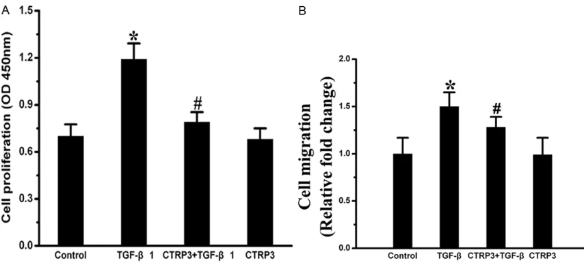
The proliferation of AFs was induced by the TGF-β1 stimulation. The OD values were signifi-cantly higher in the TGF-β1 group compared with the control group (P < 0.05). However, compared with the TGF-β1 group, CCK-8 assay showed that the OD values in the CTRP3 pre-treated group were decreased markedly (Figure
[image:4.612.90.523.71.465.2]analyzed by Trans-well migration assay (Figure
2B). CTRP3 reduced AFs migration induced by TGF-β1 than those treated with TGF-β1 alone. These results suggested a significant contribu-tion of CTRP3 to lessening TGF-β1 induced of AFs proliferation and migration.
Effect of CTRP3 on type I collagen expression of adventitial fibroblasts
The effects of CTRP3 on collagen I mRNA and protein expression were evaluated by Real-time PCR and Western blot. We found that TGF-β1 remarkably increased collagen I mRNA and pro-tein levels. In contrast, the pretreated adventi-tia fibroblasts with CTRP3 exhibited a down-regulation of collagen I mRNA and protein lev- els (Figure 3).
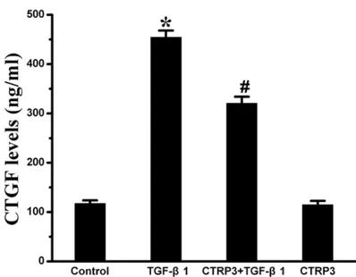
CTRP3 suppressed the expression of CTGF induced by TGF-β1
The levels of CTGF were detected by ELISA analysis. Results are depicted in Figure 4. The results demonstrated that the content of CTGF in the four groups were 117.44 ± 6.33, 454.30 ± 13.85, 320.87 ± 13.07, and 114.7 ± 8.04 ng/mL, respectively. The content of CTGF in the CTRP3 pretreatment group was markedly lower than that in TGF-β1 group (P < 0.05).
Discussion
Pathological vascular remodeling is a keystone of diverse cardiovascular diseases including
hypertension, atherosclerosis and post-angio-plasty restenosis [23]. Thus, it is necessary to find effective methods to prevent and reverse the pathological vascular remodeling. Recent study demonstrates that the adipokine CTRP3 possesses protective properties on cardiac remodeling. However, there is little known about the effect of CTRP3 on vascular remodel-ing. Our present investigation provided the first evidence that CTRP3 could directly regulate the activation of AFs stimulated by TGF-β1. The results showed that CTRP3 suppressed pheno-typic conversion of AFs to MFs and inhibited collagen synthesis and the proliferation and migration of AFs induced by TGF-β1, elucidating a protective role of CTRP3 in pathological vas-cular remodeling by modulating the responses of adventitial fibroblasts. In addition, CTRP3 exerts an effect on the secretion of CTGF, which implies a role for CTGF in vessel protection induced by CTRP3.
[image:5.612.90.517.69.263.2]involved in cardiovascular disease, including mechanical stress, high glucose [26, 27]. It regulates a variety of pathophysiological and physiological processes including cell prolifera-tion, migration and differentiation. Under the in- fluence of high levels of TGF-β1, adventitial fibroblasts can be activated and contribute to vascular remodeling. In experimental models, down expression of the TGF-β1 gene could attenuate these events and thereby prevent constrictive remodeling and neointima hyper-plasia after vascular injury [28, 29]. Therefore, antagonism of vascular TGF-β1 signaling is con-sidered as a putative therapeutic target in dis-orders of the blood-vessel interface.
The activated adventitial fibroblasts, known as myofibroblasts, play important roles in the pathological vascular remodeling [30]. The switched phenotypic of adventitial fibroblasts to myofibroblasts is considered as a hallmark of vascular remodeling following vascular injury and is critical in fibroblasts aviation induced by TGF-β1 [31, 32]. Several studies have docu-mented that the activated myofibroblasts could elevate the proliferation, migration and extra-cellular matrix deposition. Myofibroblasts are characterized by the appearance of α-SMA and production of ECM components [33, 34]. They are metabolically active cells and can perform multiple cellular functions in response to cha- nges in local environments [35]. The
accumula-tion of myofibroblasts can contribute to chang-es in the structure and tone of the vchang-essel wall under pathophysiologic conditions [36, 37]. Collagen is a major component of ECM and accounts for more than fifty percent of vascular wall with stenosis [38]. Among all the collagen subtypes, type I collagen is considered to be most relevant to arterial remodeling. Synthesis and accumulation of collagen are essential for vascular homeostasis, development, and wou- nd healing. However, excessive collagen expres-sion and deposition can lead to fibrotic disor-ders. AS an active component of the vessel wall, ECM plays an important role in the patho-physiology of vascular diseases. Considerable studies have demonstrated TGF-β1 could induce fibroblasts into myofibroblasts by stimu-lating α-SMA expression [39] and promoting the secretion of ECM proteins, which have been found to play a vital role in the process of path-ological vascular remodeling. In accordance with previous findings, the α-SMA mRNA and protein expression levels is obviously up-regu-lated in AFs stimuup-regu-lated by TGF-β1. Moreover, we observed that pretreatment of adventitial fibroblasts with CTRP3 markedly decreased the expression of α-SMA and type I collagen, which were induced by TGF-β1. These findings sug-gest that CTRP3 could blunt vascular remodel-ing by inhibitremodel-ing the differentiation of adventi-tial fibroblasts and secretion of type I collagen. Figure 3. CTRP3 weakens TGF-β1-induced collagen synthesis. A: Collagen I protein levels were assessed by Western blot. GAPDH was a loading control. B: Level of quantification of Collagen I as a ratio of GAPDH in densitometric units was presented. C: Collagen I mRNA levels were assessed by quantitative real-time PCR.
*P < 0.05 versus the control group; #P < 0.05 versus
[image:6.612.94.518.71.337.2]The proliferative and migratory responses of cells in the adventitia contribute to vascular remodeling in restenosis [24]. The proliferation and migration of myofibroblasts are critical cel-lular events in the development of constructive remodeling [40]. TGF-β1 is reported to be one of the most key factors that induce the prolif-eration and migration of AFs in response to vas-cular injury. Previous researches demonstrated blockage of TGF-β1 signaling pathway could markedly prevent fibroblast proliferation [41]. Previous studies showed that adventitia cell migration play a more vital role than cell prolif-eration in vascular remodeling [22]. Thus, we investigated the effects of CTRP3 on AFs prolif-eration and migration. Our present study revealed that TGF-β1 indeed up-regulated cell proliferation and migration in adventitial fibr- oblasts, which was consistent with former stud-ies. Interestingly, we found all these changes were noticeable attenuated by treatment with CTRP3. Taken together, these results suggest-ed a significant effect for CTRP3 in regulating fibroblasts growth and migration during vascu-lar remodeling.
To further examine whether CTGF is also involved in the process, we studied the secre-tion of CTGF in TGF-β1 induced AFs pretreated with or without CTRP3. CTGF, which is believed to be a mediator of the profibrogenic effects of TGF-β1, plays a critical role in the architectural changes, followed by the vessel injury [42]. CTGF modulates different signal transduction pathways by interacting with various molecules,
including cytokines and receptors, matrix pro-teins and growth factors, which result in differ-ent changes in cellular responses. In vitro stud-ies have shown CTGF not only induces the activation of fibroblasts and enhances extracel-lular matrix production but also regulates the activity of TGF-β1 [43], which all lead to vascu-lar remodeling and fibrosis. It has been report-ed that the maintenance of vascular function was modulated by interplay of TGF-β1 and CTGF [44]. CTGF could regulate the TGF-β1 signal pathway and contribute to the activation of AFs [42, 45]. Serving as an extracellular adapter protein, CTGF activates TGF-β signals by direct binding to TGF-β1 receptors in the extracellular space and then helps to present them to their receptors to stimulate cellular response. The previous research found that CTRP3 exerts potent anti-fibrotic and anti-inflammatory effects in Human primary colonic lamina pro-pria fibroblasts by inhibiting the expression of collagen I and CTGF. Our study showed the simi-lar results that CTRP3 obviously decreased the content of CTGF protein in cultured AFs induced by TGF-β1, although it was unable to suppress the basal level of CTGF protein expression. Added to previous findings that CTGF can induce fibroblasts activation and have a close interaction with TGF-β1, the findings above sug-gest that, CTRP3 may suppress the activation of AFs provoked by TGF-β1, at least in part, by directly inhibiting the CTGF protein secretion or by inducing genes that are involved in the sup-pression of CTGF. Our present investigation reveals a model of molecular crosstalk and support the active role for CTGF in vascular remodeling.
[image:7.612.88.287.67.222.2]In conclusion, this study first demonstrated the effect of CTRP3 on the activation of adventitial fibroblasts. CTRP3 could prevent TGF-β1 ind- uced adventitial fibroblasts proliferation, migra-tion, phenotypic conversion, collagen synthesis and the expression of CTGF. The discoveries above have raised the prospect that CTRP3 might ameliorate pathological vascular remod-eling by manipulating the responses of adventi-tial fibroblasts, in which CTGF may be involved. Collectively, the approaches aimed at enhanc-ing CTRP3 levels may be a logical strategy for therapies in the alleviation of vascular remodel-ing. However, further investigation is required to prove the viability of therapeutic approach in humans.
Figure 4. ELISA assay was used to determine the ex-pression of CTGF. Results are presented as the mean ± SD. *P < 0.05 versus the control group; #P < 0.05
Acknowledgements
This project was supported by the Science and Technology Commission of Shanghai Mun- icipality (10JC1408902).
Disclosure of conflict of interest
None.
Address correspondence to: Dr. Jing Sheng, Depa- rtment of Geriatrics, Ninth People’s Hospital Aff- iliated to Shanghai Jiao Tong University School of Medicine, 639 Zhi Zao Ju Road, Shanghai 200011, China. Tel: +86-13651763192; Fax: 86-21-631368 56; E-mail: [email protected]
References
[1] Roger VL, Go AS, Lloyd-Jones DM, Adams RJ, Berry JD, Brown TM, Carnethon MR, Dai S, de Simone G, Ford ES, Fox CS, Fullerton HJ, Gil-lespie C, Greenlund KJ, Hailpern SM, Heit JA, Ho PM, Howard VJ, Kissela BM, Kittner SJ, Lackland DT, Lichtman JH, Lisabeth LD, Makuc DM, Marcus GM, Marelli A, Matchar DB, Mc-Dermott MM, Meigs JB, Moy CS, Mozaffarian D, Mussolino ME, Nichol G, Paynter NP, Rosa-mond WD, Sorlie PD, Stafford RS, Turan TN, Turner MB, Wong ND, Wylie-Rosett J; American Heart Association Statistics Committee and Stroke Statistics Subcommittee. Heart disease and stroke statistics-2011 update: a report from the American Heart Association. Circula-tion 2011; 123: e18-e209.
[2] Gibbons GH and Dzau VJ. The emerging con-cept of vascular remodeling. N Engl J Med 1994; 330: 1431-1438.
[3] Goel SA, Guo LW, Liu B and Kent KC. Mecha-nisms of post-intervention arterial remodel-ling. Cardiovasc Res 2012; 96: 363-371. [4] Sartore S, Chiavegato A, Faggin E, Franch R,
Puato M, Ausoni S and Pauletto P. Contribution of adventitial fibroblasts to neointima forma-tion and vascular remodeling: from innocent bystander to active participant. Circ Res 2001; 89: 1111-1121.
[5] Stenmark KR, Nozik-Grayck E, Gerasimovska-ya E, Anwar A, Li M, Riddle S and Frid M. The adventitia: Essential role in pulmonary vascu-lar remodeling. Compr Physiol 2011; 1: 141-161.
[6] Sorescu D. Smad3 mediates angiotensin II- and TGF-beta1-induced vascular fibrosis: Smad3 thickens the plot. Circ Res 2006; 98: 988-989.
[7] Lan TH, Huang XQ and Tan HM. Vascular fibro-sis in atherosclerofibro-sis. Cardiovasc Pathol 2013; 22: 401-407.
[8] Maeda T, Jikko A, Abe M, Yokohama-Tamaki T, Akiyama H, Furukawa S, Takigawa M and Waki-saka S. Cartducin, a paralog of Acrp30/adipo-nectin, is induced during chondrogenic differ-entiation and promotes proliferation of chon- drogenic precursors and chondrocytes. J Cell Physiol 2006; 206: 537-544.
[9] Akiyama H, Furukawa S, Wakisaka S and Mae-da T. Cartducin stimulates mesenchymal chon-droprogenitor cell proliferation through both extracellular signal-regulated kinase and phos-phatidylinositol 3-kinase/Akt pathways. FEBS J 2006; 273: 2257-2263.
[10] Yokohama-Tamaki T, Maeda T, Tanaka TS and Shibata S. Functional analysis of CTRP3/cart-ducin in Meckel’s cartilage and developing condylar cartilage in the fetal mouse mandi-ble. J Anat 2011; 218: 517-533.
[11] Maeda T and Wakisaka S. CTRP3/cartducin is induced by transforming growth factor-beta1 and promotes vascular smooth muscle cell proliferation. Cell Biol Int 2010; 34: 261-266. [12] Peterson JM, Wei Z and Wong GW.
C1q/TNF-related protein-3 (CTRP3), a novel adipokine that regulates hepatic glucose output. J Biol Chem 2010; 285: 39691-39701.
[13] Yoo HJ, Hwang SY, Hong HC, Choi HY, Yang SJ, Choi DS, Baik SH, Bluher M, Youn BS and Choi KM. Implication of progranulin and C1q/TNF-related protein-3 (CTRP3) on inflammation and atherosclerosis in subjects with or without metabolic syndrome. PLoS One 2013; 8: e55744.
[14] Yi W, Sun Y, Yuan Y, Lau WB, Zheng Q, Wang X, Wang Y, Shang X, Gao E, Koch WJ and Ma XL. C1q/tumor necrosis factor-related protein-3, a newly identified adipokine, is a novel antiapop-totic, proangiogenic, and cardioprotective mol-ecule in the ischemic mouse heart. Circulation 2012; 125: 3159-3169.
[15] Li JM, Zhang X, Nelson PR, Odgren PR, Nelson JD, Vasiliu C, Park J, Morris M, Lian J, Cutler BS and Newburger PE. Temporal evolution of gene expression in rat carotid artery following bal-loon angioplasty. J Cell Biochem 2007; 101: 399-410.
[16] Kambara T, Ohashi K, Shibata R, Ogura Y, Maruyama S, Enomoto T, Uemura Y, Shimizu Y, Yuasa D, Matsuo K, Miyabe M, Kataoka Y, Mu-rohara T and Ouchi N. CTRP9 protein protects against myocardial injury following ischemia-reperfusion through AMP-activated protein ki-nase (AMPK)-dependent mechanism. J Biol Chem 2012; 287: 18965-18973.
ex-acerbated cardiac injury in diabetic mice. Basic Res Cardiol 2013; 108: 315.
[18] Uemura Y, Shibata R, Ohashi K, Enomoto T, Kambara T, Yamamoto T, Ogura Y, Yuasa D, Joki Y, Matsuo K, Miyabe M, Kataoka Y, Muro-hara T and Ouchi N. Adipose-derived factor CTRP9 attenuates vascular smooth muscle cell proliferation and neointimal formation. FASEB J 2013; 27: 25-33.
[19] Zhang YG, Li J, Li YG and Wei RH. Urotensin II induces phenotypic differentiation, migration, and collagen synthesis of adventitial fibro-blasts from rat aorta. J Hypertens 2008; 26: 1119-1126.
[20] Kim DK, Huh JE, Lee SH, Hong KP, Park JE, Seo JD and Lee WR. Angiotensin II stimulates prolif-eration of adventitial fibroblasts cultured from rat aortic explants. J Korean Med Sci 1999; 14: 487-496.
[21] Liu H, Lin S, Xiao D, Zheng X, Gu Y and Guo S. Evaluation of the Wound Healing Potential of Resina Draconis (Dracaena cochinchinensis) in Animal Models. Evid Based Complement Al-ternat Med 2013; 2013: 709865.
[22] Li L, Zhu DL, Shen WL and Gao PJ. Increased migration of vascular adventitial fibroblasts from spontaneously hypertensive rats. Hyper-tens Res 2006; 29: 95-103.
[23] Heeneman S, Sluimer JC and Daemen MJ. An-giotensin-converting enzyme and vascular re-modeling. Circ Res 2007; 101: 441-454. [24] Schwartz SM, Reidy MA and O’Brien ER.
As-sessment of factors important in atheroscle-rotic occlusion and restenosis. Thromb Hae-most 1995; 74: 541-551.
[25] Shi Y, O’Brien JE Jr, Fard A and Zalewski A. Transforming growth factor-beta 1 expression and myofibroblast formation during arterial re-pair. Arterioscler Thromb Vasc Biol 1996; 16: 1298-1305.
[26] Li JH, Huang XR, Zhu HJ, Johnson R and Lan HY. Role of TGF-beta signaling in extracellular matrix production under high glucose condi-tions. Kidney Int 2003; 63: 2010-2019. [27] Li JH, Huang XR, Zhu HJ, Oldfield M, Cooper M,
Truong LD, Johnson RJ and Lan HY. Advanced glycation end products activate Smad signal-ing via TGF-beta-dependent and independent mechanisms: implications for diabetic renal and vascular disease. FASEB J 2004; 18: 176-178.
[28] Nikol S, Isner JM, Pickering JG, Kearney M, Leclerc G and Weir L. Expression of transform-ing growth factor-beta 1 is increased in human vascular restenosis lesions. J Clin Invest 1992; 90: 1582-1592.
[29] Majesky MW, Lindner V, Twardzik DR, Schwartz SM and Reidy MA. Production of transforming growth factor beta 1 during repair of arterial injury. J Clin Invest 1991; 88: 904-910.
[30] Powell DW, Mifflin RC, Valentich JD, Crowe SE, Saada JI and West AB. Myofibroblasts. I. Para-crine cells important in health and disease. Am J Physiol 1999; 277: C1-9.
[31] Forte A, Della Corte A, De Feo M, Cerasuolo F and Cipollaro M. Role of myofibroblasts in vas-cular remodelling: focus on restenosis and an-eurysm. Cardiovasc Res 2010; 88: 395-405. [32] Coen M, Gabbiani G and Bochaton-Piallat ML.
Myofibroblast-mediated adventitial remodel-ing: an underestimated player in arterial pa-thology. Arterioscler Thromb Vasc Biol 2011; 31: 2391-2396.
[33] Swaney JS, Roth DM, Olson ER, Naugle JE, Meszaros JG and Insel PA. Inhibition of cardiac myofibroblast formation and collagen synthe-sis by activation and overexpression of adeny-lyl cyclase. Proc Natl Acad Sci U S A 2005; 102: 437-442.
[34] Hinz B, Phan SH, Thannickal VJ, Galli A, Bocha-ton-Piallat ML and Gabbiani G. The myofibro-blast: one function, multiple origins. Am J Pathol 2007; 170: 1807-1816.
[35] Gabbiani G. The myofibroblast in wound heal-ing and fibrocontractive diseases. J Pathol 2003; 200: 500-503.
[36] Hinz B. Formation and function of the myofibro-blast during tissue repair. J Invest Dermatol 2007; 127: 526-537.
[37] Shi-Wen X, Chen Y, Denton CP, Eastwood M, Renzoni EA, Bou-Gharios G, Pearson JD, Dash-wood M, du Bois RM, Black CM, Leask A and Abraham DJ. Endothelin-1 promotes myofibro-blast induction through the ETA receptor via a rac/phosphoinositide 3-kinase/Akt-dependent pathway and is essential for the enhanced contractile phenotype of fibrotic fibroblasts. Mol Biol Cell 2004; 15: 2707-2719.
[38] Lu P, Wang S, Cai W and Sheng J. Role of TGF-beta1/Smad3 signaling pathway in secretion of type I and III collagen by vascular smooth muscle cells of rats undergoing balloon injury. J Biomed Biotechnol 2012; 2012: 965953. [39] Zhou HY, Chen WD, Zhu DL, Wu LY, Zhang J,
Han WQ, Li JD, Yan C and Gao PJ. The PDE1A-PKCalpha signaling pathway is involved in the upregulation of alpha-smooth muscle actin by TGF-beta1 in adventitial fibroblasts. J Vasc Res 2010; 47: 9-15.
[40] Wernig F and Xu Q. Mechanical stress-induced apoptosis in the cardiovascular system. Prog Biophys Mol Biol 2002; 78: 105-137.
[41] Kuwahara F, Kai H, Tokuda K, Kai M, Takeshita A, Egashira K and Imaizumi T. Transforming growth factor-beta function blocking prevents myocardial fibrosis and diastolic dysfunction in pressure-overloaded rats. Circulation 2002; 106: 130-135.
and Kent KC. Arterial gene transfer of the TGF-beta signalling protein Smad3 induces adap-tive remodelling following angioplasty: a role for CTGF. Cardiovasc Res 2009; 84: 326-335. [43] Grotendorst GR and Duncan MR. Individual
do-mains of connective tissue growth factor regu-late fibroblast proliferation and myofibroblast differentiation. FASEB J 2005; 19: 729-738. [44] Beddy D, Mulsow J, Watson RW, Fitzpatrick JM
and O’Connell PR. Expression and regulation of connective tissue growth factor by trans-forming growth factor beta and tumour necro-sis factor alpha in fibroblasts isolated from strictures in patients with Crohn’s disease. Br J Surg 2006; 93: 1290-1296.