www.impactjournals.com/oncotarget/ Oncotarget, Vol. 7, No. 26
Schlafen2 mutation unravels a role for chronic ER stress in the
loss of T cell quiescence
Ibrahim Omar1, Antonio Lapenna1, Leonor Cohen-Daniel1, Boaz Tirosh2 and
Michael Berger1
1 The Lautenberg Center for Immunology and Cancer Research, The Biomedical Research Institute Israel-Canada of the
Faculty of Medicine, The Hebrew University Hadassah Medical School, Jerusalem, Israel
2 Institute for Drug Research, School of Pharmacy, Faculty of Medicine, Hebrew University of Jerusalem, Jerusalem, Israel
Correspondence to: Michael Berger, email: michaelb@ekmd.huji.ac.il
Keywords: ER stress, UPR, Slfn2, quiescence, XBP1, Immunology and Microbiology Section, Immune response, Immunity
Received: May 08, 2016 Accepted: May 22, 2016 Published: June 03, 2016
AbstrAct
Immunologically naïve lymphocytes are kept in a quiescent state until antigen engagement. These quiescent immune cells are characterized by small cell size, lack of spontaneous proliferation and low metabolic rate. Lymphocyte quiescence is actively enforced condition which ensures the preservation of proper differentiation and proliferation capabilities of naïve and memory lymphocytes. Previously we described a chemically induced mutation in Schlafen2 (Slfn2), termed elektra, which breaks quiescence and compromises immunity. However, the mechanism by which Slfn2 maintains quiescence remains unknown. Here we demonstrate that elektra T cells display chronic ER stress under steady state conditions. Modulation of ER stress response by depletion of either UPR mediators XBP1 or CHOP, improved viability and partially corrected the developmental abnormalities and proliferation capabilities of elektra T cells. Altogether, our results demonstrate a functional connection between Slfn2 induced quiescence in T cells and ER homeostasis, clarifying a novel mechanism by which immune cell quiescence is maintained.
IntroductIon
The endoplasmic reticulum (ER) is the entry site of proteins into the secretory pathway and a central organelle for lipid synthesis. To acquire a native state in the ER, newly translated proteins undergo post translation
modifications and are engaged by molecular chaperones. A disruption of ER homeostasis by physiological,
pathogenic or chemical insults leads to the accumulation of unfolded or misfolded proteins, conditions termed ER stress [1]. ER stress elicits an unfolded protein response (UPR), a signaling pathway that emanates from the ER, aimed to restore ER function [2]. The UPR is mediated
through the activation of three ER transmembrane stress
sensors; pancreatic ER kinase (PKR)-like ER kinase (PERK), activating transcription factor 6 (ATF6) and inositol-requiring enzyme 1 (IRE1) [3]. These proteins are maintained in an inactive state through interaction with the ER chaperone, glucose regulated protein 78 (GRP78, also known as Bip). Unfolded proteins can interact directly with IRE1 to mediate its activation or trigger the release of GRP78 from the UPR sensors allowing their activation
[4]. Activated IRE1, PERK and ATF6 promote adaptation to ER stress through the activation of transcriptional
upregulation of key ER chaperones as well as by
controlling protein synthesis through the phosphorylation
of eIF2α. The transcriptional arm of the UPR adjusts ER
folding capacity, enhances ER-associated degradation (ERAD) and drives the expansion of ER volume [5]. However, if ER stress is not alleviated in a timely manner, the UPR promotes apoptosis via multiple mechanisms. The switch from pro-survival to pro-apoptotic roles depends on the cell type and the type of insults [6]. One of the
key factors in maintaining this balance is the activation
of the transcription repressor CHOP, of which level is
controlled by all UPR arms, primarily by PERK through ATF4 induction [7]. CHOP has been shown to alter the balance between pro-survival and pro-apoptotic Bcl-2 family members [8, 9] and death receptor 5 (DR5) thus
promoting apoptosis through the mitochondrial pathway
[10]. Depletion of CHOP in both animal models and cell lines showed a significant reduction in ER stress-induced apoptosis [7]. IRE1 possess both pro-survival
and pro-death roles. Dependent on its kinase activity, IRE1
activates TNF receptor associated factor 2 (TRAF2) and JNK, which ultimately signals to apoptosis. This activity of IRE1 occurs in parallel to its nuclease activity which mediates the splicing of XBP1 mRNA to generate the transcriptionally active form of XBP1 protein. Depending on the stress strength and persistency as well as on the cell type, active XBP1 can drive either pro-survival or pro-death transcriptional networks [11-15]. XBP1 mRNA
is not the only substrate for IRE1 nuclease activity. A
growing list of mRNA and miRNA molecules was found
to be targeted for degradation by IRE1, in a manner critical
to cell survival [16]. Importantly, constitutive ER stress conditions, termed chronic ER stress, usually arise on the
background of severe disturbance of homeostasis and are
rarely seen physiologically.
Survival of naive newly thymic emigrants T cells
is dependent on their ability to maintain quiescence. Quiescent immune cells are characterized by small cell size, lack of spontaneous proliferation and low metabolic rate [17-19]. Acquisition of this state is important to
provide stress free environment and resistance to apoptosis while keeping the naïve T cells responsive to activating
stimuli [17, 19-22].
Previously, we described a mouse strain bearing a
Slfn2-mutated allele, named elektra. The elektra mutation
causes an isoleucine-to-asparagine substitution of residue
135 of the 278 amino acid of SLFN2 protein [23]. Using these mice we demonstrated an essential regulatory role
for SLFN2 in both innate and adaptive immune responses
[23].
In elektra mutant mouse, naïve newly thymic emigrant (CD44lo) fail to maintain quiescence and instead
acquire a semiactivated phenotype characterized by
activation of part of JNK and p38, higher propensity to enter cell cycle as well as downregulation of IL7Ra and CD62L [23]. As a result, upon maturation (CD44hi) or
activation signals, elektra T cells fail to acquire memory-like phenotype and to engage pro-survival machinery leading to premature apoptosis. In addition to T cells,
inflammatory monocytes are also affected by the elektra mutation, exhibiting similar fragility in the face of
signals of proliferation or activation [23]. A recent study from our group showed an essential role for Slfn2 in the progression of T cell malignancies such as T-ALL and
lymphoma as well as in other diseases evolving aberrant T cell development [24]. These findings highlight the
great potential in targeting Slfn2 and other family
members for therapeutic purposes, either to manipulate specific immune responses or to suppress blood borne malignancies. However, the mechanism by which Slfn2
maintains quiescent, stress-free environment in T cells is still unknown.
In the present study, we demonstrate that elektra monocytes and T cells exhibit chronic ER stress
conditions. By partially preventing the engagement of
the UPR response either by CHOP or XBP1 depletion,
viability of elektra cells was restored and proliferation
capabilities of elektra T cells were improved. These results
establish for the first time a functional connection between the loss of quiescence in Slfn2-deficiency to chronic
unresolved ER stress.
results
er stress regulated genes are elevated in elektra monocytes
In elektra cells both JNK and the p38 pathways are
constitutively active without the activation of the ERK1/2 pathway. This phenotype is typical to a variety of stress conditions, such as starvation, ER stress, DNA damage and oxidative stress [23].
To identify which of the stress conditions is
responsible for the aberrant activation of the MAPK
pathway in elektra cells, we performed an unbiased transcriptome profiling. To avoid possible secondary
defects, such as activation of apoptotic signaling pathway
mediated by the elektra mutation, we decided to analyze
monocyte precursors (CD11b+/ly6Chi) from the bone
marrow. These cells are phenotypically normal and
viable in elektra mice [23]. The gene expression profile of the BM monocytes precursors clearly shows elevated
levels of cell stress related genes, particularly ER stress, in elektra cells as compare to cells from wild-type mice.
Among these are genes coding for members of activating transcription factors/cAMP response element binding
protein (ATF/CREB) family; ATF3, ATF4 and ATF5 [25,
26]. Up-regulation of these genes has been strongly related
to cellular stresses, survival and cell death. Additionally, components of ER stress mediated apoptosis pathway i.e. C/EBP homologous protein (CHOP/DDIT3/GADD153)
[8] and TRIB3 [27], an Akt inhibitor, were found to be significantly enriched in elektra cells. Interestingly, during
ER stress, CHOP and TRIB3 are induced by ATF4 [27]
which is also induced in elektra cells as mentioned above. Furthermore, we also observed up regulation of several
chaperones; Hspa5 (encodes for the ER chaperone Bip),
Hspb7, Hsph1 and the co-chaperone Dnaja1 (Hsp40)
in elektra cells, emphasizing up regulation of the UPR and ER stress [28]. Finally, our results show elevated level of the protein synthesis regulator, Eif2ak2, which
phosphorylates and inhibits the translation initiation factor eIF2α leading to translation inhibition, an essential process in UPR [29]. Microarray results were validated by real
time PCR (Figure 1B).
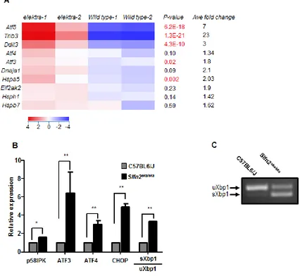
The splicing of XBP1 mRNA is a hallmark of ER stress. Semi quantitative real time-PCR analysis for XBP1
splicing (Figure 1B), which was also confirmed by PCR
spliced form, condition that is rarely seen for unstimulated cells. These results demonstrate that the elektra mutation
in Slfn2 leads to the unabated activation of stress response in BM monocytes.
chronic er stress of elektra t cells
Next we aimed to confirm that the ER stress response
is activated also in elektra T cells. For this purpose T cells were isolated from elektra spleens and the expression of ER stress genes was measured in comparison to wt cells.
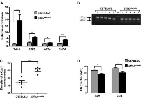
ER stress response genes were indeed elevated in elektra T cells (Figure 2A), and the ratio between the spliced and
unspliced transcripts of XBP1 was elevated (Figure 2B).
In addition, ER tracker staining shown to be decreased
in elektra T cells as compare to wt T cells (Figure 2C)
suggesting a defect in ER structure. These data confirm
[image:3.612.97.523.226.621.2]that similarly to elektra monocytes, ER stress response is activated also in elektra T cells.
Figure 1: High expression of er stress related genes in elektra inflammatory monocytes. A. Heat map of cDNA expression array shows differentially expressed genes in C57BL6/J and Slfn2eka/eka bone marrow CD11b+ ly6Chi sorted monocytes. P-values (highly significant are marked by red font)and average fold of change are included. b. Gene expression analyzed by semi-quantitative RT-PCR
of ER stress response genes in C57BL6/J and Slfn2eka/eka bone marrow CD11b+ ly6chimonocytes (n=4). c. Representative result of XBP1
Elektra t cells die via er stress induced apoptosis
Elektra mice overexpressing BCL2 in the T cell compartment, BCL2(Tg)/Slfn2eka/eka have normal numbers
of T cells. In addition, Bcl2 is downregulated in elektra
T cells [23, 24]. These results demonstrate that elektra
T cells undergo apoptosis via the intrinsic apoptotic pathway [23, 24]. Since chronically activation of ER stress promotes apoptosis in several cell types [30], we
postulated a contribution of this pathway in the demise of elektra T cells.
To test this possibility, we examined whether elektra T cell death is indeed mediated by chronic ER stress by
evaluating whether modulation of the ER stress response
by either down regulation of XBP1 or CHOP can rescue
the elektra T cell phenotype. To this end, Lck-Cre XBP1lox/ lox mice, in which XBP1 is knocked out specifically in the
T cell compartment, or CHOP knockout mice, CHOP-/-,
were crossed to elektra mice to generate Lck-Cre XBP1lox/ lox/ Slfn2eka/eka or CHOP-/-/Slfn2eka/eka transgenic mice
respectively. The percentages of CD4+ and CD8+ T cells
from spleens of Lck-Cre XBP1lox/lox/Slfn2eka/eka or CHOP -/-/Slfn2eka/eka transgenic mice were markedly elevated as
compare to those from elektra mice, in fact resembling
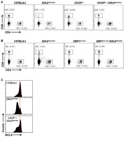
those of wild-type mice (Figure 3A and 3B). In addition and in line with these results, BCL2 expression levels were restored in CD8+ T cells from CHOP-/-/Slfn2eka/eka
mice (Figure 3C). Our data implicate the XBP1/CHOP pathway as mediator of elektra T cell death.
blocking er stress response partially restores elektra t cell semiactivated phenotype
Viability is not the only defect of elektra T cells. Previously we showed that elektra T cells exist in a
semiactivated state, characterized by down regulation of
the IL7Ra (CD127) and the homing receptor L-Selectin
(CD62L) on both naïve newly thymic emigrant (CD44lo)
[image:4.612.66.554.317.659.2]and on naïve mature (CD44hi) T cells. In addition, the
Figure 2: Activation of uPr and er stress response in slfn2eka/eka T cells. A. Gene expression of ER stress response and UPR
genes in naïve T cells purified from C57BL6/J and Slfn2eka/eka splenocytes (n=4 each, 96-98% and 95-96% purity of wt and elektra T cells
mature CD44hielektra T cells fail to acquire a
memory-like phenotype (CD122+) and instead develop into
recently-activated (CD122-) T cells [23].
BCL2 overexpression [23] or p53 deficiency [24]
rescue elektra T cell death, but does not affect their
[image:5.612.92.526.132.634.2]semiactivated phenotype, and thus does not correct their proliferation defect [23]. These results illustrate the complexity of the elektra phenotype and indicate that
Figure 3: CHOP or XBP1 deficiency rescues the death of Slfn2eka/eka cd4+ and cd8+ T cells. A., b. Flow cytometry analysis
of the expression of CD4 and CD8 by cells from the spleen of wild-type, Slfn2eka/eka , CHOP-/- and CHOP-/-/Slfn2eka/eka (A) or XBP1lox/lox
and XBP1lox/lox/Slfn2eka/eka (B) (n=9). Numbers in gates indicate percent cells in each. c. Flow cytometry of BCL2 expression in splenic
apoptosis of the elektra T cells is a consequence of their acquisition of a semiactivated state.
Therefore, we next assessed whether manipulating
the ER stress response by CHOP or XBP1 deficiency also
impinges on the acquisition of the semiactivated state of the elektra T cells and their proliferation capabilities. To
this end T cells from Lck-Cre XBP1lox/lox/Slfn2eka/eka and
CHOP-/-/Slfn2eka/eka mice were further immunophenotyped
by flow cytometry.
Similar to elektra T cells, both Lck-Cre XBP1lox/ lox/Slfn2eka/eka and CHOP-/-/Slfn2eka/eka CD44hi CD8+ and
[image:6.612.68.544.134.656.2]CD4+ T cell populations failed to gain a memory-like
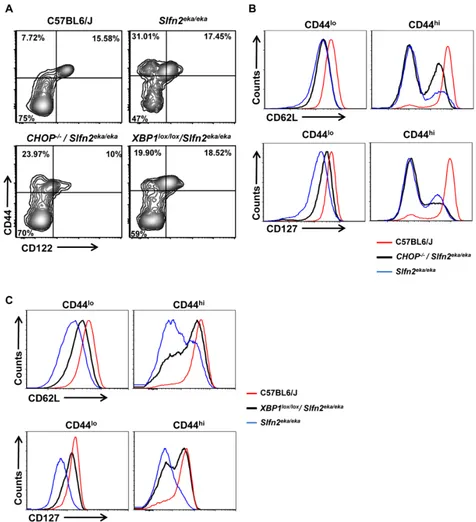
Figure 4: CHOP or XBP1 deficiency partially rescues the semiactivated phenotype of Slfn2eka/eka cd8+ T cells. A. Flow
cytometry analysis of the staining for CD122 (IL-2Rβ) and CD44 in splenic CD8+ T cells from C57BL6/J, Slfn2eka/eka, CHOP-/-/Slfn2eka/eka
or XBP1lox/lox/Slfn2eka/eka mice (n =9). Numbers in quadrants indicate percent cells in each. b., c. Flow cytometry analysis for the expression levels of CD62L (upper panel) and IL-7Rα (CD127) (lower panel) on surface of both CD8+CD44lo (left) and CD8+CD44hi (right) T cells
phenotype of CD44hi/CD122+, and were mostly CD122−
or CD122lo (Figure 4A and Supplementary Figure 1A).
Comparable to elektra, in CHOP-/-/Slfn2eka/eka most of this
CD44hi/CD122−/lo population display a complete shedding
of CD62L (L-selectin) (Figure 4B and Supplementary Figure 1B, right upper panel), and no surface expression of
IL-7 receptor α-chain (IL-7Rα or CD127) (Figure 4B and
Supplementary Figure 1B, right lower panel). In Lck-Cre XBP1lox/lox/Slfn2eka/eka mice most of the CD44hiCD122lo/− T cell population showed partial restoration of both CD62L
(Figure 4C and Supplementary Figure 1C, right upper panel) and CD127 (Figure 4C and Supplementary Figure 1C, right lower panel) expression. In addition, the CD44lo
(naive) population of CD8+ and CD4+ T cells from both,
CHOP-/-/Slfn2eka/eka and Lck-Cre XBP1lox/lox/Slfn2eka/eka,
mice partially restored the expression of both CD62L
(Figure 4B and 4C and Supplementary Figure 1B and C, left upper panels) and CD127 (Figure 4B and 4C and Supplementary Figure 1B and C, left lower panels). These results suggest that the UPR through XBP1, and to lesser
extent by CHOP, compromise peripheral T cell maturation and ability to maintain quiescence.
blocking er stress response partially restores elektra t cell proliferation capacity
Next we examined the impact of ER stress on the proliferative capacity of elektra T cells. Initially, we examined the proliferation capacity of CHOP-/-/Slfn2eka/ eka or Lck-Cre XBP1lox/lox/Slfn2eka/eka T cell in response to
activation signals. For this end, wt, CHOP-/-/Slfn2eka/ eka, Lck-Cre XBP1lox/lox/Slfn2eka/eka and elektra splenic T
cells were labeled with the cytosolic dye Cell Trace and
were stimulated for 72 h in vitro with a combination of antibodies to CD3ε and CD28. In line with our published
data [23], elektra T cells completely failed to proliferate in response to activation stimuli (Figure 5A and 5B and, Supplementary Figure 2A and B). However, as opposed to elektra, both CD8+ and CD4+ T cells from CHOP-/-/
Slfn2eka/eka and Lck-Cre XBP1lox/lox/Slfn2eka/eka had undergone
proliferation, although less than T cells from CHOP-/- and
Lck-Cre XBP1lox/lox that proliferated comparably to the T
cells from wt mice (Figure 5A and 5B and, Supplementary Figure 2A and B). These results demonstrate that the proliferation defect of elektra T cells upon activation ex
vivo is mediated, at least in part, by the chronic ER stress.
To further test the impact of the elevated ER stress on the proliferative capacity of elektra T cells, we examined CHOP-/-/Slfn2eka/eka and Lck-Cre XBP1lox/ lox/Slfn2eka/eka T cell response to homeostatic proliferation
signals. For this end, we labeled wild-type, Slfn2eka/eka,
CHOP-/-/Slfn2eka/eka and Lck-Cre XBP1lox/lox/Slfn2eka/eka
(CD45.2) splenocytes with the cytosolic dye Cell Trace
and adoptively transferred them into sublethally irradiated
wild-type (CD45.1) recipients. As expected wild-type T
cells underwent proliferation and we detected no elektra
T cells in the spleen of recipient mice 7 d after infusion (Figure 5C, left panels and Supplementary Figure 3). T
cells from both CHOP-/-/Slfn2eka/eka and Lck-Cre XBP1lox/ lox/Slfn2eka/eka failed to proliferate, however were still
detectable in a pretty high percentages (Figure 5C,
right panels and Supplementary Figure 3). These results demonstrate that in vivo, under lymphopenic environment,
elektra T cell death is mediated by the XBP1/CHOP
pathway. However, the proliferation defect of elektra T
cells cannot be attributed only to these proteins.
dIscussIon
The outcome of an immune response is dependent
on the balance between stimulatory and inhibitory signals. This delicate balance is critical for the ability of
the immune system to defend the host, while maintaining tolerance to self so to prevent autoimmunity and immunopathology. To achieve this, the immune system maintains a vast repertoire of immunologically naïve
lymphocytes in a quiescent state characterized by inactive
cell cycle (arrest in G0), small cell size and relatively
low basal metabolic activity [17]. Intuitively, it may be
expected that elimination of a regulator of quiescence would have led to hyperactive immune responses and
even autoimmunity. However, several recent findings
demonstrated that the exact opposite can also happen;
disruption of quiescence leads to immunodeficiency due to loss of proliferation capabilities and susceptibility to
cell death of T cells [23, 31-34]. These results suggest that
quiescence programming has a broader role in immunity
than previously recognized, and that quiescence rather than a passive process is an active process that enforces passiveness. Nevertheless, most of the factors and pathways that maintain immune cell quiescence have yet
to be identified.
Naïve quiescent T cells favor energy production
over biosynthesis. To accommodate for this need, naïve
T cells rely predominantly on the high-energy-yielding
mitochondrial metabolism in which metabolites are
oxidized via the TCA cycle [18, 35]. Upon activation, T
cells undergo dramatic shift in cell metabolism switching from oxidative metabolism to aerobic glycolysis, to
support their expansion and effector functions [18, 35]. The Forkhead Box O1 (FOXO1) [31, 33] and
tuberous sclerosis 1 (Tsc1) [32, 34] are both well-established quiescence maintaining factors in T cells. Both proteins play a major role in controlling the metabolic
shift that is needed for the transition from quiescence
to activation. In addition to their role in metabolism, FOXO1 and Tsc1 have a major role in protecting cells
from oxidative stress [36] or ER stress [37] respectively.
Figure 5: CHOP or XBP1 deficiency partially rescues Slfn2eka/eka cd8+ t cells proliferation capacity and death in
lymphopenic environment in vivo. A., b. Cell trace dilution of splenic CD8 T cells obtained from C57BL6/J, Slfn2eka/eka , CHOP-/-/ Slfn2eka/eka (A) or XBP1lox/lox/Slfn2eka/eka (B) mice stimulated for 72 hours with plate bounded anti-CD3ε (2µg/ml) plus anti-CD28 (1µg/ml)
and IL-2 (20ng/ml) (n=6 each genotype). Histograms are gated on CD8+. c. Dot plot of CD45.2 versus CD8 (left panels) and histograms
presenting Cell trace dilution of splenic CD8+ (right panels) obtained from CD45.2 C57BL6/J, Slfn2eka/eka, CHOP-/-/Slfn2eka/eka or XBP1lox/lox/ Slfn2eka/eka adoptively transferred into irradiated (400 rad) CD45.1 mice for 7 days (n=5). Numbers on plots indicate percentages of donor
[image:8.612.65.551.74.626.2]accompanied by development of stress conditions to be met by compensatory responses. This balance can have a substantial influence on cell fate decisions.
Here we showed that elektra T cells display chronic ER stress under steady state conditions (Figure
2). Modulation of the ER stress response by depletion of either XBP1 or CHOP restored the viability (Figure 3) and partially improved the developmental abnormalities and proliferation capabilities of elektra T cells (Figures
4 and 5). These results establish a functional connection between the loss of quiescence in elektra T cells and chronic unresolved ER stress.
Previous attempts to rescue elektra T cell viability either by over-expression of BCL2 [23] or p53 deficiency
[24] demonstrated that although the propensity to undergo apoptosis was reduced, elektra T cells displayed a
semiactivated phenotype and were unable to proliferate.
These results strongly indicate that the elektra T cell death is a consequence of loss of quiescence. The results presented here demonstrate that knockout of CHOP or XBP1 not only prevented elektra T cell death, but also
partially prevented the semiactivated phenotype and partially restored the T cell proliferative capacity upon activation (Figures 3 and 5).
Deletion of the tuberous sclerosis complex 1 (Tsc1) gene leads to a substantial reduction in peripheral T cell numbers [32, 37], which correlates with increased propensity to apoptosis that can be rescued by expression of Bcl-2 [37]. Moreover, Tsc1 deficiency in T cells leads to acquisition of a semiactivated phenotype caused by loss of quiescence [37]. This phenotype highly resembles the phenotype observed in elektra T cells.
Tsc1, together with its partner Tsc2, restrains the activation of mammalian target of rapamycin (mTOR), an
important regulator of metabolism and translation that is
typically induced following PI3K signaling [38]. In the context of tumors and liver cells, ER stress and activation of the UPR pathway are important pathological features of
TSC deficiency; TSC1 and TSC2 deficiency induces ER
stress and activates the UPR [37]. The resulting ER stress
increases the vulnerability of various cell lines and tumors
to apoptosis [37]. Although not validated in TSC1 KO
T cells, ER stress may also contribute to the quiescence
defect in this model.
The functional connection between the loss of quiescence in Slfn2-deficiency and chronic unresolved ER stress is not known. As noted above, the elektra phenotype is caused by a point mutation in Slfn2 gene.
Therefore, one may argue that the elektra mutation
possibly results in misfolding and aggregation of the mutated Slfn2 protein leading to the observed ER stress.
However, elektra heterozygous mice do not demonstrate
any of the defects observed in elektra homozygous mice [23]. In addition, other cells than T cells and monocytes, which also express Slfn2 (e.g. granulocytes, B cells and
NK cells), are not influenced by the elektra mutation [23].
Furthermore, Slfn2 overexpression in elektra homozygous completely rescues the entire phenotypes of the elektra mice [23]. Finally, knockdown of Slfn2 by shRNA
recapitulate the elektra phenotype in EL4 cells [24]. These evidences strongly suggest that the ER stress, as well as other aspects of the elektra phenotype, is caused by the loss-of-function of the Slfn2 rather than by a gain-of-function caused by misfolding of the protein. Therefore,
we propose two options for the underlying mechanism;
one possibility suggests that Slfn2 directly regulates ER homeostasis. According to this possibility the UPR should be a main defect in elektra cells. In this regard, Slfn2 can play a role in protein folding in the ER (chaperone),
protein translocation into the ER and protein trafficking within and downstream to the ER. A second possibility is that the observed chronic ER stress in elektra cells is
a consequence of an overall much broader phenotype caused by Slfn2 deficiency. For example; an unbalanced translational regulation [39], impaired lipid and cholesterol biosynthesis and trafficking [40], disturbances of redox homeostasis [41], glucose deprivation [42] and aberrant
Ca+2 regulation [43]. All of these conditions also result
in ER stress; however, UPR is only a part of the effect.
Interestingly, the mechanism by which TSC deficiency results in ER stress induced cell death was attributed to
increased mTOR activity [37], which triggers uncontrolled
protein synthesis. Moreover, Li, M. et al. demonstrated
that the human SLFN family member, Slfn11, inhibits
retroviral protein translation in a codon-usage dependent
manner [44]. Considering these results and our findings,
it is appealing to suggest that in elektra cells the chronic ER stress is mediated due to loss of protein translational control which leads among several other defects also to
inability to match protein load to the folding capacity
of the ER, supporting the second option. This scenario suggests a potential regulatory role for Slfn2 in protein translation. Therefore, models that directly explore the
role of protein translation in T cell quiescence should be
developed.
MAterIAls And MetHods
Mice
Slfn2eka/eka mice were previously generated as
described in Berger et al. [23] . The C57BL/6J (wild-type), CHOP−/−, C57BL/6.SJL (PtprcaPep3b; Ly5.1) (CD45.1) and B6.Cg-Tg(Lck-icre)3779Nik/J (Lck-Cre) mice were from The Jackson Laboratory. The T cell specificXBP1 KO mice were generated by crossing mice containing a conditional floxed allele of XBP-1 (XBP-1lox/ lox, described in [45]) with transgenic mice expressing Cre
under the control of the Lck gene promoter (Lck-Cre).
free conditions in the Hebrew University animal facilities
according to Institutional Animal Care and Use Committee regulations. All mice were maintained on the C57BL/6
background and used for experiments at 8–12 weeks of
age.
real time Pcr
Total RNA from purified monocytes or T cells was extracted with Direct-zol RNA MiniPrep Plus (Zymo
Research). cDNA was synthesized using ProtoScript First
Strand cDNA Synthesis Kit (Neb). Quantitative
real-time PCR was then performed using QuantStudio 12K Flex Real Time PCR system with a Power SYBR green PCR master mix kit (Applied Biosystems). The primers
sequences used in this study are previously described [46].
Flow cytometry
Spleen cells were stained with various conjugated mAbs against cell-surface markers. In some of the
experiments, following the cell surface staining, cells
were fixed, permeabilized and stained with intracellular antibody using Leucoperm-kit (AbD Serotec) according
to manufacturer instructions. ER tracker staining was
performed by incubating cells with 1µM ER-Tracker blue-white DPX (Molecular Probes) diluted in Hankʼs Balanced Salt Solution for 30 minutes in 37˚C. Stained cells were analyzed by Gallios flow cytometer with Kaluza
software (Beckman Coulter).
Inflammatory monocytes and T cells isolation
For monocytes isolation total bone marrow cells were harvested and inflammatory monocytes were isolated by EasySep mouse monocyte isolation kit (Stem Cell Technologies). For further purification, CD11b+ Ly6Chigh
monocytes were sorted by flow cytometry. Total T cells were isolated from spleen by EasySep mouse T cell
isolation kit (Stem Cell Technologies).
Adoptive transfer of t cells
A total of 1×10⁷ CD45.2 splenocytes were labeled
with Cell-trace violet (Molecular Probes c34557) and IV injected into CD45.1 C57BL/6J recipient mice that had been sublethally irradiated (400 rads) 24 h earlier. 7
days after adoptive transfer, spleen cells were harvested,
stained for CD45.2, CD8 and CD4 and analyzed by
flow-cytometry for cell-trace dilution.
t cell proliferation assay
A total of 2.5 × 10⁶ cell-trace violet-labeled spleen
cells were activated in 24-flat-well plates previously bounded with anti-CD3ε (2µg/ml) and anti-CD28 (1µg/
ml) and IL-2 (20ng/ml). The cells were analyzed 72 h after
activation by flow cytometry as described in the adoptive
transfer section.
Antibodies
The following antibodies were used for flow cytometry: CD8α (53-6.7), CD4 (L3T4),
anti-CD45.1 (A20), anti-CD45.2 (104), anti-Bcl-2 (10C4),
anti-CD44 (IM7), CD122 (IL2RB) (5H4), CD127 (IL7R) (SB/199), anti-CD11b (M1/70), and anti-Ly6C (AL-21; Biolegend). Purified CD3ε (145–2C11) and anti-CD28 (37.51; both from Biolegend) were used at the
appropriate concentration for T cell activation.
AcknowledgMents
This work was supported by grants from the
ISRAEL SCIENCE FOUNDATION grant No.1275/12, ISRAEL CANCER RESEARCH FUND grant No.
13/726/RCDA, Marie Curie People grant No. 322006 and
Concern Foundation
conFlIct oF Interest
The authors declare no conflict of interest.
Authorship contributions
Conceived and designed the experiments: I.O. and
M.B. Performed the experiments: I.O. A.L. BT. and L.C. Analyzed the data: I.O and M.B. Wrote the manuscript: I.O. B.T. and M.B.
reFerences
1. Gaut JR and Hendershot LM. The modification and assembly of proteins in the endoplasmic reticulum. Curr Opin Cell Biol. 1993; 5:589-595.
2. Healy SJM, Gorman AM, Mousavi-Shafaei P, Gupta S
and Samali A. Targeting the endoplasmic reticulum-stress
response as an anticancer strategy. Eur J Pharmacol. 2009;
625:234-246.
3. Hetz C. The unfolded protein response: controlling cell fate
decisions under ER stress and beyond. Nat Rev Mol Cell Biol. 2012; 13:89-102.
5. Rutkowski DT and Kaufman RJ. A trip to the ER: coping with stress. Trends Cell Biol. 2004; 14:20-28.
6. Szegezdi E, Logue SE, Gorman AM and Samali A. Mediators of endoplasmic reticulum stress-induced apoptosis. EMBO Rep. 2006; 7:880-885.
7. Zinszner H, Kuroda M, Wang X, Batchvarova N, Lightfoot
RT, Remotti H, Stevens JL and Ron D. CHOP is implicated in programmed cell death in response to impaired function
of the endoplasmic reticulum. Genes Dev. 1998; 12:982-995.
8. McCullough KD, Martindale JL, Klotz LO, Aw TY and Holbrook NJ. Gadd153 sensitizes cells to endoplasmic reticulum stress by down-regulating Bcl2 and perturbing the cellular redox state. Mol Cell Biol. 2001; 21:1249-1259. 9. Puthalakath H, O’Reilly LA, Gunn P, Lee L, Kelly PN,
Huntington ND, Hughes PD, Michalak EM, McKimm-Breschkin J, Motoyama N, Gotoh T, Akira S, Bouillet P and Strasser A. ER stress triggers apoptosis by activating BH3-only protein Bim. Cell. 2007; 129:1337-1349. 10. Lu M, Lawrence DA, Marsters S, Acosta-Alvear D,
Kimmig P, Mendez AS, Paton AW, Paton JC, Walter P and
Ashkenazi A. Opposing unfolded-protein-response signals converge on death receptor 5 to control apoptosis. Science.
2014; 345:98-101.
11. Li B, Yi P, Zhang B, Xu C, Liu Q, Pi Z, Xu X, Chevet
E and Liu J. Differences in endoplasmic reticulum stress signalling kinetics determine cell survival outcome through
activation of MKP-1. Cell Signal. 2011; 23:35-45. 12. Yoshida H, Matsui T, Yamamoto A, Okada T and Mori
K. XBP1 mRNA is induced by ATF6 and spliced by
IRE1 in response to ER stress to produce a highly active
transcription factor. Cell. 2001; 107:881-891.
13. Song Y, Shen H, Du W and Goldstein DR. Inhibition of x-box binding protein 1 reduces tunicamycin-induced
apoptosis in aged murine macrophages. Aging Cell. 2013;
12:794-801.
14. Zeng L, Zampetaki A, Margariti A, Pepe AE, Alam S, Martin D, Xiao Q, Wang W, Jin Z-G, Cockerill G, Mori
K, Li Y-SJ, Hu Y, Chien S and Xu Q. Sustained activation of XBP1 splicing leads to endothelial apoptosis and
atherosclerosis development in response to disturbed flow.
Proceedings of the National Academy of Sciences of the
United States of America. 2009; 106:8326-8331.
15. Margariti A, Li H, Chen T, Martin D, Vizcay-Barrena G, Alam S, Karamariti E, Xiao Q, Zampetaki A, Zhang Z, Wang W, Jiang Z, Gao C, Ma B, Chen Y-G, Cockerill G, et
al. XBP1 mRNA splicing triggers an autophagic response in endothelial cells through BECLIN-1 transcriptional
activation. The Journal of biological chemistry. 2013; 288:859-872.
16. Upton J-P, Wang L, Han D, Wang ES, Huskey NE, Lim L, Truitt M, McManus MT, Ruggero D, Goga A, Papa FR and
Oakes SA. IRE1alpha cleaves select microRNAs during ER stress to derepress translation of proapoptotic Caspase-2.
Science. 2012; 338:818-822.
17. Hamilton SE and Jameson SC. CD8 T cell quiescence revisited. Trends Immunol. 2012; 33:224-230.
18. Pearce EL and Pearce EJ. Metabolic pathways in immune
cell activation and quiescence. Immunity. 2013; 38:633-643.
19. Yusuf I and Fruman DA. Regulation of quiescence in
lymphocytes. Trends Immunol. 2003; 24:380-386.
20. Sprent J and Surh CD. Normal T cell homeostasis: the conversion of naive cells into memory-phenotype cells. Nature immunology. 2011; 12:478-484.
21. Chechlinska M, Siwicki JK, Gos M, Oczko-Wojciechowska M, Jarzab M, Pfeifer A, Jarzab B and Steffen J. Molecular
signature of cell cycle exit induced in human T
lymphoblasts by IL-2 withdrawal. BMC Genomics. 2009;
10:261.
22. Coller HA, Sang L and Roberts JM. A new description of
cellular quiescence. PLoS Biol. 2006; 4:e83.
23. Berger M, Krebs P, Crozat K, Li X, Croker BA, Siggs OM, Popkin D, Du X, Lawson BR, Theofilopoulos AN, Xia Y, Khovananth K, Moresco EM, Satoh T, Takeuchi O, Akira
S, et al. An Slfn2 mutation causes lymphoid and myeloid
immunodeficiency due to loss of immune cell quiescence.
Nat Immunol. 2010; 11:335-343.
24. Goldshtein A MZS, Omar I, Cohen-Daniel L, Popkin D, and Berger M. Loss of T-cell quiescence by targeting Slfn2
prevents the development and progression of T-ALL.
Oncotarget. 2016; doi: 10.18632/oncotarget.9390.
25. Jager R, Bertrand MJM, Gorman AM, Vandenabeele P and
Samali A. The unfolded protein response at the crossroads of cellular life and death during endoplasmic reticulum
stress. Biol Cell. 2012; 104:259-270.
26. Teske BF, Fusakio ME, Zhou D, Shan J, McClintick JN, Kilberg MS and Wek RC. CHOP induces activating
transcription factor 5 (ATF5) to trigger apoptosis in
response to perturbations in protein homeostasis. Mol Biol Cell. 2013; 24:2477-2490.
27. Ohoka N, Yoshii S, Hattori T, Onozaki K and Hayashi H.
TRB3, a novel ER stress-inducible gene, is induced via ATF4-CHOP pathway and is involved in cell death. Embo
J. 2005; 24:1243-1255.
28. Liu Y and Chang A. Heat shock response relieves ER stress.
Embo J. 2008; 27:1049-1059.
29. Koromilas AE. Roles of the translation initiation factor
eIF2alpha serine 51 phosphorylation in cancer formation
and treatment. Biochim Biophys Acta. 2015; 1849:871-880. 30. Maurel M, McGrath EP, Mnich K, Healy S, Chevet E
and Samali A. Controlling the unfolded protein response-mediated life and death decisions in cancer. Semin Cancer Biol. 2015; 33:57-66.
31. Ouyang W, Beckett O, Flavell RA and Li MO. An essential role of the Forkhead-box transcription factor Foxo1 in
control of T cell homeostasis and tolerance. Immunity.
32. Yang K, Neale G, Green DR, He W and Chi H. The tumor
suppressor Tsc1 enforces quiescence of naive T cells to promote immune homeostasis and function. Nature
immunology. 2011; 12:888-897.
33. Kerdiles YM, Beisner DR, Tinoco R, Dejean AS, Castrillon DH, DePinho RA and Hedrick SM. Foxo1 links homing and survival of naive T cells by regulating L-selectin, CCR7 and interleukin 7 receptor. Nature immunology. 2009;
10:176-184.
34. O’Brien TF, Gorentla BK, Xie D, Srivatsan S, McLeod IX, He Y-W and Zhong X-P. Regulation of T-cell survival and mitochondrial homeostasis by TSC1. Eur J Immunol. 2011;
41:3361-3370.
35. MacIver NJ, Michalek RD and Rathmell JC. Metabolic
regulation of T lymphocytes. Annual review of
immunology. 2013; 31:259-283.
36. Tothova Z, Kollipara R, Huntly BJ, Lee BH, Castrillon DH, Cullen DE, McDowell EP, Lazo-Kallanian S, Williams IR,
Sears C, Armstrong SA, Passegue E, DePinho RA and Gilliland DG. FoxOs are critical mediators of hematopoietic stem cell resistance to physiologic oxidative stress. Cell.
2007; 128:325-339.
37. Ozcan U, Ozcan L, Yilmaz E, Duvel K, Sahin M, Manning BD and Hotamisligil GS. Loss of the tuberous sclerosis
complex tumor suppressors triggers the unfolded protein
response to regulate insulin signaling and apoptosis. Mol Cell. 2008; 29:541-551.
38. Shimobayashi M and Hall MN. Making new contacts: the mTOR network in metabolism and signalling crosstalk. Nat Rev Mol Cell Biol. 2014; 15:155-162.
39. Muaddi H, Majumder M, Peidis P, Papadakis AI, Holcik M, Scheuner D, Kaufman RJ, Hatzoglou M and Koromilas AE.
Phosphorylation of eIF2alpha at serine 51 is an important determinant of cell survival and adaptation to glucose
deficiency. Mol Biol Cell. 2010; 21:3220-3231.
40. Chandran S and Machamer CE. Inactivation of ceramide transfer protein during pro-apoptotic stress by Golgi disassembly and caspase cleavage. Biochem J. 2012; 442:391-401.
41. Delaunay-Moisan A and Appenzeller-Herzog C. The
antioxidant machinery of the endoplasmic reticulum:
Protection and signaling. Free Radic Biol Med. 2015;
83:341-351.
42. Leon-Annicchiarico CL, Ramirez-Peinado S,
Dominguez-Villanueva D, Gonsberg A, Lampidis TJ and Munoz-Pinedo C. ATF4 mediates necrosis induced by glucose deprivation and apoptosis induced by 2-deoxyglucose in the same cells.
FEBS J. 2015; 282:3647-3658.
43. Li G, Mongillo M, Chin KT, Harding H, Ron D, Marks AR and Tabas I. Role of ERO1-alpha-mediated stimulation of
inositol 1,4,5-triphosphate receptor activity in endoplasmic
reticulum stress-induced apoptosis. J Cell Biol. 2009; 186:783-792.
44. Li M, Kao E, Gao X, Sandig H, Limmer K, Pavon-Eternod M, Jones TE, Landry S, Pan T, Weitzman MD and David M. Codon-usage-based inhibition of HIV protein synthesis by human schlafen 11. Nature. 2012; 491:125-128. 45. Goldfinger M, Laviad EL, Hadar R, Shmuel M, Dagan A,
Park H, Merrill AH, Jr., Ringel I, Futerman AH and Tirosh
B. De novo ceramide synthesis is required for N-linked
glycosylation in plasma cells. J Immunol. 2009;
182:7038-7047.
46. Uzi D, Barda L, Scaiewicz V, Mills M, Mueller T, Gonzalez-Rodriguez A, Valverde AM, Iwawaki T, Nahmias Y, Xavier R, Chung RT, Tirosh B and Shibolet
O. CHOP is a critical regulator of acetaminophen-induced