The role of Cas8 in type I CRISPR interference
Simon D.B. Cass*1, Karina A. Haas†1, Britta Stoll†, Omer S. Alkhnbashi‡, Kundan Sharma§,
Henning Urlaub§∥, Rolf Backofen‡¶, Anita Marchfelder†2 and Edward L. Bolt*2
*School of Life Sciences, University of Nottingham, Queen’s Medical Centre, Nottingham NG7 2UH, U.K. †Biology II, Ulm University, 89069 Ulm, Germany
‡Bioinformatics group, Department of Computer Science, University of Freiburg, Georges-K¨ohler-Allee 106, 79110 Freiburg, Germany §Max Planck Institute of Biophysical Chemistry, Am Faßberg 11, 37077 G¨ottingen, Germany
∥Bioanalytics Research Group, Institute of Clinical Chemistry, University Medical Center G¨ottingen, G¨ottingen, Germany ¶Centre for Biological Signalling Studies (BIOSS), Cluster of Excellence, University of Freiburg, Germany
Synopsis
CRISPR (clustered regularly interspaced short palindromic repeat) systems provide bacteria and archaea with adapt-ive immunity to repel invasadapt-ive genetic elements. Type I systems use ‘cascade’ [CRISPR-associated (Cas) complex for antiviral defence] ribonucleoprotein complexes to target invader DNA, by base pairing CRISPR RNA (crRNA) to protospacers. Cascade identifies PAMs (protospacer adjacent motifs) on invader DNA, triggering R-loop formation and subsequent DNA degradation by Cas3. Cas8 is a candidate PAM recognition factor in some cascades. We analysed
Cas8 homologues from type IB CRISPR systems in archaeaHaloferax volcanii(Hvo) andMethanothermobacter
ther-mautotrophicus(Mth). Cas8 was essential for CRISPR interference in Hvo and purified Mth Cas8 protein responded to PAM sequence when binding to nucleic acids. Cas8 interacted physically with Cas5–Cas7–crRNA complex, stimulating
binding to PAM containing substrates. Mutation of conserved Cas8 amino acid residues abolished interferencein vivo
and altered catalytic activity of Cas8 proteinin vitro. This is experimental evidence that Cas8 is important for targeting
Cascade to invader DNA.
Key words: archaea, CRISPR-associated (Cas)8, CRISPR-associated complex for antiviral defence (Cascade), clustered regularly interspaced short palindromic repeat (CRISPR), nuclease.
Cite this article as: Bioscience Reports (2015)35, e00197, doi:10.1042/BSR20150043
INTRODUCTION
CRISPR (clustered regularly interspaced short palindromic re-peat) systems were discovered in Streptococcus thermophilus
[1], by providing adaptive immunity to invasive genetic ele-ments, recently reviewed in [2,3]. Immunity arises from base pairing of host encoded CRISPR RNA (‘crRNA’) with invader DNA/RNA, promoting nucleolytic degradation of the invader, processes called ‘interference’. DNA sequences that are targeted by crRNA are called ‘protospacers’ and can be identified from an archive of previously encountered protospacers arrayed in a CRISPR locus as ‘spacers’, separated from one another by repeat sequences. ‘Adaptation’ or ‘spacer acquisition’ processes fur-nish CRISPR with new spacer-repeat units requiring two highly conserved CRISPR-associated (‘Cas’) proteins, Cas1 and Cas2. Cas1–Cas2 adaptation may be functionally linked to interference
...
Abbreviations:ACBB, amylose column-binding buffer; Cas, CRISPR-associated; Cascade, CRISPR-associated complex for antiviral defence; CRISPR, clustered regularly interspaced short palindromic repeat; crRNA, CRISPR RNA; His6–Cas8′, N-terminal hexahistidine-tagged Mth Cas8′; MBP, maltose-binding protein; PAM, protospacer adjacent motif; TBE, Tris-Borate-EDTA; Trp, tryptophan.
1These authors contributed equally to the research.
2Correspondence may be addressed to either author (email [email protected] or [email protected]).
(‘primed’) or not (‘na¨ıve’), in each case by mechanisms unclear, reviewed in [3–5].
Cas proteins that catalyse interference show substantial di-versity, with current classification into three major groups, types I, II and III [6–8], characterized by distinct effector complexes that manoeuvre crRNA to base pair with target DNA or RNA. Type I, II and IIIA systems target DNA catalysed by respectively, ‘Cas-cade’ (Cas complex for antiviral defence) [9,10], Cas9 [11,12] or CSM complex [13,14]. In contrast, CMR complexes target RNA in type IIIB CRISPR systems [15–17].
A–F [6], with further refinement into types A–G to accommod-ate variants in types IA, ID and IG [18]. Common structural features are observed in Cascades from the different CRISPR sub-types in bacteria and archaea [9,10,19–22]. crRNA is de-livered into Cascade as a single spacer element sequence after truncation of crRNA transcripts by Cas5 and Cas6 nucleases [10,19,20,23–28], reviewed in [29,30]. Sulfolobus solfataricus
[19] andThermoproteus tenax[21] Cascades (both type IA) and
Escherichia coli(type IE) [10] use multiple copies of Cas7 protein to form a backbone filament with crRNA, functionally analog-ous to the Csy3-crRNA backbone ofPseudomonas aeruginosa
Cascade (Type IF) [20]. Another variation of Cascade subunit type with common function is observed in type IC systems that use Csd2 to form Cas7-like crRNA filaments [31]. Interference is established by Cascade base pairing crRNA with protospacer (invader) DNA through ‘seeding’ [20,32–35] and further into an R-loop [9], reviewed in [36] that promotes nucleolytic de-gradation of invader DNA probably by interaction of Cascade with Cas3 helicase-nuclease. Recent atomic structures ofE. coli
Cascade complex have provided detailed insight into the arrange-ment of protein subunits relative to one another and to crRNA to provide a mechanism for interference in type IE CRISPR [37–39].
Cascade can identify invader DNA by interaction with PAM (protospacer adjacent motif) sequences. PAMs are short (2–5 nt) sequences located on invader DNA upstream of the protospacer that trigger Cascade–Cas3 interference [40]. A mechanism for Cascade–PAM recognition described inE. coliinvolves a CasA ‘large subunit’ contacting target DNA via its ‘L1 loop’ [41,42], as part of multiple interactions with Cas5 [37–39]. CasA is the ‘signature’ protein of type IE CRISPR systems, essential for Cas-cade function [6]. Atomic resolution structures of Cascade [37– 39] show CasA interlocking with CasD, contributing to binding of the crRNA 5′-handle. CasA also contacts PAM as part of
the tight association with CasD. In other type I CRISPR sys-tems Cas8 is predicted to be functionally analogous to CasA [18], as a ‘signature’ protein for type IA, IB and IC systems, referred to respectively, as Cas8a2, Cas8b and Cas8c [6]. A more recent analysis highlighted diversity of Cas8 proteins leading to their renaming as Cas8, Cas8′ and Cas8′′proteins and
cre-ating new type-I CRISPR variants based on Cas8 protein se-quences and positioning ofcas8genes relative to othercasgenes [18]. An important role for Cas8 in interference was previously demonstrated in the euryarchaeonHaloferax volcanii(Hvo) [43]. Here we report genetic and biochemical analyses of Cas8 homo-logues from Hvo andMethanothermobacter thermautotrophicus
(Mth).
EXPERIMENTAL
Cultivation ofHaloferax volcaniistrains
H. volcanii strains H119 (!leuB, !pyrE2, !trpA) [44] and
!cas8[22] were grown aerobically at 45◦C in Hv-YPC medium
[45].H. volcaniistrains!cas8containing plasmids with mutated
cas8genes were cultivated in Hv-Ca medium supplemented with
0.25 mM tryptophan (Trp).E. colistrains DH5α(Invitrogen) and
GM121 were grown aerobically at 37◦C in 2YT medium [46].
Transformation ofH. volcaniiand generation of strain!cas8
For transformation of H. volcanii, plasmids were passaged through methylase deficientE. coliGM121 cells and introduced intoH. volcaniiby the PEG method [44]. Transformants were plated on selective media. Gene deletion inH. volcaniiwas per-formed as described previously [47]. Briefly, an integrative plas-mid carrying flanking regions of the gene to be deleted and the
pyrE2 gene as an auxotrophic marker, was incorporated into the genome by homologous recombination. Removal of this plasmid was forced by supplementing the media using 5-fluoroorotic acid (5-FOA, final concentration 50µg/ml) that is converted to toxic
5-fluorouracil by orotate phosphoribosyltransferase encoded by
pyrE2 gene. Positive clones were selected by colony PCR and gene deletion was subsequently confirmed by Southern blot hy-bridization. Using this methodcas8was deleted resulting in strain
!cas8.
Plasmids forHaloferax volcanii
Primers and plasmids are detailed in Supplementary Informa-tion. For generation of the integrative plasmid for cas8 dele-tion, acas8fragment with up- and downstream flanking regions (546 and 501 bp respectively) was amplified from genomic DNA by PCR using the oligonucleotides Csh1KOup, Csh1KOdo and Phusion DNA polymerase (Biozym). This fragment was ligated in pTA131 digested with EcoRV. With this plasmid, an inverse PCR was performed using the oligonucleotides IPCsh1KOup and IPCsh1KOdo, followed by ligation of the PCR product to obtain the integrative plasmid pTA131-cas8updo withcas8flanking re-gions only [48].
For complementation of cas8, pTA927-N-FLAG-cas6 [22] was digested with HindIII and BamHI to remove cas6 and to subsequently insert the cas8 gene. The insert was gener-ated by a PCR on genomic DNA with oligonucleotides 5-HindIII-cas8 and 3-cas8-NcoI-BamHI and subsequent diges-tion of the PCR product with HindIII and BamHI. Mutadiges-tions were introduced into the cas8 gene using the QuikChange®
II-Site Directed Mutagenesis Kit (Agilent Technologies). Both pTA927-N-FLAG-cas8and pTA927-cas8-mutX were used for complementation of deletion strain !cas8. For interference
tests, the four PAM sequences that have been identified for
Northern blot analysis of crRNA formation in
Haloferax volcanii
FromH. volcaniicultures grown to exponential growth phase, total RNA was isolated using TRIzol®Reagent (Life
Techno-logies) and remaining DNA was digested with RQ1 RNase-Free DNase (Promega). Ten micrograms RNA was separated on 8 % urea-polyacrylamide gels and transferred to a nylon membrane (Hybond-N+, GE Healthcare). For detection of crRNA, the oligonucleotide probes against spacer 1 from locus P1 and 5S RNA (control) were labelled radioactively using
γ-32P-ATP and T4 polynucleotide kinase (Thermo Scientific).
Signals were detected with a radiosensitive photofilm (GE Healthcare).
Interference tests inHaloferax volcanii
A plasmid based invader assay [43] was performed to test functionality of !cas8 x pTA927-N-FLAG-cas8 and !cas8
x pTA927-cas8-mutX in the interference reaction. Plasmid invaders pTA352-PAM3 [43], pTA352-PAM9 [43], pTA352-PAM25 (present study), pTA352-PAM26 (present study), pTA352-PAM27 (present study) and pTA352-PAM54 (present study) were used and the vector pTA352 (without any in-sert) served as control. Transformants were plated on Hv-Min+Trp medium without leucine and uracil. Interfer-ence tests were performed at least three times to obtain statistically relevant data and activity in interference was defined for minimum 100-fold reduction in transformation rate.
Co-purification of Cas proteins with N-FLAG-Cas7 and identification by MS
H119 was transformed with pTA927-N-FLAG-cas7 and cells grown to a D650 of 0.6 in medium containing 0.25 mM Trp to induce protein expression. To further induce protein expression additional Trp was added to a final concentration of 3 mM. The culture was incubated for further 3 h, cells were pelleted and washed once with salt-enriched PBS buffer [2.5 M NaCl, 150 mM MgCl2, 1×PBS (137 mM NaCl, 2.7 mM KCl, 8 mM Na2HPO4,
2 mM K2HPO4, pH 7.4)]. Cells were resuspended in lysis
buf-fer [100 mM Tris/HCl, pH 7.5, 10 mM EDTA, 10 mM MgCl2,
1 mM CaCl2, 8 units/µl DNase RQ1 (Promega), 13µl/ml
pro-tease inhibitor cocktail (Sigma)], incubated for 30 min at 4◦C
and subsequently lysed by sonication. Cell lysate clarified by ul-tracentrifugation (15 min at 100,000g) and 0.03 volume of 5 M NaCl was added to the resulting supernatant. For subsequent FLAG-affinity purification, the supernatant was incubated over night at 4◦C with anti-FLAG M2 affinity gel (Sigma) equilibrated
with precooled washing buffer (0.2 M Tris/HCl, pH 7.4, 0.5 M NaCl). After washing, FLAG-tagged Cas7 was eluted by adding 3×FLAG peptide (150 ng/µl in washing buffer; Sigma). Proteins
of the elution fraction were separated by SDS/PAGE (8 % poly-acrylamide) which was subsequently stained with Coomassie. The proteins were in-gel digested with trypsin as described in [50]. Peptides extracted from the in-gel digestion were analysed
by LC–MS/MS on an Orbitrap XL instrument (Thermo Fischer Scientific) under standard conditions. The fragment spectra ob-tained for peptides were searched againstH. volcaniidatabase (www.halolex.mpg.de) [51] using MASCOT as a search engine. Peptides with the peptide score lower than 20 were considered unspecific.
MethanothermobacterCas8′cloning, gene
expression and protein purification
DNA primer sequences for cloning and mutagenesis are listed in Supplementary Material. Cas8′(ORF Mth 1090) was
ampli-fied by PCR fromM. thermautotrophicus!H genomic DNA.
The gene fragment cloned into pET14b facilitated expression of N-terminal hexahistidine-tagged Mth Cas8′(His6–Cas8′).
Site-directed mutagenesis of Cas8′ was based on the Quick-change
protocol, with mutations verified by DNA sequencing. His6–
Cas8′ protein was expressed in Escherichia coli strain BL21
Codon Plus, at 37◦C with expression induced by IPTG (0.5 mM)
atD600 0.6 for 2–4 h at 30◦C. Harvested cells were
resuspen-ded in buffer B (20 mM Tris/HCl, pH 8.0, 500 mM NaCl, 5 mM imidazole) containing PMSF (0.1 mM) and freeze-thawed prior to lysis by sonication, followed by centrifugation at 39 000gfor 20 min. His6–Cas8′was purified on an AKTA–FPLC with each
step followed by SDS/PAGE. Soluble proteins were loaded on to a 5 ml of His Trap FF column charged with nickel chloride and equilibrated in buffer B. His6–Cas8′eluted into fractions within
a gradient of 5 – 500 mM imidazole in buffer B were pooled and loaded on to a HI Load Superdex 200 26/60 column equilib-rated in buffer C (20 mM Tris/HCl, pH 8.0, 150 mM NaCl, 1 mM DTT and 0.1mM PMSF) followed by elution in the same buffer in one column volume. His6–Cas8′ fractions were pooled and
loaded on to 5 ml of heparin HP column equilibrated in buffer C. His6–Cas8′eluted in a gradient of 150–1500 mM NaCl and
fractions containing His6–Cas8′ were pooled and dialysed into
buffer D [20mM Tris/HCl, pH 8.0, 500 mM KoAc, 1 mM DTT, 0.1 mM PMSF and 40 % (w/v) glycerol] for storage in aliquots at −80◦C. Mutant Cas8′proteins were purified using the same
methods.
Nucleic acid substrates for analysis of Cas8′in vitro Oligonucleotides were purchased from MWG and are listed in Supplementary Materials. Labelling of oligonucleotides and their annealing into substrates followed standard methods, summar-ized briefly: oligonucleotide (300 ng) was 5′-end-labelled with 32P fromγ32P-ATP using T4 polynucleotide kinase (NEB).
La-belled oligonucleotide was purified from unincorporatedγ32
EMSA
EMSAs mixed protein(s) with substrate in buffer HB [100 mM Tris/HCl, pH 7.5, 10 mM DTT, 500µg/ml BSA and 30 % (v/v)
glycerol], typically incubated at 44.8◦C for 10 min. Reactions
were then mixed by pipetting and loaded directly into wells of a gel comprising 7 % polyacrylamide in 1×TBE buffer. Protein– nucleic acid complexes were separated by electrophoresis at 105 V for approximately 170 min in 1×TBE running buffer and detected by gel drying and phosphorimaging. Protein–nucleic acid complex formation was quantified compared with a no-protein control, using AIDA software to calculate the percentage of substrate bound and plotting in Prism to determine binding affinity expressed asKD.
Nuclease assays
His6–Cas8′ proteins were mixed with substrates (2 nM) in HB
buffer supplemented with either 10 mM MgCl2, 5 mM EDTA or
nothing and incubated at 44.8◦C for 10 min. Reactions were
terminated by addition of 3µl of stop solution [2.5 % (w/v)
SDS, 200 mM EDTA and 10 mg/ml proteinase K] and loaded into 10 % TBE non-denaturing gels or 15 % polyacrylamide/urea denaturing gels. Gels were dried, imaged and analysed as for EMSAs.
Protein–protein interactions
The gene encodingcas5(ORF Mth1087) was amplified from
M. thermautotrophicus(Mth)!H genomic DNA by PCR and
the gene fragment cloned into pMal-C2x for expression of Mth Cas5 fused at its N-terminus to E. colimaltose-binding pro-tein (MBP–Cas5). MBP tagging of Mth Cas5 greatly improved its solubility and stability for expression inE. coli.cas7(ORF Mth1088) was amplified similarly tocas5,for cloning into pCDF-1b generating a non-tagged Cas7 protein. Co-expression of MBP-Cas5 and Cas7 inE. colistrain BL21 Codon Plus was in broth containing additional glucose (0.2 % w/v), protein expression being induced by addition of IPTG (1 mM) at D600 between 0.4–0.5. Cas5–Cas7 was purified as a complex through mul-tiple steps on an AKTA–FPLC, followed using SDS/PAGE. Clarified soluble proteins were loaded into a column contain-ing 5 ml of amylose sepharose resin and equilibrated in amyl-ose column-binding buffer (ACBB; 20mM Tris/HCl, pH 8.0, 100 mM NaCl, 1 mm DTT and 0.1 mM PMSF). MBP–Cas5 and Cas7 co-eluted within a gradient of 0–5 mM maltose in ACBB and fractions containing MBP–Cas5–Cas7 were pooled and loaded on to 5 ml of Heparin HP column equilibrated in buf-fer C. MBP–Cas5–Cas7 co-eluted in a gradient of 150–1500 mM NaCl and fractions containing MBP–Cas5–Cas7 were pooled and dialysed into buffer D [20 mM Tris/HCl, pH 8.0, 500 mM KoAc, 1 mM DTT and 40 % (w/v) glycerol] for storage in aliquots at −80◦C.
MBP–Cas5–Cas7 was used to test for physical interaction with Cas8′. Fifty microlitres of amylose resin slurry was
equi-librated in 100µl of wash buffer (W; 20 mM Tris/HCl, pH 8.0,
150 mM NaCl, 1 mM EDTA and 1 % Tween) and centrifuged at
700gfor 30 s, supernatant removed and washing repeated five times. Twenty micrograms of MBP–Cas5–Cas7, His6–Cas8′ or
MBP–Cas5 and –Cas7 and His6–Cas8b′we added to the resin
to a final volume of 500µl and end-to-end mixed for 2–4 h at
4◦C. Resin was pelleted as before and washed three times as
pre-viously. SDS/PAGE disruption buffer was added to resin pellet and boiled. First wash and pellet analysed via SDS/PAGE. Two identically loaded SDS/PAGE gels were used for electroblot-ting on to PVDF and western blotelectroblot-ting to detect the presence of MBP–Cas5 or His6–Cas8′proteins via their affinity tags.
Mem-branes were incubated overnight at 4◦C in western blocking
buf-fer (WBB; 50 mM Tris/HCl, pH 7.6, 150 mM NaCl and 0.1 % Tween, supplemented with 5 % milk powder), before probing each separately with monoclonal antibodies against MBP (NEB) or His6 (Sigma). Washed membranes were then probed with
HRP-conjugated anti-mouse antibody (against His6) or anti-goat
antibody (against MBP) to develop using an ECL detection kit and imaged using FujiFilm LAS300 machine.
RESULTS
Mutations in Cas8 that inactivate CRISPR interference
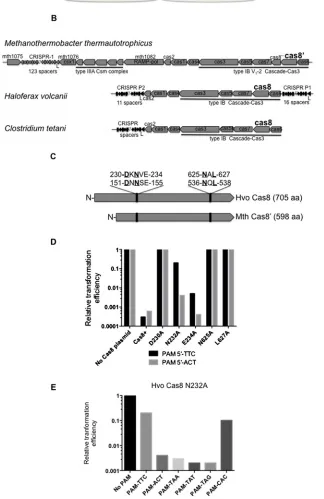
Plasmid protection assays inH. volcanii(Hvo) identified multiple PAM sequences and that disruption ofcas8abolished interfer-ence [43]. To investigate Cas8 further,!cas8Hvo cells were analysed in plasmid protection assays when expressing mutant or wild-type Cas8 from a second plasmid (Figure 1A). Single amino acid substitutions were introduced into Hvo Cas8 based on the alignment with Cas8 homologues from archaea and a bacterium (summarized inFigures 1B and 1C and a full align-ment in Supplealign-mentary Figure S1). Cas8 proteins are diverse [18] with low overall sequence identity, but conserved amino acids were identified to investigate Cas8 function (Figure 1C). Mutation in Hvo Cas8 Asp230, Asn625 and Leu627abolished
in-terference, equivalent to cells lackingcas8(Figure 1D). Mutated Asn232showed reduced interference, by∼20 %, toward plasmids
with PAM 5′-TTC, but had little effect on interference toward
plasmid with PAM 5′-ACT (Figure 1D). Additional assays on
!cas8Hvo pN232A-Cas8 using six Hvo PAMs highlighted a
PAM bias, with interference reduced when PAM 5′-TTC or 5′
-CAC was used, but with no effect of N232A on the other PAMs
(Figure 1E). These genetic assays identified regions of Cas8 that
are essential for interference and suggest that Cas8 is sensitive to PAM sequences.
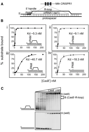
Cas8′binding to DNA and R-loop nucleic acid
substrates
binding to DNA/RNA substrates in EMSAs. Therefore, we pur-ified the Cas8 homologue fromM. thermautotrophicus (Mth) (Supplementary Figure S2), an organism amenable for ana-lysis of its DNA-binding proteins. Mth Cas8 is called Cas8′
[18] because it is 119 amino acids shorter than Hvo Cas8, but it has the conserved amino acid residues required for CR-ISPR interference by Hvo Cas8, summarized inFigures 1C–1E. We constructed nucleic acid substrates for Cas8′ binding that
centred on duplex DNA or R-loop shown inFigure 2(A). Sub-strates were either + or − PAM and contained a 5′-crRNA
handle known to be important for interference in type IB CR-ISPR systems, whereas the 3′-handle is dispensable for
interfer-ence in the same systems [52]. To predict Mth PAM, we ana-lysed 123 spacers in Mth CRISPR-1, identifying protospacers from seven mobile genetic elements to deduce a PAM of 5′
-CCN-3′, detailed in Supplementary Results and
Supplement-ary Figure S3. CC dinucleotide PAM for Mth had been iden-tified in a previous analysis [53], although reported as GG from the reverse complement of the Mth genome. Therefore we in-corporated 5′-CCC into substrates for +PAM or 5′-AAA for −PAM.
Results of Cas8′ EMSAs are in Figures 2(B) and 2(C).
Cas8′ bound to a +PAM R-loop or duplex DNA with
highest affinity (Figure 2B respectively, Kd 5.3+−0.7 nM and Kd 9.1 +−0.2 nM), compared with the same substrates −PAM (Figure 2B respectively, Kd 40.7+−1.0 nM and Kd
18.3+−0.8 nM). It was significant that Cas8′ bound to
R-loops, + or −PAM, as distinct in-gel protein–DNA com-plexes, compared with in well aggregates of protein–DNA observed for Cas8′ mixed with duplex DNA (Figure 2C).
These EMSAs suggested that Cas8′ in isolation can recognize
PAM sequence and may have structural preference for bind-ing stably to branched DNA or R-loops compared with duplex DNA.
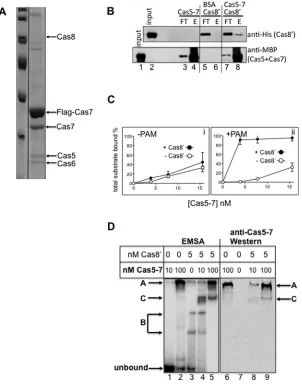
Physical interaction of Cas8′with Cas5–Cas7 and
PAM-dependent stimulation of nucleic acid binding
Cas5 and Cas7 are integral to bacterial and archaeal Cas-cades [9,19] and are predicted to function with Cas8 during CRISPR interference. Previous studies identified a Cas5–Cas7 complex in Hvo [22] and physical association of Cas8′ with
Cas7 during fractionation of Methanothermobacter cell bio-mass [54]. In the present work, FLAG-tagged Cas7 was ex-pressed in Hvo cells to detect protein interactors, identifying Cas8 when FLAG–Cas7 affinity enriched cell extracts were analysed by MS (Figure 3A). To test for physical interaction of Cas8′ with Cas5–Cas7, we first co-purified Mth Cas7 with
Cas5 (Supplementary Figure S4A), the latter an N-terminal fusion to E. coli MBP, giving soluble MBP–Cas5–Cas7 that bound crRNA1. MBP–Cas5–Cas7 interacted physically with
Cas8′ (Figure 3B), with the same results observed either with
or without pre-incubation of Cas5–Cas7 with crRNA1. Cas8′
bound to MBP–Cas5–Cas7 pre-incubated with amylose resin
(Figure 3B, top panel, lane 8), but did not bind to control amylose
resin in BSA (Figure 3B, top panel, lane 6). MBP–Cas5–Cas7 was detected, as expected, inFigure 3(B), lane 8, but not in lane 6 containing BSA. The reciprocal reaction (MBP–Cas5– Cas7 to Ni2+-NTA bound Cas8′) was not effective because
MBP–Cas5–Cas7 bound Ni2+-NTA even when Cas8′ was
ab-sent. These assays indicated physical interaction of Cas8′ with
Cas5–Cas7, although a maximum of only 10 % of Cas8′
in-put could be detected as bound to MBP–Cas5–Cas7 in these conditions.
Cas8′ was tested for any influence effect on binding by
Cas5–Cas7 to duplex+−PAM in EMSAs. Cas8′stimulated total
substrate binding when +PAM but had little effect on bind-ing to −PAM (Figure 3C and representative gels in Supple-mentary Figure S4B). Significantly, EMSAs mixing Cas5–Cas7
+ Cas8′ showed a novel complex that was not present when
either Cas5–Cas7 or Cas8 were alone (Figure 3D): Cas5–Cas7 at 10 nM or 100 nM formed in-well protein–DNA aggregates with, respectively, 8 % and 67 % of substrate (labelled A,
Fig-ure 3D, lanes 1 and 2). Cas8′ alone (5 nM) bound 55 % of
the substrate in distinct complexes (labelled B inFigure 3D, lane 3). A new complex (complex C) was defined when pre-mixing Cas8′(5 nM) with Cas5–Cas7 (10 or 100 nM) and 90 %–
100 % of substrate was bound (Figure 3D, lanes 4 and 5). West-ern blotting of identical EMSA using antibodies against MBP identified MBP–Cas5–Cas7 in complex C (Figure 3D, lanes 8 and 9), confirming that Cas5–Cas7 can form a distinct com-plex in EMSAs that is not an aggregate but dependent on Cas8′.
(A) Summary ofH. volcanii(Hvo) plasmid protection assays. See also parts (D) and (E). Transformation efficiency was measured for a plasmid containing PAMs into Hvo!cas8cells complemented by pCas8+ or pCas8MUTANTto determine their effects on interference. Efficient transformation of plasmid containing a PAM [43] and protospacer indicated loss of interference, manifest as colony growth supported byleuanduracomplementation from the plasmid. (B) Type IB CRIS-PR-Cas systems of the euryarchaeaH. volcanii(Hvo) andM. thermautotrophicus(Mth) and the bacteriumClostridium tetani
(Cte). All have conserved gene order Cas8-Cas7-Cas5-Cas3. Mth contains an additional open reading frame (mth1089) called Cas8′′leading to its classification as a CRISPR type IB variant V1-2 and renaming of Cas8b [6] to Cas8′[18]. The
predicted amino acid sequence of Cas8′′shows no significant homology to any protein in database searches. (C) Cartoon
illustrating two conserved amino acid patches in Hvo Cas8 and Mth Cas8′homologues, which were targeted for
mutagen-esis in data presented in parts (D) and (E) and in the subsequent data Figures. A full alignment of Cas8/Cas8′/Cas8b is
Figure 2 EMSAs for binding Cas8′to duplex and R-loop with +and−PAM
(A) Cartoon of R-loop/duplex model substrates used; sequences of DNA and RNA strands are given in supplemental data. +PAM 5′-CCN (CCC) or−PAM (AAA) sequences and a 5′RNA handle were incorporated into duplex or R-loop as indicated.
For R-loop, crRNA oligonucleotide (shown in red) was synthesized with sequence complementary to a spacer from Mth CRISPR-1. (B). Graphs comparing measurements of Cas8′binding to duplex and R-loop substrates+
−PAM, as indicated in each panel i–iv. Asterisks denote the32P-end labelled DNA strand. EMSAs were in triplicate for plotting as mean values with bars for standard error. Substrate (2.0 nM) was mixed with Cas8′at concentrations (nM): 0, 1.56, 3.125, 6.25, 12.5,
25.0, 50.0, 100, 120, 200. (C) Cas8′gave distinct in gel complex when binding to R-loop (labelled B), compared with in
well aggregates of protein–nucleic acid (complex A) with a duplex DNA.
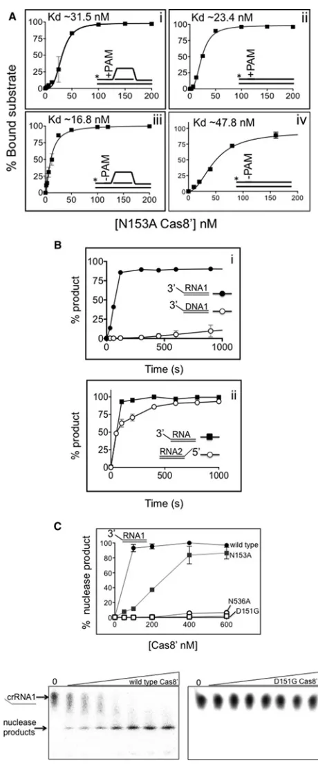
Cas8 amino acid residues that are essential for interferencein vivoare also essential for a Cas8′
nuclease activityin vitro
Mutant Mth Cas8′ proteins D151G, N153A and N536A were
purified (Supplementary Figure S2), corresponding to mutations in Hvo Cas8 that had reduced or abolished interference activ-ity in plasmid protection assays (Asp230, Asn232 and Asn625
Table 1A). Cas8′ mutant proteins were proficient in binding
to R-loop +PAM (Supplementary Figure S5) and other sub-strates (result not shown) and interacted with MBP–Cas5-Cas7 (Supplementary Figure S6). We investigated N153A substrate binding in more detail because the corresponding Hvo muta-tion (Asn232) caused reduced interference to PAMs 5′-CAC or
Figure 3 Interaction of Cas8 and Cas8′with Cas5–Cas7
(A) Coomassie stained SDS/PAGE profile of co-purifying proteins with Flag-Tagged Cas7 expressed inHaloferaxcells. Cas8 was detected by MS. (B) Reconstitution of physical interaction between purifiedMethanothermobacterCas8′(20µg)
with purified complex of affinity taggedMethanothermobacterCas5–Cas7 (20µg). Upper panel shows western blot using anti-(His)6antibody to detect (His)6Cas8′and the lower panel used anti-MBP antibody to detect MBP in MBP–Cas5–Cas7.
‘Input’ is a duplicate loading of total amount of used Cas8′(upper panel) or Cas5–Cas7 (lower panel). Cas8′was detected
in the elution (E) after binding to amylose – MBP–Cas5–Cas7 (lane 8) but did not bind to amylose pre-bound with BSA (lane 6). (C) Measurements of duplex DNA binding+−PAM byMethanothermobacterCas5–Cas7 either with or without Cas8′, as
labelled. Data values for total DNA binding were calculated for each concentration of Cas5–Cas7 (4, 8, 15 nM)+−Cas8′
(5 nM). (D) Corresponding EMSA and western blots for detection of Cas5–Cas7 in a Cas8′ dependent in-gel complex.
Lanes 1–5 (left panel) show phosphorimaged EMSA complexes arising from reactions binding of Cas5–Cas7 (complex A) or Cas8′(B complexes). A new complex C was observed when Cas5–Cas7 and Cas8′are present. Western blotting
detected Cas5–Cas7 in complex C (lanes 8 and 9), as well as complex A (lanes 6, 8 and 9).
5′-CCC were not bound by N153A appreciably better than
sub-strates−PAM in EMSAs (Figure 4A), contrasting with the bind-ing behaviour of wild-type Cas8′(Figure 2). It is possible that
this observation might account for subtly reduced interference, discussed later.
To test for Cas8′catalytic activity correlating to lack of
inter-ference from the aspartic acid/asparagine mutants we re-visited previous work that had identified Mth Cas8′nuclease activity
tar-geting ssDNA flaps [54]. We now compared this DNase activity to equivalent RNase activity. Cas8′was much more efficient as a
nuclease, measured as a function of time, on an ssRNA flap of the same sequence as ssDNA (Figure 4Bi). In these reactions, RNA was present in a RNA–DNA hybrid, but RNA nuclease activity was not detected in RNA–RNA duplex, even though Cas8′
binding to DNA–DNA, RNA–DNA and RNA–RNA substrates was similar (Supplementary Figure S7). Cas8′nuclease activity
was detected on RNA with 3′- and 5′-ends (Figure 4Aii), but
only on the strand with ssRNA overhang. Cas8′ D151G and
Figure 4 Cas8′substrate binding and nuclease activities (A) Binding of N153A Cas8′to substrates+
−PAM as indicated in panels i–iv, for comparison with wild-type Cas8′data inFigure 2(B). Asterisks
denote the32P-end labelled DNA strand. EMSAs were in triplicate for plotting as mean values with bars for standard error. Substrate (2 nM) was mixed with Cas8′N153A at concentrations of (nM); 0, 1.56, 3.125,
6.25, 12.5, 25.0, 50.0, 100, 150 and 200 for panels i and ii; 0, 1.56,
but binding was intact (Supplementary Figure S8B). N153A had intermediate nuclease activity (Figure 4C). The catalytic activity of Cas8′ and inactivating mutations in conserved residues that
are required for interference in Hvo, is evidence that Cas8′is an
RNA nuclease in cells, discussed more below.
DISCUSSION
Cas8 proteins are candidates for guiding Cascade to invader DNA in some type I CRISPR systems [18,21]. Mutation of Cas8 had been implicated in loss of interference inHaloferax(Hvo) [43] and we investigated this using genetic analysis of Hvo Cas8 in CRISPR interference and nucleic acid binding and processing by Cas8′ from Methanothermobacter (Mth). We propose that
Cas8/Cas8′is part of Cascade, contributing to PAM and structure
specific nucleic acid binding, influencing interaction of Cas5– Cas7 with nucleic acids. An interesting ssRNA nuclease activity of Cas8′was detectedin vitro, requiring conserved amino acids
that are essential for interferencein vivo.
Three lines of evidence indicated nucleic acid binding and PAM sensing by Cas8′, in isolation and when mixed with Cas5–
Cas7. Firstly, isolated Cas8′formed distinct complexes with
R-loop substrates in EMSAs and predicted PAM 5′-CCN
stimu-lated its binding to duplex and R-loop substrates (Figure 2). Second, the subtle PAM induced behaviour of Cas8 or Cas8′
mutated in respectively, Asn232 (Tables 1A and 1B) or Asn153
(Figure 4A) supports interaction with PAM either alone or when
bound with Cas5–Cas7 in Cascade, an interaction that is per-turbed by the asparagine mutation. Third, we observed enhanced substrate binding from Cas8′–Cas5–Cas7 +PAM, compared
with either Cas5–Cas7 or Cas8′alone, which was not observed
when −PAM (Figure 3). Cas8′in these assays converted Cas5–
Cas7 protein aggregates into a distinct binding complex suggest-ing that Cas8′modulates how Cas5–Cas7 can precisely assemble
on the substrate, thereby controlling its aggregation. Based on
E. coliCascade structures detailing precise positioning for CasA relative to CasD (Cas5) [37–39], it is likely that interaction of Cas8 with Cas5–Cas7 is important for PAM sensing and for the choreography of Cascade binding to nucleic acids that leads to stable R-loop formation.
Mutation of Hvo Asp230and Asn625abolished Cas8
interfer-ence (Table 1A) and mutation of the corresponding residues in
3.125, 6.25, 12.5, 25.0, 50.0, 100, 120 and 200 for panel iii and 0, 6.25, 12.5, 20.0, 40.0, 80.0 and 160 nM in panel iv. (B) Graphs comparing Cas8′(100 nM) nuclease activity as a function of time on;
panel i, comparing DNase to RNase activity on 3′ssDNA or ssRNA flaps
(1 nM of each), panel ii, comparing RNase activity on 3′or 5′ssRNA
flaps (1 nM of each). In each case the strand labelled was that with the single stranded region and assays were analysed on denaturing gels in triplicate for plotting as mean values with bars for standard error. (C) Comparative RNase activity of Cas8′and mutants on 5′-ssRNA flap
(1 nM) containing protein at 0, 50, 100, 200, 400 and 600 nM. In each panel the data points are means of assays in duplicate showing stand-ard error and representative gels for wild-type Cas8′and a catalytically
Mth Cas8′, Asp151and Asn536, abolished ssRNA nuclease
activ-ity (Figure 4). Cas8′degraded ssRNA with either 3′- or 5′-ends.
However, we were unable to detect RNase activityin vitrofrom Hvo Cas8, despite using a wide range of high and low salt assay conditions, possibly because of instability of the purified protein that appeared during storage. Therefore we cannot conclude that abolished nuclease activity of Cas8′D151G and N536A mutants
explains the loss of genetic interference by Hvo Cas8 D230A and N625A mutants, but the correlation is interesting. Northern blotting for crRNA in Hvo!cas8cells showed that Cas8 is not
needed for processing crRNA into pre-crRNA or crRNA in cells (Supplementary Figure S9). Also, Hvo cells do contain a nuclease that removes nts from the 3′-end of crRNA after processing from
pre-crRNA but this RNase function is not altered if cells lack Cas8 (Anita Marchfelder, personal communication). 5′-crRNA
handles are essential for interference in Hvo cells and are there-fore not processed after crRNA formation [52]. Therefore the role of Cas8 RNase activity, if any, in Cascade-mediated CRISPR in-terference is undetermined. We cannot exclude the possibility that Cas8 RNase activity may be needed for some other aspect of RNA metabolism and processing in these organisms that has an indirectly important role for some type I CRISPR systems.
AUTHOR CONTRIBUTION
Simon Cass, Karina Hass and Britta Stoll did experiments. Omer S. Alkhnbashi and Rolf Backofen analysed and determined the CRISPR-Cas types of Haloferax volcanii and Methanothermobac-ter thermautotrophicususing bioinformatics, determining the Mth Cas8 as Cas8′and the Hvo as Cas8. Edward Bolt and Anita March-felder organized the project and wrote the manuscript. Kundan Sharma and Henning Urlaubdid mass spectrometry analyses to identify the proteins co-purifying with the FLAG-tagged Cas7.
ACKNOWLEDGEMENTS
We thank Thorsten Allers (Nottingham, U.K.) forHaloferax plas-mids.
FUNDING
This work was supported by the Biotechnology and Biological Sci-ences Research Council PhD studentship and the German Re-search Council (Deutsche Forschungsgemeinschaft) [grant num-bers DFG MA1538/16-1, UR225/1-1 and BA2168/5-1].
REFERENCES
1 Barrangou, R., Fremaux, C., Deveau, H., Richards, M., Boyaval, P., Moineau, S., Romero, D.A. and Horvath, P. (2007) CRISPR provides acquired resistance against viruses in prokaryotes. Science315, 1709–1712CrossRef PubMed
2 Barrangou, R. and Marraffini, L.A. (2014) CRISPR-Cas systems: prokaryotes upgrade to adaptive immunity. Mol. Cell54, 234–244 CrossRef PubMed
3 van der Oost, J., Westra, E.R., Jackson, R.N. and Wiedenheft, B. (2014) Unravelling the structural and mechanistic basis of CRISPR-Cas systems. Nat. Rev. Microbiol.12, 479–492 CrossRef PubMed
4 Heler, R., Marraffini, L.A. and Bikard, D. (2014) Adapting to new threats: the generation of memory by CRISPR-Cas immune systems. Mol. Microbiol.93, 1–9
CrossRef PubMed
5 Westra, E.R., Swarts, D.C., Staals, R.H., Jore, M.M., Brouns, S.J. and van der Oost, J. (2012) The CRISPRs, they are a-changin’: how prokaryotes generate adaptive immunity. Annu. Rev. Genetics46, 311–339CrossRef
6 Makarova, K.S., Haft, D.H., Barrangou, R., Brouns, S.J., Charpentier, E., Horvath, P., Moineau, S., Mojica, F.J., Wolf, Y.I., Yakunin, A.F. et al. (2011) Evolution and classification of the CRISPR-Cas systems. Nat. Rev. Microbiol.9, 467–477 CrossRef PubMed
7 Chylinski, K., Le Rhun, A. and Charpentier, E. (2013) The tracrRNA and Cas9 families of type II CRISPR-Cas immunity systems. RNA Biol.10, 726–737CrossRef PubMed
8 Koonin, E.V. and Makarova, K.S. (2013) CRISPR-Cas: evolution of an RNA-based adaptive immunity system in prokaryotes. RNA Biol. 10, 679–686CrossRef PubMed
9 Jore, M.M., Lundgren, M., van Duijn, E., Bultema, J.B., Westra, E.R., Waghmare, S.P., Wiedenheft, B., Pul, U., Wurm, R., Wagner, R. et al. (2012) Structural basis for CRISPR RNA-guided DNA recognition by Cascade. Nat. Struct. Mol. Biol.18, 529–536 CrossRef
10 Brouns, S.J., Jore, M.M., Lundgren, M., Westra, E.R., Slijkhuis, R.J., Snijders, A.P., Dickman, M.J., Makarova, K.S., Koonin, E.V. and van der Oost, J. (2008) Small CRISPR RNAs guide antiviral defense in prokaryotes. Science321, 960–964
CrossRef PubMed
11 Charpentier, E. and Marraffini, L.A. (2014) Harnessing CRISPR-Cas9 immunity for genetic engineering. Curr. Opin. Microbiol.19C, 114–119
CrossRef
12 Jinek, M., Jiang, F., Taylor, D.W., Sternberg, S.H., Kaya, E., Ma, E., Anders, C., Hauer, M., Zhou, K., Lin, S. et al. (2014) Structures of Cas9 endonucleases reveal RNA-mediated conformational activation. Science343, 1247997
CrossRef PubMed
13 Rouillon, C., Zhou, M., Zhang, J., Politis, A., Beilsten-Edmands, V., Cannone, G., Graham, S., Robinson, C.V., Spagnolo, L. and White, M.F. (2013) Structure of the CRISPR interference complex CSM reveals key similarities with cascade. Mol. Cell52, 124–134 CrossRef PubMed
14 Marraffini, L.A. and Sontheimer, E.J. (2008) CRISPR interference limits horizontal gene transfer in staphylococci by targeting DNA. Science322, 1843–1845
CrossRef PubMed
15 Staals, R.H., Agari, Y., Maki-Yonekura, S., Zhu, Y., Taylor, D.W., van Duijn, E., Barendregt, A., Vlot, M., Koehorst, J.J., Sakamoto, K. et al. (2013) Structure and activity of the RNA-targeting Type III-B CRISPR-Cas complex ofThermus thermophilus. Mol. Cell52, 135–145CrossRef PubMed
16 Zebec, Z., Manica, A., Zhang, J., White, M.F. and Schleper, C. (2014) CRISPR-mediated targeted mRNA degradation in the archaeonSulfolobus solfataricus. Nucleic Acids Res.42, 5280–5288CrossRef PubMed
17 Spilman, M., Cocozaki, A., Hale, C., Shao, Y., Ramia, N., Terns, R., Terns, M., Li, H. and Stagg, S. (2013) Structure of an RNA silencing complex of the CRISPR-Cas immune system. Mol. Cell 52, 146–152CrossRef PubMed
19 Lintner, N.G., Kerou, M., Brumfield, S.K., Graham, S., Liu, H., Naismith, J.H., Sdano, M., Peng, N., She, Q., Copie, V. et al. (2011) Structural and functional characterization of an archaeal clustered regularly interspaced short palindromic repeat (CRISPR)-associated complex for antiviral defense (CASCADE). J. Biol. Chem.286, 21643–21656
CrossRef PubMed
20 Wiedenheft, B., van Duijn, E., Bultema, J.B., Waghmare, S.P., Zhou, K., Barendregt, A., Westphal, W., Heck, A.J., Boekema, E.J., Dickman, M.J. and Doudna, J.A. (2011) RNA-guided complex from a bacterial immune system enhances target recognition through seed sequence interactions. Proc. Natl. Acad. Sci. U.S.A.108, 10092–10097CrossRef PubMed
21 Plagens, A., Tripp, V., Daume, M., Sharma, K., Klingl, A., Hrle, A., Conti, E., Urlaub, H. and Randau, L. (2014)In vitroassembly and activity of an archaeal CRISPR-Cas type I-A Cascade interference complex. Nucleic acids Res.42, 5125–5138
CrossRef PubMed
22 Brendel, J., Stoll, B., Lange, S.J., Sharma, K., Lenz, C., Stachler, A.E., Maier, L.K., Richter, H., Nickel, L., Schmitz, R.A. et al. (2014) A complex of Cas proteins 5, 6, and 7 is required for the biogenesis and stability of clustered regularly interspaced short palindromic repeats (crispr)-derived rnas (crrnas) inHaloferax volcanii. J. Biol. Chem.289, 7164–7177CrossRef PubMed 23 Nam, K.H., Haitjema, C., Liu, X., Ding, F., Wang, H., DeLisa, M.P.
and Ke, A. (2012) Cas5d protein processes pre-crRNA and assembles into a cascade-like interference complex in subtype I-C/Dvulg CRISPR-Cas system. Structure20, 1574–1584 CrossRef PubMed
24 Garside, E.L., Schellenberg, M.J., Gesner, E.M., Bonanno, J.B., Sauder, J.M., Burley, S.K., Almo, S.C., Mehta, G. and MacMillan, A.M. (2012) Cas5d processes pre-crRNA and is a member of a larger family of CRISPR RNA endonucleases. RNA18, 2020–2028 CrossRef PubMed
25 Punetha, A., Sivathanu, R. and Anand, B. (2014) Active site plasticity enables metal-dependent tuning of Cas5d nuclease activity in CRISPR-Cas type I-C system. Nucleic Acids Res.42, 3846–3856CrossRef PubMed
26 Haurwitz, R.E., Jinek, M., Wiedenheft, B., Zhou, K. and Doudna, J.A. (2010) Sequence- and structure-specific RNA processing by a CRISPR endonuclease. Science329, 1355–1358
CrossRef PubMed
27 Richter, H., Zoephel, J., Schermuly, J., Maticzka, D., Backofen, R. and Randau, L. (2012) Characterization of CRISPR RNA processing inClostridium thermocellumandMethanococcus maripaludis. Nucleic Acids Res.40, 9887–9896CrossRef PubMed
28 Carte, J., Wang, R., Li, H., Terns, R.M. and Terns, M.P. (2008) Cas6 is an endoribonuclease that generates guide RNAs for invader defense in prokaryotes. Genes Dev.22, 3489–3496 CrossRef PubMed
29 Reeks, J., Naismith, J.H. and White, M.F. (2013) CRISPR interference: a structural perspective. Biochem. J.453, 155–166 CrossRef PubMed
30 Niewoehner, O., Jinek, M. and Doudna, J.A. (2014) Evolution of CRISPR RNA recognition and processing by Cas6 endonucleases. Nucleic Acids Res.42, 1341–1353CrossRef PubMed
31 Nam, K.H., Kurinov, I. and Ke, A. (2011) Crystal structure of clustered regularly interspaced short palindromic repeats (CRISPR)-associated Csn2 protein revealed Ca2+-dependent double-stranded DNA binding activity. J. Biol. Chem.286, 30759–30768CrossRef PubMed
32 Semenova, E., Jore, M.M., Datsenko, K.A., Semenova, A., Westra, E.R., Wanner, B., van der Oost, J., Brouns, S.J. and Severinov, K. (2011) Interference by clustered regularly interspaced short palindromic repeat (CRISPR) RNA is governed by a seed sequence. Proc. Natl. Acad. Sci. U.S.A.108, 10098–10103
CrossRef PubMed
33 Wiedenheft, B., Lander, G.C., Zhou, K., Jore, M.M., Brouns, S.J., van der Oost, J., Doudna, J.A. and Nogales, E. (2011) Structures of the RNA-guided surveillance complex from a bacterial immune system. Nature477, 486–489CrossRef PubMed
34 Kunne, T., Swarts, D.C. and Brouns, S.J. (2014) Planting the seed: target recognition of short guide RNAs. Trends Microbiol.22, 74–83 PubMed
35 Maier, L.K., Lange, S.J., Stoll, B., Haas, K.A., Fischer, S., Fischer, E., Duchardt-Ferner, E., Wohnert, J., Backofen, R. and Marchfelder, A. (2013) Essential requirements for the detection and
degradation of invaders by theHaloferax volcaniiCRISPR/Cas system I-B. RNA Biol.10, 865–874
CrossRef PubMed
36 Ivancic-Bace, I., Al Howard, J. and Bolt, E.L. (2012) Tuning in to interference: R-loops and cascade complexes in CRISPR immunity. J. Mol. Biol.422, 607–616CrossRef PubMed
37 Zhao, H., Sheng, G., Wang, J., Wang, M., Bunkoczi, G., Gong, W., Wei, Z. and Wang, Y. (2014) Crystal structure of the RNA-guided immune surveillance Cascade complex inEscherichia coli. Nature 515, 147–150CrossRef PubMed
38 Mulepati, S., Heroux, A. and Bailey, S. (2014) Crystal structure of a CRISPR RNA-guided surveillance complex bound to a ssDNA target. Science345, 1479–1484
CrossRef PubMed
39 Jackson, R.N., Golden, S.M., van Erp, P.B., Carter, J., Westra, E.R., Brouns, S.J., van der Oost, J., Terwilliger, T.C., Read, R.J. and Wiedenheft, B. (2014) Crystal structure of the CRISPR RNA-guided surveillance complex fromEscherichia coli. Science345, 1473–1479CrossRef PubMed
40 Shah, S.A., Erdmann, S., Mojica, F.J. and Garrett, R.A. (2013) Protospacer recognition motifs: mixed identities and functional diversity. RNA Biol.10, 891–899
CrossRef PubMed
41 Sashital, D.G., Wiedenheft, B. and Doudna, J.A. (2012) Mechanism of Foreign DNA selection in a bacterial adaptive immune system. Mol. Cell46, 606–615
CrossRef PubMed
42 Hochstrasser, M.L., Taylor, D.W., Bhat, P., Guegler, C.K., Sternberg, S.H., Nogales, E. and Doudna, J.A. (2014) CasA mediates Cas3-catalyzed target degradation during CRISPR RNA-guided interference. Proc. Natl. Acad. Sci. U.S.A.111, 6618–6623 CrossRef PubMed
43 Fischer, S., Maier, L.K., Stoll, B., Brendel, J., Fischer, E., Pfeiffer, F., Dyall-Smith, M. and Marchfelder, A. (2012) An archaeal immune system can detect multiple protospacer adjacent motifs (PAMs) to target invader DNA. J. Biol. Chem.287, 33351–33363
CrossRef PubMed
44 Allers, T., Ngo, H.P., Mevarech, M. and Lloyd, R.G. (2004) Development of additional selectable markers for the halophilic archaeonHaloferax volcaniibased on the leuB and trpA genes. Appl. Environ. Microbiol.70, 943–953
CrossRef PubMed
45 Allers, T., Barak, S., Liddell, S., Wardell, K. and Mevarech, M. (2010) Improved strains and plasmid vectors for conditional overexpression of His-tagged proteins inHaloferax volcanii. Appl. Environ. Microbiol.76, 1759–1769
CrossRef PubMed
46 Miller, J. (1972) Experiments in Molecular Genetics, Cold Spring Harbour Laboratory, New York
47 Bitan-Banin, G., Ortenberg, R. and Mevarech, M. (2003) Development of a gene knockout system for the halophilic archaeonHaloferax volcaniiby use of the pyrE gene. J. Bacteriol. 185, 772–778CrossRef PubMed
49 Norais, C., Hawkins, M., Hartman, A.L., Eisen, J.A., Myllykallio, H. and Allers, T. (2007) Genetic and physical mapping of DNA replication origins inHaloferax volcanii. PLoS Genetics3, e77 CrossRef PubMed
50 Shevchenko, A., Wilm, M., Vorm, O. and Mann, M. (1996) Mass spectrometric sequencing of proteins silver-stained polyacrylamide gels. Anal. Chem.68, 850–858
CrossRef PubMed
51 Pfeiffer, F., Broicher, A., Gillich, T., Klee, K., Mejia, J., Rampp, M. and Oesterhelt, D. (2008) Genome information management and integrated data analysis with HaloLex. Arch. Microbiol.190, 281–299
CrossRef PubMed
52 Maier, L.K., Stachler, A.E., Saunders, S.J., Backofen, R. and Marchfelder, A. (2015) An active immune defense with a minimal CRISPR (clustered regularly interspaced short palindromic repeats) RNA and without the Cas6 protein. J. Biol. Chem.290,
4192–4201CrossRef PubMed
53 Mojica, F.J., Diez-Villasenor, C., Garcia-Martinez, J. and Almendros, C. (2009) Short motif sequences determine the targets of the prokaryotic CRISPR defence system. Microbiology155, 733–740 CrossRef PubMed
54 Guy, C.P., Majernik, A.I., Chong, J.P. and Bolt, E.L. (2004) A novel nuclease-ATPase (Nar71) from archaea is part of a proposed thermophilic DNA repair system. Nucleic Acids Res.32, 6176–6186CrossRef PubMed