MECHANISTIC STUDIES ON GLUTAMATE
DECARBOXYLASE AND SERINE
HYDROXMETHYLTRANSFERASE
Janet Elizabeth Rose
A Thesis Submitted for the Degree of PhD
at the
University of St Andrews
1993
Full metadata for this item is available in
St Andrews Research Repository
at:
http://research-repository.st-andrews.ac.uk/
Please use this identifier to cite or link to this item:
http://hdl.handle.net/10023/14295
MECHANISTIC STUDIES ON
I"
GLUTAMATE DECARBOXYLASE
AND
SERINE HYDROXMETHYLTRANSFERASE
a thesis presented by Janet Elizabeth Rose
to the
UNIVERSITY OF ST. ANDREWS
in application for
THE DEGREE OF DOCTOR OF PHILOSOPHY
ProQuest Number: 10167256
All rights reserved
INFORMATION TO ALL USERS
The quality of this reproduction is dependent upon the quality of the copy submitted.
In the unlikely event that the author did not send a com plete manuscript
and there are missing pages, these will be noted. Also, if material had to be removed, a note will indicate the deletion.
uest
ProQuest 10167256
Published by ProQuest LLO (2017). Copyright of the Dissertation is held by the Author.
All rights reserved.
This work is protected against unauthorized copying under Title 17, United States C ode Microform Edition © ProQuest LLO.
ProQuest LLO.
789 East Eisenhower Parkway P.Q. Box 1346
1 was admitted to the Faculty of Science of the University of St. Andrews under Ordinance General No. 12 on lêfe^..Q.^^fcç^teer.(^Oand as a candidate for the degree of Ph. D. on .lSt..Q.CfcC>ifiS<:...J.S0.O
Signed . Date ..lEfc..Ssptetvte‘..lîï)2
DECLARATION
I, Janet Elizabeth Rose, hereby certify that this thesis has been composed by myself,
that it is a record of my own work, and that it has not been accepted in partial or if complete fulfilment of any other degree or professional qualification.
Signed ... Date
I hereby certify that the candidate has fulfilled the conditions of the Resolution and
Regulations appropriate to the degree of Ph. D.
Signature of supervisor
Copyright
In submitting this thesis to the University of St. Andrews I understand that I am giving permission for it to be made available for use in accordance with the regulations of the
University library for the time being in force, subject to any copyright vested in the work not being affected thereby. I also understand that the title and abstract will be
published, and that a copy of the work may be made and supplied to any bona fide
TO
Acknowledgements
I would like to thank my supervisor Prof. David Gani for his continual enthusiasm and
encouragement throughout the course of this work, and for giving me the opportunity to work in such a beautiful part of the country.
Thanks to the SERC and MSD for financial assistance during my studies.
I must thank all of the staff, both at St. Andrews and Southampton University for their help, particularly John Langley, Melanja Smith and Dr. Mackie.
Thank you to those in the group who have kept me going over the last 3 years, Catherine and Nigel for their company when Richard kept vanishing back to the
Southampton Physics Department, Andrew and Mahmoud for proof-reading my thesis
and help with all things chemical, and Paul for being here! I must also thank Kev for helping me find my way around 211a and Catherine for making sure my glassware
arrived in Scotland and wasn’t “borrowed” before I arrived.
Finally I must thank Richard for his never ending support, for coming to St. Andrews
with me, and for completing the last year of his physics Ph.D in a room surrounded by organic chemists.
ABSTRACT
(2S)- and (2R)-Serine O-suIphate have been synthesised and shown to inactivate
glutamate decarboxylase (GAD) from E. Coli. Novel methodology was developed to enable the stereospecific synthesis of (2S) and (2R)-a-deuteriated serine in order to
probe the mechanism of inactivation. The rates of inactivation of glutamate decarboxylase by (2S)-, (2S)-[2-^H]-, (2R)- and (2R)-[2-^H]-serine 0-sulphate have
been measured for each of the isotopomers at a range of concentrations. From the
data obtained the deuterium isotope effects were determined for each enantiomer. The inactivation by the (2S)-enantiomer was shown to involve C“-H bond cleavage while inactivation by the (2R)-isomer involves C“-decarboxylation. Both processes were
shown to occur on the 4’-re-face of the coenzyme, the opposite face to that utilised in the physiological decarboxylation reaction.
The methodology developed for the synthesis of the deuteriated serines involved the
regiospecific introduction of deuterium to the C-6 centre of (3R)- and
(3S)-2,5-dimethoxy-3-isopropyl“3,6-dihydropyrazine. Schollkopf chemistry was then exploited
for the stereospecific alkylation at C-6 of the dihydropyrazines. This chemistry was versatile and enabled the synthesis of other a-deuteriated a-amino acids. For example
(2S)-[2-^H]-phenylalanine, (2S)-[2-^H]-allylglycine and (2S)-[2-^H]-aspartic acid were synthesised using this chemistry.
The decarboxylation of 2-aminomalonic acid by cytosolic serine
hydroxymethyltransferase (SHMT) was studied. Contrary to previous reports, the reaction was found to be stereospecific and the newly Introduced hydrogen was shown
to occupy the 2-pro-S position of the glycine product.
Contents
Acknowledgements
Abstract List of Figures List of Tables Abbreviations
1.0 Pyridoxal 5’-phosphate dependent enzymes
1.1 The metabolic role of PLP dependent enzymes
1.2 Structure and mechanism of PLP dependent enzymes 1.2.1 Transamination
1.2.2 Decarboxylation
1.3 Mechanism-based inhibitors of PLP enzymes
1.3.1 Transaminase Inhibitors
1.3.2 Decarboxylase Inhibitors
1.3.3 Other PLP Inhibitors 1.4 Research objectives
Page i ii iii iv V 1 1 6 8 15 19 20 25 27 31
1
2.0 Glutamate Decarboxylase (GAD) 32
2.1 Properties and Structure of GAD 32
2.1.1 Properties and structure of mammalian GAD 32
2.1.2 Properties and structure of plant GAD 34 2.1.3 Properties and structure of bacterial GAD 35
2.1.4 Glutamate decarboxylase as a chemotherapeutic target 35
2.1.5 Substrate and reaction specificity 36
2.1.7 Stereochemical studies 39 2.1.8 Active-site structures of PLP-dependent decarboxylases 44
2.1.9 Optical properties of bacterial GAD 46
2.1.10 Pyrroloquinoline quinone (PQQ) 48
2.2 Inhibition of Glutamate Decarboxylase by (2S)-serine O-sulphate 51
2.1.1 Introduction 51
2.2.2 Synthesis of O-sulphate esters 58
2.2.3 Preliminary studies with GAD 60
2.2.4 Studies with (2S)-serlne O-sulphate 63
2.3 Deuteriated (2S)-serine O-sulphate as a mechanistic probe 64
2.3.1 Synthesis of (2S)-[2-^H]-serine O-sulphate
from (2S)-aspartic acid 65
2.3.2 An alternative route to deuteriated serines
via cyclic sulphamidates 72
2.3.2 Synthesis of (2S)-[2-^H]-serine O-sulphate
using Schollkopf chemistry 77
2.4 (2R)-Serine O-sulphate as a mechanistic probe 83
2.4.1.Synthesis of (2R)-[2-^H]-serine O-sulphate 83 2.5 Synthesis of other deuteriated a-amino acids 84
2.6 Mathematical analysis of the data from the
inactivation of GAD by serine O-sulphates 88
2.7 Results and interpretation of the
inactivation experiments 93
2.7.1 (2S)-serine O-sulphates 93
2.7.2 (2R)-serine O-sulphates 96
2.7.3 Incubations of (2S)- and (2R)-homocysteic
2.7.4 Uv/vis measurements on the inactivation mixture 103
2.8 Conclusions 102
Î
3.0 Serine hydroxymethyltransferase (SHMT) 109
3.1 Properties and structure of SHMT 110
3.1.1 Properties and structure of mammalian SHMT 110 3.1.2 Properties and structure of plant SHMT 112
3.1.3 Properties and structure of bacterial SHMT 116
3.1.4 SHMT as a chemotherapeutic agent 118
3.1.5 Substrate and reaction specificity 121
3.1.6 Role of PLP 123
3.1.7 Role of tetrahydrofolic acid 126
3.1.8 Stereochemical studies 128
3.1.9 Active-site structure 132
3.2 The decarboxylation of aminomalonic acid by SHMT 137 3.2.1 Re-examination of the decarboxylation of
2-aminomalonic acid by SHMT 142
3.2.2 Re-examination of the decarboxylation
reaction in the light of the findings of Thomas 144
3.3 Conclusions 150
4.0 Experimental 152
Appendix 220
List of Figures
1.1 The two base mechanism for aspartate 6-decarboxylase
2.1 The PLP-Schiff’s base of (2S)-glutamic acid and (2S)-serine O-sulphate 2.2 The proton nmr spectra of serine O-sulphate camphanamides
2.3 The proton nmr spectra of the dihydropyrazine and
the C-6 dideuteriated dihydropyrazine
2.4 kapp vs [I] for inhibition of GAD by (2S)- and (2S)-[2-^H]-serine O-sulphate
Page 18 57 62
81
94 2.5 1/kapp vs 1/[l] for inhibition of GAD by (2S)- and (2S)-[2-^H]-serine O-sulphate 94 2.6 The proton nmr spectra of serine camphanamide methyl esters
3.1 Inhibitors of SHMT
3.2 Disposition of cysteine residues in three forms of SHMT
3.3 The proton nmr spectra of glycine camphanamides 3.4 The mass spectra of glycine camphanamides
101
119
137 147
148/149
I
■
I
III
1
i' • V ■ I. Ji».? ’
List of Tables
1.1 Comparison of the amino acid sequences for selected regions of AAT isozymes
1.2 The Schiff’s base forming regions of several AAT isozymes
1.3 Some therapeutically important target enzymes dependent on PLP
2.1 Active-site residues of several AAT isozymes
2.2 Schiff’s base forming region of PLP-dependent decarboxylase enzymes
2.3 Amino acids synthesised using Schollkopf chemistry 2.4 Kj and kjnact for inactivation of GAD by (2S)- and
(2S)-[2-^H]-serine O-sulphate
2.5 Average rates of inactivation of GAD by (2R)- and (2R)-[2-^H]-serine O-sulphate
3.1 SHMT protein properties
3.2 The reaction and substrate specificity of SHMT
Page
14
15 19
45 46
87
93
96
111
121
A bbreviation AAT c. AAT m. AAT AET BOG n-BuU DDC DOPA DMSO E2 EG
E coli
e.e.
FH4
5,10-CH2FH4
5-methyl-FH4
5-hydroxymethyi-FH 4
F-MOG GABA GABA-T GAD HOC I.EP. kapp Km M eaning
aspartate aminotransferase
cytosolic aspartate aminotransferase
mitochondrial aspartate aminotransferase aminoethylisothiuronium bromide
benzyloxycarbonyl
norma/butyllithium dopa decarboxylase dihydrophenylalanine
dimethylsulphoxide bimoiecular elimination
Enzyme Catalogue
Escherichia coll enantiomeric excess tetrahydrofolate 5,10-methylenetetrahydrofolate 5-methyltetrahydrofolate 5-hydroxymethyltetrahydrofolate 9-fluorenylmethyloxycarbonyl y-aminobutyric acid
y-aminobutyric acid transaminase glutamate decarboxylase
histidine decarboxylase Isoelectric point
apparent first order rate constant Michaelis constant
1
Ki
kcat
kjnact
kinact(H)/kinact(D)
Kh/Kd
Mr NAD+ NADH NADP+ NaTMS NMR ODC PLP PMP PO-PQQ SAM SDM SDS SHMT TBDMSCI
enzymic inhibition constant
enzymic catalytic constant/turnover no.
catalytic constant for inhibition
ratio of kjnact’s for protiated compound and deuteriated compound
ratio of inhibition constants for protiated compound and deuteriated compound
relative molecular mass
nicotinam ide adenine dinucleotide (oxidised form)
nicotinam ide adenine dinucleotide (reduced
form)
nicotinamide adenine dinucleotide phosphate (oxidised form)
3-(trimethylsilyl)propionic-2,2,3,3-^H4 sodium salt nuclear magnetic resonance
ornithine decarboxylase
pyridoxal 5' phosphate pyridoxamine 5’-phosphate
phosphate
pyrroloquinone quinoline
S-adenosylmethionine site-directed mutagenisis sodium dodecylsulphate
serine hydroxymethyltransferase ^Butyldimethylsliylchloride
VII
""■T
:
:THF tetrahydrofuran
tic thin layer chromatography T
TMS tetramethylsilane
Vmax maximum rate of substrate turnover at
saturation
V enzymic reaction rate
Vjnact inactivation rate
V/K first order rate constant for enzymic reaction at
low substrate concentration
'V
VhA/d°(V/K) (VhA/o)/(Kh/Kd)
Proline Pro P
Serine Ser S
Threonine Thr T
Tryptophan Trp W
Tyrosine Tyr Y
Valine Val V
I
Amino acid Three letter code Single letter code
Alanine Ala A
Arginine Arg R
Asparagine Asn N
Aspartic acid Asp D
Cysteine Cys C
Glutamic acid Glu E
Glutamine Gin Q
Glycine Gly G
Histidine His H
Isoleucine Ile I
Leucine Leu L
Methionine Met M
J
Phenylalanine Phe F
I
VIIII
CHAPTER ONE
INTRODUCTION TO
1
OH G
CH
H
OH
OH HO
CH
H
1.0 Pyridoxal 5’-phosphate dependent enzymes
Enzymes requiring pyridoxal 5’-phosphate (PLP) (1) as acoenzyme are ubiquitous in nature, and have been isolated from bacterial and mammalian sources. PLP, the
biologicaiiy active form of vitamin Be (pyridoxol) (2), serves as a cofactor for a myriad of enzyme catalysed reactions, many of which are essential for amino acid metabolism. In the mid 1940's, SnelP identified PLP as the coenzyme for transaminases, and since
then PLP has been established as the coenzyme for over 20 different types of enzyme
catalysed reactions inciuding decarboxylases.
0 1 O—
P-II
o
(1) (2)
1.1 The metabolic role of PLP dependent enzymes
The mechanisms of action of the PLP dependent enzymes are of great scientific
interest. The PLP dependent enzymes play a major role in the regulation of
physiological processes. Some of the more important processes are detailed below.
Two important transaminases are y-aminobutyric acid transaminase (GABA-T) and co
ornithine transaminase. GABA-T is a key enzyme involved in the reguiation of the GABA-ergic system (Scheme 1.1) in the mammalian central nervous system.
-NHc
COgH
(2S)-glutamic acid
glutamate decaitKJxylase
^
NHc
COgH H
Y-aminobutyric acid (GABA)
Receptor occupation
O
HOgC
J
COgH
a-ketoglutarate O
via
intermediates in the citric
acid cycle
N
HOgC,succinate
H ' ^ "COgH succinate semialdehyde ><^xidation
COgH reduction
HO^ "COgH 4-hydroxy butanoic acid
Scheme 1.1. The GABA-ergic system.
Y-Aminobutyric acid (GABA) is a major inhibitory neurotransmitter and there is much evidence to suggest that high cerebral concentrations prevent convulsions.^
m-Ornithine transaminase not only interconnects the citric acid cycle and the urea cycle
but also couples ornithine to proline metabolism (Scheme 1.2).
-I
NHc
H
1
COgH
Arginine
Y
+NH, NHc COgH Proline Urea cycle NK H.ACOoH Ornithine
NHc (o-ornithine
transaminase COgH 5-caitKDxylic acid
reduction
NHr
H
COgH
Citrulline
Y
o
NHc
O
NHcHOgC
a-ketoglutarate, an Intermediate in the citric acid cycle
COgH H' J "COgH
,H
Glutamic acid
Scheme 1.2. The metabolic significance of ornithine transaminase.
The decarboxylases are one of the most important classes of PLP dependent enzymes. The physiological role of the amino acid decarboxylases in bacteria is not fully understood, but inducible decarboxylases for glutamate, arginine and lysine have
been purified to homogeneity.^ One of the most important physiological roles of the decarboxylases in mammals is the conversion of amino acids to pharmacologically
active amines. The formation of dopamine, histamine, GABA, and putrescine all depend on PLP-dependent decarboxylation of their respective parent amino acids.
The biosynthesis of the higher polyamines, spermine and spermidine, from putrescine involves the pyruvate-dependent decarboxylation of S-adenosylmethionine (SAM)
(Scheme 1.3).
Mammalian brain glutamate decarboxylase is directly responsible for the biosynthesis
3
1
of GABA in the GABA-ergic system (Scheme 1.1). Knowledge of the mechanism of action will enable the synthesis of inhibitors to control the level of GABA in the brain.
Ornithine decarboxylase (ODC) is the first and rate-limiting enzyme in the biosynthetic
pathway leading to putrescine and higher polyamines (Scheme 1.3)."^ The activity of the enzyme in vivo increases dramatically in response to cellular stimulation which
promotes regeneration and replication. Cellular levels of the enzyme are high during growth, and low at other stages.® The association of high polyamine levels with rapid cellular proliferation, and protein biosynthesis, led to the idea that polyamines may be
required for RNA/DNA biosynthesis.® The enzyme has been identified as a target for cancer chemotherapy."^ NHc ornithine decarboxylase, PLP Huw^ COgH ornithine NHo NHc putrescine S I Ad yCHg
H\^
COgH S-adenosylmethionineSAM NHg
decaitKDxylase
S'
I Ad S-adenosyl-(methylthio) -propylamine Higher polyamines spermidineScheme 1,3. The synthesis of higher polyamines.
Histidine decarboxylase (HOC) catalyses the decarboxylation of histidine to give
histamine. In mammals, histidine is important as the major receptor agonist.^ Overproduction of histamine is associated with many biological responses including
antihistamines, antagonists for the receptors, has been an important area in medicinal
chemistry. Histamine is also active as a regulator of peripheral blood circulation.^
The aromatic amino acid decarboxylases catalyse the formation of many pharmacologically important amines. For example, DOPA decarboxylase (DDC) is
able to convert many aromatic substrates to the corresponding amines including, phenylalanine to phenyethylamine, tyrosine to tyramlne, tryptophan to tryptamine, histidine to histamine, 3,4-dlhydroxyphenylalanine (3) to
3,4-dihydroxy-phenylethylamine (dopamine) and 5-hydroxytryptophan
(4)
to serotonin.® Dopamine and serotonin act on smooth muscles and the cardiovascular system. Dopamine is a precursor in the biosynthesis of adrenaline, and serotonin is itself neuroactive.®"{
?I
I
■i Aÿ % «(3)
OH OH NH H H NH OH(4)
Serine hydroxymethyltransferase (SHMT) catalyses the formation of
5,10-methylenetetrahydrofolic acid and glycine from tetrahydrofolic acid and (2S)-serine. The enzyme is a key enzyme in single carbon (0^) metabolism at all oxidation levels
and will be discussed further in Chapter 3.
Another important class of PLP dependent enzymes, the racemases, catalyse the interconversion of (2R)- and (2S)-amino acids. These enzymes are common in
prokaryotes and several are involved in the biosynthesis of peptidoglycan in bacterial cell walls.® Alanine racemase is an important enzyme In peptidoglycan biosynthesis
and as such the enzyme Is a target for the action of many antibacterial agents,
including (2R)-cyc/oserine (5),^^ 0-carbamyl-{2R)-serine (6)^^ and phosphoalanine (7)JG
NH
NH H
(6)
NH
CHc
PO 2
-(7)
1.2 Structure and mechanism of PLP dependent enzymes
The coenzyme, PLP, is bound at the enzyme active site as a Schiff's base complex
with the e-amino group of an active site lysine residue (Scheme 1.4, 8).^^'^'*The nature
of the coenzyme binding at the active site has been shown by chemical and X-ray crystallographic studies. Introduction of the substrate leads to a transaldimination
reaction which frees the active-site lysine residue and leads to the formation of another Schiff's base (Scheme 1.4, (9)). When the Schiff’s base is bound at an enzyme active
site it can undergo a range of reactions as a result of labilisation of the C“ bonds.
For some enzymic systems the transaldimination reaction causes a large
conformational change. After the formation of the substrate aldimine (9) specific cleavage of one of the C“ bonds of the amino acid occurs.
Dunathan^^ proposed that the bond to be cleaved is orientated perpendicularly to the
plane of the conjugated pyridinium ring system by the apoenzyme, so that the maximal
orbital overlap is achieved between the developing negative charge at C “ and the
extended 7c-electron system.
Lys
PO
CH
(8)
^ Transaldimination
R H,
PO
CH.
(9)
Products
eg. 0 - H cleavage
.OH PO
CH.
1.2.1 Transamination
Transaminases {or aminotransferases) are the best studied of the PLP-dependent enzymes. They catalyse the reversible interconversion of amino acids and a-ketoacids using the coenzyme as an ammonia carrier. In the first half-reaction the proton of
the aldimine (10) is transferred to the ketimine (11) on the C-4’-s/-face of the coenzyme giving pyridoxamine 5'-phosphate (PMP) (12) and an a-ketoacid (Scheme
1.5a). When a new a-ketoacid is added to the solution it condenses with the PMP
(12)
to give a second ketimine(13).
1,3-suprafacial proton transfer from C-4’ to C*on the 4’-s/-face of the coenzyme gives the aldimine(14)
which undergoes transaldimination to yield a new (2S)-amino acid and the internal PLP-aldimine (8) (Scheme 1.5b).1
s
Enz NH
â^V^Oo
(10)
HO2C
CO2H
o
NH
,OH PO
CH PMP. (12)
Enz -NH2
H
PO
CH
Enz NH
PO
CH
(11)
1
i
Scheme 1.5a. The first haif-reaction catalysed by aspartate aminotransferase.
9
Enz— NHg
HOgC^ ^COgH
OpC
NH. Enz NH.
.OH
PO PC
CH
Enz
I
-ly":
H (13)
Enz— NH.
H
CH.
PC PC
CH. H
(8) (14)
HOpC. i COgH
Scheme 1.5b. The second half-reaction catalysed by aspartate aminotransferase.
%
Of all the PLP-dependent enzymes, aspartate aminotransferase (AAT) has been studied the most i n t e n s e l y X - r a y structures of the active holoenzyme-substrate
complex have been published for chicken mitochondrial AAT,^° pig cytosolic AAT,^
and chicken heart cytosolic AAT^^ at 2.8Â. More recently a crystal structure of £ coli i AAT has been elucidated,and the overall structure was found to be similar to that of
higher animals.
Arigoni and Besmer,^® and Dunathan et have independently studied the 4 aminotransferase reaction. It was discovered that pyridoxal was a coenzyme for the
reaction, but that It did not bind to the apoenzyme as tightly as PLP. Both groups of researchers demonstrated that the removal of the proton from C-4' occurs from the
4*-s/-face of the pyridoxamine. For the transamination catalysed by pyridoxamine pyruvate transaminase it was shown that direct internal hydrogen transfer occurred between 0 “
of (2S)-alanine and 0-4' of the coenzyme suggesting that the protonation and
deprotonation are suprafacial processes mediated by a single base.
X-ray crystallographic studies of mitochondrial chicken heart AAT has allowed the
identification of the active-site residues that are important for binding PLP and the substrate (Scheme 1.6). The protonated nitrogen of the pyridinium ring forms a
hydrogen bond with Asp222, whilst the deprotonated phenolic hydroxyl is hydrogen |
bonded to Tyr225. The 2-methyl group lies in a pocket defined by eight amino acids. ] The 5’-phosphate ester is hydrogen bonded within a hydrophilic pocket to six amino
acids, including Ser255 and Arg266. Arg266 neutralises the dianionic phosphate. The
proximal and distal carboxylate groups of the physiological substrates are bound by Arg386 and Arg292 respectively. These studies have improved the understanding of the conformational changes which occur in the protein binding.
The protein consists of two identical subunits each consisting of two domains. The coenzyme is bound to the larger domain and is situated in an open pocket near the
subunit interface which is made up of residues from both subunits. The two carboxylate groups of the dicarboxylic acid substrates are bound to Arg386 and Arg292 from
adjacent subunits, and the substrate specificity is determined by these interactions.
The charge stabilisation provided by these interactions not only positions the substrate correctly for efficient catalysis but also shifts the conformational equilibrium to the closed form which brings Arg386 3Â closer to the coenzyme which is rigidly positioned
in the smaller domain. Transaldimination of the e-amino group of Lys258 by the
a-amino group of the substrate to form the external aldimine is accompanied by a tilting
of the coenzyme by «30°, Scheme 1.6. The released e-amino group of Lys258 then
serves as a proton donor/acceptor in the 1,3-prototropic shifts accompanying transamination. At this stage or after formation and hydrolysis of the Initial ketimine the coenzyme rotates back by «20°. Throughout this process, the pyridinium ring is
hydrogen bonded to the p-carboxylate group of Asp222.
Two genetically distinct isoenzymes exist in animal tissue, cytosolic (cAAT) and
mitochondrial (mAAT).^® The primary structures of a number of a number of AAT's have been completely or partly elucidated.^® The amino acid sequence of mAAT's from
chicken, pig, rat,^ mouse,^ horse,® and human® and of cAAT from chicken, pig,®
mouse, ^ and horse^® are known as well as those from £ coli and £ coli K12.®^ In
general the mAATs from different sources show «85% sequence homology.
TyrTO Ser255 Ser107
Arg266 ' ' / }
+ * I
Thr109 /
GlylOS
^g292 Arg386
Lys258 -Q Q + QQ
-. I W - H
NHg
Ù O ' His193
I Asn194
vL Ala224
O ^ A sp 222
Tyr70
H-- N_^Lys258
Hr Ar
■
Arg266 ^ |
^Arg292
Arg386
CO2
VN CH3
H o
HO
I
Tyr225 oI
Asp222 Lys258A:g292 -OgC.. HgN
Arg266 2-q pQ
'H HO
N X H3
^Lys258
Arg292 O g C .^ ^
Arg266 S-Q^pQ
H O
V(225
Asp222
Scheme 1.6. The active site of chicken mitochondrial aspartate aminotransferase.
Similar homology is also found between cAAT’s. However the sequence homology
between two isozymes of the same species is only about 45% .^ It should be noted that in all the above examples there is almost 100% homology for the regions of the protein
corresponding to substrate or coenzyme binding sites. The active enzyme, as stated
above is a dimer of two identical sub-units (Mr 45 000 Daltons)^^ and contains two independent active sites.^*^® A comparison of the amino acid sequence for selected regions of AAT isoenzymes^ and the amino acid sequences for the Schiff’s base
forming region of several AATs^^are shown in Tables 1.1. and 1.2.
13
Table 1.1. Comparison of the amino acid sequences for selected regions of AAT isozymes.
Isoenzyme Amino acid residue number
Source 7 0 10 8 14 0 190 222
* * * * *
Human (mit.) EYL SGTG TWGNH LHACAHNPTG FFDMAYQGF Chicken (mit.) EYL SGTG SWGNH LHACAHNPTG YFDMAYQGF Pig (mit.) EYL SGTG SWGNH LHACAHNPTG FFDMAYQGF Rat (mit.) EYL SGTG SGGNH LHACAHNPTG FFDMAYQGF Chicken (cyt.) EYL GGTG TWENH LHACAHNPTG FFDSAYQGF Pig (cyt.) EYL GGTG TWENH LHACAHNPTG FFDSAYQGF
E. coli NYL GGTG SWPNH FHGCCHNPTG LFDFAYQGF
Isoenzyme
Source 258 266 292 360 38 6
* * * * * Human (mit.) QSYAKN ERV IRP MFC GRI Chicken (mit.) QSYAKN ERA IRP MFC GRI Pig (mit.) QSYAKN ERV IRP MFC GRI Rat (mit.) QSYAKN ERV IRP MFC GRI Chicken (cyt.) QSFSKN ERV VRT MFS GRI Pig (cyt.) QSFSKN ERV VRV MFS GRI
E. coli SSYSKN ERV IRA DFS GRV
* Corresponds to the numbered residue, the residues are numbered according to the sequence for cytsolic pig AAT.
Table 1.2. The Schiff’s base forming regions of several AAT isozymes.
Isoenzyme Source
Amino acid sequence
1
E. coli
Chicken (mitochondrial) Turkey (mitochondrial) Pig (mitochondrial) Rat (mitochondrial) Human (mitochondrial) Chicken (cytosolic) Pig (cytosolic)
I V A S S Y S K N F G L Y V L S Q S Y A K N M G L Y V L S Q S Y A K N M G L Y C L C Q S Y A K N M G L Y C L C Q S Y A K N M G L Y C L C Q S Y A K N M G L Y F C A Q S P S K N F G L Y F C A Q S F S K N F G L Y
i
1.2.2 Decarboxylation
Specific a-decarboxylases are known for more than ten amino acids. For example
glutamate, histidine, tyrosine, lysine, ornithine, arginine, methionine, valine, DOPA and phenylalanine decarboxylase. In each case the a-carboxyl group is cleaved to give
CO2 and an amine as the products (Scheme 1.7). All of the above decarboxylases require PLP as a coenzyme. (For the physiological roles of the decarboxylase enzymes see section 1.1).
Enz— Enz NH
R H-"
PLP-Holoenzyme PO PO
CH H
H H A
Enz— B: H
H ^ =
R R
NHc
PO
CH PLP-Holoenzyme
Scheme 1.7. Mechanism of decarboxylation.
However only one ^-decarboxylase is known. Aspartate p-decarboxylase catalyses the a-decarboxylation of amlnomalonic acid^ and the p-decarboxylatlon of (2S)-aspartic
acid to give (2S)-alanine and carbon dioxide.®® Enzymes also exist which catalyse the
condensation of an amino acid with an electrophile and subsequent decarboxylation of
the product. Almost all of these enzymes require PLP as a coenzyme. Two exceptions are Lactobacillus histidine decarboxylase"*®’^^ and E. coll S-adenosylmethionine decarboxylase,®’^® as these require pyruvate as a prosthetic group.
;
All of the a-amino acid decarboxylases studied to date, except two, show retention of
stereochemistry at 0®. These include glutamate,^® "*® lysine,"*^"*® tryptophan,®®-®*
tyrosine,®® phenylalanine,®® histidine,®* methyldopa,®® a-methylglutamic acid,"*® 16
1
ornithine/®*®®'^® and arginine/^'^^'^^ The two decarboxylases which show Inversion of
configuration are wheat®® and bacterial®^ meso-a.co-diaminopimeiic acid
f
decarboxylases which decarboxylate at the (R)-amino acid centre of the substrate (15) to give (2S)-lysine (16) with inversion of stereochemistry at C“, Scheme 1.8.
jecarooxyiase, i ^ i ^
— --- H H
COgH ^ COgH COgH H'
(15) (16)
Scheme 1,6. The decarboxylation of a,co-diaminoplmelic acid (15).
Meso-a,m-diaminopimelic acid decarboxylase is the only known a-amino acid
decarboxylase which acts on a D-amino acid. However only the (2R,6S)-isomer is a substrate for the enzyme from Bacillus sphaericus, the (2R,6R)- and (2S,6S)-isomers
are not substrates or inhibitors.®^ There is evidence that the conformation of the coenzyme-substrate complex in L-specific decarboxylases is controlled by binding of distal groups in the extended side chain of the substrates.®® For the decarboxylases
studied it has been shown that PLP binds to the protein in the presence of the
substrate with the C-4'-sHace of the coenzyme exposed to the solvent.
It was reported in the literature®"^ that aminomalonate decarboxylase [an activity of serine hydroxymethyltransferase (SHMT)] catalysed the non-stereospecific
decarboxylation of aminomalonate. We have shown®® that the glycine formed in the earlier experiments®^ was racemic, however the racemic nature of the glycine was not
due the enzyme catalysed reaction but due to a non-enzymic exchange of the
a-hydrogen which occurred prior to the decarboxylation reaction (see Chapter 3 for more details).
In addition to the a-decarboxylation of aminomalonic acid^ and the p-decarboxylation of (2S)-aspartic acid,® the enzyme aspartate p-decarboxylase also catalyses the
desulphination of (2S)-cysteine sulphinate, p-elimination of {2S)-3-chloroalanine, and a variety of transaminations between several (2S)-amino acids and a-ketoglutarate,
pyruvate and oxaloacetate.®® The decarboxylation of aminomalonate occurs with retention of configuration.^®-®^ However the stereochemical course of
p-decarboxylation of (2S)-aspartate occurred with inversion of configuration at C-3,®® A
single base mechanism seems unlikely due to the fact that when the enzyme was incubated with C“-tritiated (2S)-aspartic acid 17% of the from the a position of aspartate was incorporated at the C-4' pro-S position of the PMP, whereas less than
1% was found in the alanine, A two base mechanism® has now been proposed for the
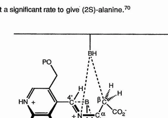
enzyme (Figure 1.1). The two base mechanism would explain the stereochemistry of the aminomalonate decarboxylation, and the unusual observation that
aspartate-p-decarboxylase, modified by the use of N-methyl-PLP, can decarboxylate (2R)-aspartate at a significant rate to give (2S)-alanine.^®
BH PO
[image:37.617.139.421.453.651.2]HN +
Figure 1.1. The two base mechanism for aspartate p-decartx)xylase.
1.3 Mechanism-based inhibitors of PLP enzymes
Many mechanism-based or suicide inhibitors have been prepared and tested for specific time-dependent inhibition of PLP-dependent enzymes. There has been a wide
interest in such inactivators as potential drug candidates, because of the pharmacological and therapeutic utility of inhibiting specific PLP enzymes (Table 1.3), The suicide inhibitors are substrate analogues and as such are useful tools for
investigating the mechanism of specific enzymes because they act at the active site of
[image:38.618.78.462.334.487.2]the enzyme.
Table 1.3. Some therapeutically important target enzymes dependent on PLP.
Enzyme Potential Effect
bacterial alanine racemase brain GABA transaminase
mammalian ornithine decarbo^q^'lase mammalian DOPA decarboxylase
antibacterial antiepileptic antineoplastic ant ihypertensive
In general, enzymes that require PLP, catalyse some chemical change at the a-, p-, or y-carbon of the common a - or y-amino acids. In every case the role of the PLP moiety is
to stabilise the carbanionic intermediates that develop during the catalytic process. The
suicide inhibitors all have latent functional groups that become activated by their proximity to the site of an enzyme-generated carbanion. Such an intermediate can breakdown to yield a reactive species that may react with an active-site amino acid
side chain, or with tightly bound PLP coenzyme. Each of these processes leads to the
inactivation of the enzyme, in the first case the active site is blocked by an unreactive
species and in the second the coenzyme is no longer available to bind to the substrate. i
Mechanism-based inactivating functional groups in substrates that inhibit PLP
enzymes include, acetylenic, olefinic, p-halo substituents and other leaving groups, nitriles, aryl sulphoxides, dihydroaromatics and phosphonoamino a c id s .T h e most common functional groups used for suicide inhibitors, and the mechanisms of
inactivation are presented in the next three sections.
1.3.1 Transaminase inhibitors
In the catalysis of transamination reactions, there are two half-reactions for each
complete catalytic cycle (see Section 1.2). In the first half-reaction catalysis proceeds through the substrate-PLP anion and the product a-imino acid-PMP enzyme complex (Scheme 1.5a, (11)). Both of these intermediates are species allowing for activation of
latent functional groups in suicide substrates. Activation by net oxidation to the ketoacid equivalent is observed in both olefinic and acetylenic amino acid analogues. The olefin analogues include vinyl glycine ( 1 7 ) , (E)-methoxy-vinyl glycine (18),^'*
and p-methylene aspartate (19).^® The generally accepted inactivation mechanism is as outlined in Scheme 1.9.
NHg
COgH
(18)
The acetylenic analogues include the natural product (2S)-propargyl glycine,^ and the mechanism of inactivation is depicted in Scheme 1.10.
2 0
Enz-NHg
H
â ^COgH
NH. (17)
H A^COc
PO
CH
Enz tNH
N
HC
W r
PO
CH
wi
'=1
Enz-Lys-NH HOgC,
O
+
PLP
Enz-Lys-NH Enz-Lys-NH;
Scheme 1.9. The mechanism of inactivation of AAT by vinyigiycine (17).
21
i
J
Enz
Enz Enz
Enz
COo-PO PO
PO
CH CH
CH
Enz— B
Scheme 1.10. Mechanism of Inactivation of AAT by (2S)-propargylglycine.
Some of the more common types of inhibitor contain good leaving groups at QP. These compounds are able to undergo facile elimination to generate olefinic intermediates.
These include, 3-chloro-(2S)-alanine (2 0)^^*^® an inhibitor of alanine
aminotransferase, and {2S)-serine O-sulphate (2 1) ^ which inhibits aspartate aminotransferase and glutamate decarboxylase (see Chapter 2 for more details).
NHg NHg
COgH (20)
COgH
(21)
2 2
The accepted mechanism had been that the killing species was the aminoacryi-PLP intermediate, which undergoes a putative Michael reaction with an enzymic
nucleophile. Although a Michael reaction does occur with some enzymes (aspartate-p-decarboxylase, serine hydroxymethyl-transferase(SHMT)), Metzier and coworkers® have shown that the inactivation of aspartate aminotransferase is not due to the
occurrence of a Michael reaction. Bright and coworkers®^ have used the nitro group of 3-nitroalanine as an inactivator of aspartate aminotransferase and alanine aminotransferase.® The nitroalanine undergoes a-proton abstraction followed by
p-nitro elimination. Alanine aminotransferase is also inhibited by cyc/oserine (5),® while
aspartate aminotransferase is inhibited to varying extents by a - and y-cyciog\u\am\c
acids.®"^'®® a-Cyc/oglutamate®"^'®® (Scheme 1.11, (22)) is believed to acylate an
active-site bound nucleophile to give a stable inactivated complex. y-Cyc/ogiutamate is thought to form an oxime of p-aminooxyglutamate with PLP by ring opening of the
isoxazolidone ring. Various analogues of GABA inhibit GABA-transaminase. These
include p-chloro, 3-phenyl, y-acetylenic and y-vlnyi GABA and ethanolamine O sulphate.® Finally the mammalian GABA transaminase is inhibited by the naturally occurring product gabaculine (23) via an interesting aromatisation mechanism to give
an aromatic PMP adduct®®*®® (Scheme 1.12, (24)).
1
1
O -N
—Enz HOgC
+ PLP
O NHg O— N
—Enz
Scheme 1.11. Mechanism of inactivation of AAT by a-cyc/oglutamate.
,COp-Enz
COgH
GABA-T PLP
NH NH
(23)
PO PO
CH CH.
(24)
Scheme 1.12. Mechanism of inactivation of GABA-T by gabaculine (23).
24
'•'I
1.3.2 Decarboxylase Inhibitors
These inhibitors are particularly important because of the role of the decarboxylases in the biosynthesis of many pharmacologically active amines (see section 1.1). Many examples with alkynyl, olefinic, and mono-, di-, or trihalomethyl groups have been
reported. Decarboxylases bind C-2 alkylated a-amino acids in the active-site pocket and the decarboxylation step occurs as for the natural substrate. When the alkyl groups are a-ethynyl, a-vinyl, a-fluoromethyl, and a,a-difluoromethyl amino acids, loss of CO2
generates a reactive intermediate which can react with an active-site nucleophile or remain bound to the PLP and so these compounds act as suicide substrates. The
inactivation of DOPA decarboxylase by a-vinyl DOPA (25)®®*®^ and a,a-difluoromethy! DOPA (26)^ are shown in Scheme 1.13.
5-Hexyne-1,4-diamine (2 7 ), a putrescine analogue, inactivates ornithine
decarboxylase;®® in view of the microscopic reversibility principle, presumably via a similar mechanism to that for a-ethynylornithine.
(27)
Enz
a)
COgH (25)
b)
NHg FgHG
COgH (26)
OH
OH
PO
I A
inactivation
FgHC>^
inactivation
Scheme 1.13. The Inactivation of DORA decarboxylase by a) a-vlnyl DORA (25) and b) a,a-dlfluoromethyl DORA (26).
1
Inhibitors of ornithine decarboxylase include p.ydehydroornithine (28),^ a
-fluoromethylputrescine (29)® and a,a-difiuoromethylornithine (30).® Haiomethyl and olefinic groups have been incorporated into a single molecule by Bey and collègues.®® a-Fluoromethyldehydroornithine (31) and a,a-difluoromethyldehydroputrescine (32)
have been tested as ornithine decarboxylase inhibitors. The dehydroornithine is more
effective as an inhibitor than the saturated analogues and its Kj is 30 times lower.
1
4 '%
26
CO2H H CO2H
(28) (29) (30)
NH. NH.
NH.
NH
FCH
(32)
NH2 NHg NHg
%
1.3.3 Other PLP inhibitors
Other types of PLP Inhibitors include those for racemases, and for enzymes that f catalyse reactions at the p-carbon.
PLP-dependent racemase enzymes, in particular alanine and glutamate racemase are #
important because they are involved in the biosynthetic pathway leading to
peptidoglycan (see section 1,1). Several suicide substrates, both natural and synthetic are known, including p-substituted alanines®^®® (for alanine racemase) and phosphoalanine (for gram-positive alanine racemase only). In the latter case the
inactivation mechanism is thought to involve the initial reversible formation of a weak
complex (competitive inhibition) which slowly isomerises to a stoichiometric complex which then dissociates extremely slowly. The complex is not reducible by
borohydride and shows a non-perturbed fluorescence spectrum for the bound coenzyme.
Enzymes that catalyse a,p-elimination are not susceptible to inhibition initiated by
many of the typical suicide substrates containing halogens, acetylenic or olefinic functionalities. However, occasionally an abortive reaction occurs with a physiological
2 8
substrate. For example, threonine deaminase is very slowly inactivated by (2S)-serine (and also by 3-choroalanine^°^) about once in every 10"^ turnovers.^However
(2S,3R)-threonine does not cause inactivation, presumably because the intermediate
is less reactive to Michael addition or enamine condensation (which gives the inhibited Schnackerz-type p r o d u c t(S c h e m e 1,14).
£ coli tryptophanase is irreversibly inactivated by trifluoroalanine.^°® The hypothesis is that the difluoroaminoacrylyl-PLP-aldimine adduct is more susceptible to nucleophilic
attack at C-3 by an enzyme bound base than hydrolysis at the aldimine carbon atom.
The enzyme p-cystathionase is irreversibly inactivated by the fungal toxin rhizobiotoxin^*^® (Scheme 1.15, (33)). The alkoxyvinylglyclne analogue, rhizobiotoxin,
may undergo initial C“-H abstraction, and then an addition and elimination sequence,
similar to that for (E)-methoxyvinylglycine with aspartate transaminase, p- j Cystathionase is also irreversibly inhibited by trifluoroalanine.^®^ Tryptophan synthase
is also irreversibly inhibited by trifluoroalanine, trichloro- and dichloroalanine.^®^ £ coll
tryptophan synthase also reacts with cyanoglycine, causing reversible inactivation on |
dialysis or gel-fiitration. The inactivated coenzyme is presumed to be a stable
a-aminomalomononitrile anion.
H-N Enz
H
J
+
H^N--HX
PO PO
CH
CH
Enz
Enz NH
Enz— NH
PO PO
CH
CH
COpH Enz
OH
PO PO
CH
CH
•Enz
Schnackerz adduct
Scheme 1.14. Possible mechanisms for the inactivation of enzymes that catalyse a,p-elimination reactions.
29
1) p"Cystathionase
COpH
+ PLP (33)
Enz— Nu
Enz— Nu
inactive enzyme
J
Scheme 1.15. The inactivation of p-cystathionase by rhizobiotoxin.
1.4 Research objectives
The aim of our work was to clarify two reports in the literature. These reports were
inconsistent with the current models for reactions catalysed by PLP dependent enzymes.
The first report^^ concerned the inactivation of GAD by (2S)-serine O-sulphate (21 ).
The mechanism of which did not involve the loss of CO2 from the serine O-sulphate
(Chapter 2).
NH2
COgH (21)
The second^ reported that the decarboxylation of 2-aminomalonic acid
(34)
catalysed by SHMT appeared to be non-stereospecific (Chapter 3).NHo
COgH
(34)
CHAPTER TWO
GLUTAMATE DECARBOXYLASE
’’I
2.0 Glutamate Decarboxylase
Glutamate decarboxylase (GAD) (EC 4.1.1,15) is one of the most widely studied of the PLP-dependent decarboxylases. The enzyme catalyses the conversion of (2S)-glutamic acid to y-aminobutyric acid (GABA)
(35)
(Scheme 2.1).(35)
NHg NHg
COgH H
Scheme 2.1. The physiological reaction catalysed by glutamate decait>oxylase (GAD).
2.1 Properties and Structure of GAD
2.1.1 Properties and Structure of mammalian GAD
Mammalian GAD is of considerable physiological importance. The enzyme has been isolated from several species, Including mouse,rat,^^^ h u m a n a n d p i g . T h e
most common source of mammalian GAD is from brain tissue. The enzyme has also been cloned from fruit-fly^ and cat.^^®
The mouse enzyme was found to be unstable with respect to the conditions used for the purification processes that were originally developed. Stabilisation of the enzyme
preparation with PLP and aminoethylisothiuronium bromide (AET) enabled the completion of a 200-fold purification process in 1966.^^® In 1973 a 700-fold purification
of the mouse enzyme was achieved using a combination of ammonium sulphate $
fractionation, size exclusion, calcium phosphate gel and DEAE-Sephadex chromatography.^^® The pure mouse GAD is stable for 1-2 hours at 30-40 °C, but at
higher temperatures a rapid loss of activity occurs.117
1
The determination of the molecular weight of the various mammalian glutamate decarboxylases has been a contentious issue as there are multiple forms of the
enzyme in several species including rat^^® and pig.^^® The apparent molecular weight
of native rat brain GAD has been determined to be 110 000 ± 10 000 Daltons by size exclusion chromatography. SDS-polyacrylamide gel electrophoresis of the purified enzyme indicated a subunit molecular weight of 60 000 D a l t o n s a n d
SDS-polyacrylamide gel electrophoresis of the purified enzyme after cross linking with
dimethylsuberimidate indicated a dimeric subunit structure. ^^® The omission of #
mercaptoethanol from the SDS cocktail enabled the molecular weight to be determined using SDS-polyacrylamide gel electrophoresis under non reducing conditions. The molecular weight of the enzyme determined in this manner was found
to be 120 000 Daltons. The results of all three studies suggested that the enzyme is dimeric. The results from parallel studies indicated that human and pig brain GAD are
also dimeric. Human brain GAD has a subunit molecular weight of 67 000 ± 5 000
Daltons.^Pig brain GAD has a subunit molecular weight of 60 000 Daltons, and the native enzyme molecular weight is 120 000 Daltons.
Due to the problems encountered during the purification of the mammalian enzymes,
I
the amount of kinetic data available is limited. The Michaelis constant, Km, for the
natural substrate, (2S)-glutamic acid, has been determined for the three forms of pig brain GAD.^^^ The relative rates of the abortive décarboxylation-transamination reaction (see section 2.1.6) leading to the formation of inactive enzyme catalysed by
the three forms of porcine brain GAD have also been determined. The value of V^ax for the three forms of pig brain GAD have been determined to be 0.337, 0.73 and 0.7
33
j
unit/mg for the a-, (3- and 7-forms respectively. The effect of pH on the value of Vmax of the three forms of enzyme was found to be quite similar. In each case the highest
relative value occurred at pH 6.2-6.5. The rates of the abortive decarboxylation-transamination reaction of the three forms of porcine enzyme with the pseudo substrate (2S)-aspartic acid have also been studied.^^®
2.1.2 Properties and Structure of plant GAD
GAD activity has been detected in various plants including s q u a s h , t e a leaves,^®® potato tubers,^®® barley/®^ radish leaves,^®® wheat^®® and lupin s e e d s .H o w e v e r,
there are only a few reports of the purification of the enzyme from higher plants, for
example, from radish,^®® w h e a t a n d lupin seeds.^®® Efficient purification has not
been achieved largely due to instability and the resulting loss of enzyme activity during |
the purification process. Prior to 1985 the only report of homogeneous GAD from a higher plant, a 2 2 0-fold purification in 10% yield from squash tissue came from the
laboratory of Melius.^®'* However the subunit structure and the PLP content of the
enzyme were not determined. At that time the first molecular weight for a plant GAD was determined, 310 000 Daltons for barley root GAD. ^®® The molecular weight of the
potato tuber isozyme^®® was found to be 91 000 Daltons where SDS-polyacrylamide
gel electrophoresis gave a single band of 43 000 Daltons, consistent with a dimeric structure. More recent work with squash GAD^®® indicates that the enzyme is
homohexameric with subunits of 58 000 Daltons. Apparently, at pH 7.2, the enzyme dissociates to active dimers which appear to be less stable than the hexamer isolated at pH 5.8.
34
i-:-2.1.3 Properties and Structure of Bacterial GAD
35
i
The discovery of the bacterial protein enabled the first detailed study of the enzyme 4
m e c h a n i s m G A D has been isolated from E co//^®® and Clostridium perfringens}^ f
The Clostridium perfringens enzyme has a H of 290 000 Daltons, an unknown subunit J structure and contains 2 molecules of PLP. The E coli enzyme has been extensively
studied and is a hexameric species of H 300 000 Daltons which is readily purified, and contains one molecule of PLP per subunit, ft shows a high degree of stability and has a relatively high turnover number (kcat ~ 1-875 x 10® s'^ at pH 4.6),^®^ In several
cases the mechanistic and stereochemical features of some suicide inactivation processes have been difficult to rationalise within the context of the known properties
of pyridoxai dependent systems. In certain cases bonds connected to C“ were
apparently cleaved on the wrong and unexpected 4’-re-face of the coenzyme.®® Some
:i
of these observations have now been rationalised (see section 2.1.7).?
2.1.4 Glutamate Decarboxylase as a chemotherapeutic target
The mammalian brain enzyme is directly responsible for the biosynthesis of GABA in
the GABA-ergic system (section 1.1).^®® GABA, the product of the decarboxylase reaction is a major neurotransmitter and there is much evidence to suggest that high
cerebral concentrations prevent convulsions.® Reduced levels of GABA not only trigger convulsive attacks but are also associated with a variety of maladies including
Parkinson’s Disease, Alzheimer's Disease and Huntington’s Chorea.
2.1.5 Substrate and Reaction Specificity
{2S)-Glutamic acid is the only naturally occurring amino acid that Is decarboxylated by GAD at a significant rate. The Michaelis constant (Km) for E coli GAD was determined
to be 0.5-1.0 mM at pH 4.6 by Shukuya and Schwert in 1960.^®®
Several pseudo substrates, for example glutaric acid, (2S)-aspartic acid,
a-methyl-(2RS)-glutamic acid and (2S)-serine O-sulphate, have been incubated with the
enzyme. They were all found to inhibit the enzyme. (2S)-Aspartic acid and a-methyl-(2RS)-aspartic acid were decarboxylated but at 10® times and 10"^ times more slowly than the natural substrate. GAD is substrate and reaction specific unlike SHMT (see Chapter 3).
2.1.6 Mechanistic features of decarboxylation
During decarboxylation there are two competing reactions that occur (Scheme 2.2).
Following the transaldimination of the internal aldimine by the enzyme-bound
substrate, the protein orients the substrate so that the C^-COgH bond is orthogonal to the plane of the pyridlnium ring, the optimal geometry for the delocalisation of the
developing negative charge.^"*® Loss of the carboxylate group leads to the formation of
the quinoid intermediate (36), which is reprotonated at C® on the 4’-s/-face of the coenzyme to give the corresponding amine (37) and PLP (Scheme 2.2, a). The
second reaction occurs when the quinoid (36) is occasionally reprotonated atC -4’, GAD is one of the few enzymes that catalyse this unusual r e a c t i o n . T h i s second reaction leads to the formation of an aldehyde (38) and PMP (12) (Scheme 2.2, b).
The second reaction pathway is called an abortive decarboxylative-transamination.
"‘S
37
I
The abortive reaction results in the inactivation of the enzyme as the PLP is converted
to PMP. T i l l e y h a s studied the pH dependence of the abortive reaction and found
that at pH 4.6 approximately 1 in every 30 000 turnovers leads to an abortive reaction.
It is known that when substrate analogues, for example, (2S)-aspartic acid, are decarboxylated, pyridoxamine phosphate (PMP) (12) is produced as a result of the
abortive décarboxylation-transamination reaction (see above). The process leads to 1 ■f
inactivation of the enzyme.
I
The mechanism of inactivation by (2S)-serine O-sulphate (2 1) is more complex and is discussed in detail below (Section 2.2).
•Ï
-I
■I? 4


![Table 2.4. Values of K| and kjnact for the Inactivation of GAD by (2S)- and (2S)-[2-2H]-serine O-](https://thumb-us.123doks.com/thumbv2/123dok_us/8577622.369362/113.619.62.459.524.619/table-values-k-kjnact-inactivation-gad-s-serine.webp)
