ISSN Online: 2168-5460 ISSN Print: 2168-5452
DOI: 10.4236/ijohns.2019.86024 Nov. 14, 2019 217 Int. J. Otolaryngology and Head & Neck Surgery
Thyroid Cancer
Rodrigo Arrangoiz
*, Fernando Cordera, David Caba, Eduardo Moreno,
Enrique Luque-de-Leon, Manuel Muñoz
Sociedad Quirúrgica S.C., American British Cowdray Medical Center, Mexico City, Mexico
Abstract
Thyroid tumors include those that originate from follicular cells and those that arise from parafollicular cells (C cells). Differentiated thyroid cancer, which originates from follicular cells, includes papillary carcinoma, follicular carci-noma, oncocytic cell carcinoma (Hürthle), poorly differentiated carcicarci-noma, and anaplastic carcinoma. The incidence of thyroid cancer has been increas-ing significantly, with an estimated incidence in the United States of America of 53,990 cases by the year 2018. This neoplasm is listed as the most common endocrine tumor and represents approximately 3% of all malignant tumors in humans, with 75% of cases occurring in women, and two-thirds of cases oc-curring in people under 55 years. The increase in the prevalence/incidence of low-risk thyroid cancer over the last 10 to 20 years has required a re-appraisal of the standard one-size-fits-all approach to differentiated thyroid cancer. This adaptation to a more individualized management of the patient with thyroid cancer has led to a much more risk-adapted approach to the diagnosis, initial therapy, adjuvant therapy, and follow-up of patients with differentiated thy-roid cancer. This paper with review the current understanding of the clinical presentation, diagnostic workup, and management of thyroid cancer centered on evidence-based and personalized medicine.
Keywords
Thyroid Nodules, Thyroid Cancer, Thyroid FNA, Thyroid Nodule Workup, Thyroid Cancer Treatment, Molecular Studies for Thyroid Cancer
1. Introduction
Thyroid nodules are a major public health problem. Epidemiological studies have shown that the prevalence of palpable thyroid nodules is approximately 5% in women and 1% in men living in parts of the world with sufficient iodine [1] How to cite this paper: Arrangoiz, R.,
Cordera, F., Caba, D., Moreno, E., Luque-de- Leon, E. and Muñoz, M. (2019) Thyroid Can-cer. International Journal of Otolaryngolo-gy and Head & Neck Surgery, 8, 217-270.
https://doi.org/10.4236/ijohns.2019.86024
Received: October 15, 2019 Accepted: November 11, 2019 Published: November 14, 2019
Copyright © 2019 by author(s) and Scientific Research Publishing Inc. This work is licensed under the Creative Commons Attribution International License (CC BY 4.0).
http://creativecommons.org/licenses/by/4.0/
DOI: 10.4236/ijohns.2019.86024 218 Int. J. Otolaryngology and Head & Neck Surgery [1] [2] [3] [4]. In contrast, high-resolution neck and thyroid ultrasound can detect thyroid nodules in approximately 19% to 68% of randomly selected people, with higher frequencies in women and the elderly [3] [4]. The clinical importance of thyroid nodules lies in the need to exclude thyroid cancer, which occurs between 7% and 15% of cases, depending on age, sex, radiation exposure history, family history, among other factors [5] [6].
Thyroid neoplasms include those that originate from follicular cells and those that arise from parafollicular cells (C cells). Differentiated thyroid cancer, which originates from follicular cells, includes papillary carcinoma, follicular carcino-ma, oncocytic cell carcinoma (Hürthle), poorly differentiated carcinocarcino-ma, and anaplastic carcinoma. These thyroid tumors comprise the vast majority (more than 90% of cases) of all thyroid cancers [7]. Of these subtypes, anaplastic carci-noma is rare and is characterized by its extremely poor prognosis. Similarly, poorly differentiated carcinoma is characterized by its aggressive behavior and its unfavorable prognosis. Between 2010 and 2014, 63,229 patients per year were diagnosed with thyroid carcinoma. Of these 63,229 patients, 89.4% had papillary carcinoma, 4.6% had follicular carcinoma, 2.0% had oncocytic cell carcinoma, 1.7% had medullary carcinoma, and 0.8% had anaplastic carcinoma [8].
The incidence of thyroid cancer has been increasing significantly since the mid-1990s, with an estimated incidence in the United States of America of 53,990 cases by the year 2018 [9]. This cancer is listed as the most common endocrine neoplasm and represents approximately 3% of all malignant tumors in humans, with 75% of cases occurring in women [9] [10], and two-thirds of cases occur-ring in people under 55 years [9]. Less aggressive forms of these tumors are more common in women and younger people [8]. The thyroid cancer mortality rate has remained stable in women but has increased by approximately 1% per year since 1983 in men and will be responsible for approximately 2060 deaths in 2018 [9]. The relatively low mortality rate compared to the incidence is due, in part, to the indolent nature of the vast majority of thyroid tumors. Patients with differentiated thyroid cancer generally have an excellent long-term prognosis, with five-year survival rates close to 100% for localized disease [8]. Despite the low mortality rates, local recurrence occurs in approximately 20% of patients, and distant metastases occur in about 10% of patients 10 years after diagnosis
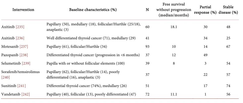
[11]. Mortality from thyroid cancer has been increasing in the last 18 years [8], which is why progress in the development of new systemic therapies for thyroid cancer refractory to iodine is extremely important. We know that medical de-velopment in this field has been delayed compared to the progress observed in the treatment of other solid tumors, however, data from emerging clinical stu-dies suggest that thyroid cancer can be treated with targeted agents, particularly kinase inhibitors, with promising results that overshadow those previously seen with cytotoxic agents [12].
DOI: 10.4236/ijohns.2019.86024 219 Int. J. Otolaryngology and Head & Neck Surgery
thyroid cancer [8][9][13]. 25% of new thyroid tumors diagnosed between 1988 and 1989 were equal to or less than 1 cm in diameter compared to 39% of new thyroid tumors diagnosed between 2008 to 2009 [13]. This may be due to the in-creasing use of high-resolution neck ultrasound and other diagnostic imaging techniques leading to finding asymptomatic thyroid lesions (incidentalomas), trends that are changing the initial treatment and follow-up of many patients with thyroid cancer [14].
The detection and diagnosis of differentiated thyroid cancer has evolved over the years with increased use of high-resolution neck and thyroid ultrasound, fine needle aspiration biopsy (FNAB), molecular tests, and thyroglobulin as a serum marker. This evolution has led to greater controversy regarding the appropriate medical and surgical management of this cancer. The type of surgical resection (lobectomy vs. total thyroidectomy), the role of lymphadenectomy (central proph-ylactic vs. therapeutic compartment), and adjuvant medical treatment for diffe-rentiated thyroid cancer are currently debated and present unique challenges in the treatment of these patients.
2. Risk Factors
In-depth knowledge of the risk factors that may predispose to developing thyro-id cancer is required when a patient is being assessed with complaints related to the thyroid gland such as thyroid nodules, voice changes, or symptoms of dysp-nea, dysphagia, or sensation of suffocation These risk factors include a personal or family history of thyroid cancer, certain diseases with a genetic predilection towards the development of thyroid cancer, and previous radiation exposure. Most thyroid cancers are idiopathic. However, the thyroid gland is very sensitive to radiation-induced oncogenesis, and radiation is the main environmental cause of thyroid cancer [15] [16] [17].
Differ-DOI: 10.4236/ijohns.2019.86024 220 Int. J. Otolaryngology and Head & Neck Surgery
ent forms of radiation have been linked to different genetic alterations associated with thyroid cancer, resulting in variable aggressiveness. Therefore, radiation exposure plays a critical role in the development of thyroid cancer, especially in patients younger than 15 years, and can play a role in its aggressiveness based on acquired genetic alterations and radiation dose [16][18] [19] [20].
Having a personal history of thyroid cancer increases the risk of developing subsequent or recurrent thyroid tumors substantially. Most differentiated thy-roid cancers are sporadic, and at least 5% of these patients will have family dis-ease [21]. There is evidence of a family predisposition, with several inherited syndromes that demonstrate an increased risk of developing thyroid cancer. The mechanisms underlying these associations are not well known. Certain histo-logical subtypes of thyroid cancer should raise the suspicion of family syn-dromes that have a genetic predisposition to develop said cancer. As for exam-ple, the cribriform-morular variant of papillary thyroid cancer is associated with familial adenomatous polyposis and should raise concerns about a ger-mline mutation of the APC gene and a predisposition to colon and rectum cancer
[22]. Familial adenomatous polyposis has been associated with the development of all different subtypes of differentiated thyroid cancer [23] [24]. Families re-lated to familial adenomatous polyposis with cases of thyroid cancer should be-gin surveillance/screening at age 15, or earlier if family members are affected at younger ages [22][23] [24] [25].
The Carney complex is a rare genetic condition associated with mutations in the PRKAR1A gene that manifests with skin pigmentation, myxomas, schwan-nomas and thyroid abnormalities, including differentiated thyroid cancer [26]. Cowden syndrome is caused by a mutation in the PTEN germ line and is asso-ciated with the development of benign and malignant breast and thyroid lesions
[27]. Peutz-Jeghers syndrome is due to germline defects in STK11 (LKB1) and is associated with gastrointestinal hamartomatous polyps, pigmented mucocuta-neous lesions, and differentiated thyroid cancer [28].
DOI: 10.4236/ijohns.2019.86024 221 Int. J. Otolaryngology and Head & Neck Surgery
thyroid tumors, and therefore appear to be an early event in thyroid tumorige-nesis [30] [31]. Some studies suggest that Ras mutations are more prevalent in follicular thyroid carcinomas, in the follicular variant of papillary thyroid cancer, and in follicular adenomas [32]. Ras mutations may result in allelic loss or in chromosomal rearrangements that lead to an increase in thyroid follicular can-cer formation rates [32]. Chromosomal rearrangements have also been observed in the formation of RET/PTC oncogenes and imply an unfavorable prognosis
[33]. There are variable data regarding the usefulness of BRAF, TP53 and TERT mutations tests in risk stratification of patients with thyroid cancer [34] [35]. BRAF V600E mutations have been associated with worse results in papillary thy-roid cancer, with higher recurrence and death rates [35].
Thyroid cancers are highly vascularized and elevated levels of vascular endo-thelial growth factor have been identified in these tumors, suggesting that angi-ogenic pathways may be a potential target for treatment [29]. In addition, during the past 30 years, thyroid cancers have been shown to be associated with genetic mutations that lead to aberrant intracellular signaling (Table 1). Preclinical and clinical data suggest that inhibition of intracellular signaling cascades, including mitogen-activated protein kinase (MAPK) and phosphatidylinositol-4,5-bispho- sphate 3-kinase (PI3K) pathways may be effective in cancer treatment of thyroid
[image:5.595.209.539.530.734.2][7] [33] [36] [37] [38] [39] [40]. RET kinase activation by a germline mutation is associated with the development of familial medullary thyroid cancer. Similar mu-tations have been detected in somatic cells that produce greater RAS/RAF activa-tion in approximately 50% of sporadic thyroid medullary cancers [41] [42]. MAPK activation in papillary thyroid cancers can occur through RET/PTC translocations or mutations in RAS or BRAF [32]. The PI3K pathway is also activated by muta-tions in PAX8/PPARγ in follicular thyroid cancers [43]. This greater understand-ing of the mutations involved in thyroid tumorigenesis will likely lead to new systemic therapies for the treatment of advanced disease.
Table 1. Prevalence of mutations in different pathological subtypes of thyroid cancer.
Type of Thyroid Cancer Mutation Prevalence (%)
Papillary
BRAF V600E RET/PTC Copy gain PI3KCA
RAS PI3KCA Copy gain BRAF
PTEN
45 20 12 10 3 3 2
Follicular
RAS PAX8/PPARγ Copy gain BRAF Copy gain PI3KCA
PTEN PI3KCA
45 35 35 12 <10 <10
DOI: 10.4236/ijohns.2019.86024 222 Int. J. Otolaryngology and Head & Neck Surgery
The RET proto-oncogene is a tyrosine kinase receptor that is primarily ex-pressed in tumors of neural crest/neuroectoderm origin, which explains the high incidence of these mutations in medullary thyroid carcinomas that originate in para-follicular cells (C cells) [44]. The RET gene is found on chromosome 10 and germline mutations produce activating mutations that change direction that are responsible for 95% of hereditary medullary thyroid carcinomas, in-cluding those associated with multiple endocrine neoplasia 2A (Sipple syndrome ) and 2B (Wagenmann-Froboese syndrome) and familial medullary thyroid can-cer [44] [45]. In 80% of cases of medullary thyroid carcinoma, the disease is spo-radic, without an inherited etiology, but a somatic mutation is identified in the RET gene in 40% to 70% of these sporadic cases [41] [42]. In these sporadic cas-es, mutations are found most frequently in codon 918 that results in the consti-tutive activation of the RET tyrosine kinase receptor [46]. Almost all patients with multiple endocrine neoplasia 2A or multiple endocrine neoplasia 2B that is transmitted in an autosomal dominant manner will develop medullary thyroid cancer and the detection of germline RET gene mutations has been of great val-ue in the early identification of patients who have a genetic basis for their dis-ease. Even in patients with sporadic medullary thyroid cancer, 6% to 10% of these patients will have a mutation in the RET proto-oncogene germline, which reveals a new family of patients with previously undiagnosed medullary thyroid cancer [41] [42] [45]. The discovery of the RET proto-oncogene has had a signifi-cant clinical impact, which affects the scrutiny and prophylactic treatment of pa-tients who are members of the families of papa-tients with multiple endocrine neop-lasia or with familial medullary thyroid carcinoma [47].
Anaplastic thyroid carcinoma develops from the dedifferentiation of thyroid tumors, although the specific reason for this transformation has not been well clarified. Mutations in the p53 suppressor gene are frequently found in anaplas-tic thyroid carcinoma and are absent in well-differentiated thyroid neoplasms
[48] [49]. This observation suggests that p53 mutations play a role later in the pathogenesis of the thyroid tumor, specifically, in the transition from dediffe-rentiation to the anaplastic phenotype. A large number of mutations in other pathways, including the PI3K/Akt and Ras/MAPK pathways have also been im-plicated in the formation of ATC [48] [49].
3. Pathology
DOI: 10.4236/ijohns.2019.86024 223 Int. J. Otolaryngology and Head & Neck Surgery Table 2. Pathological classification of malignant thyroid tumors [8] [50].
Subtype Histologic Variants Incidence
Papillary (89.4%)
Conventional/Classic Follicular Variant
Tall Cell Solid Diffuse sclerosing Papillary Micro-Carcinoma
Oncocytic Columnar Cell
Clear Cell Morular Cribriforme
Marco-follicular Papillary with HobnailCharateristics Papillarywith stroma similar to fascitis Combined Papillary and Medullary Carcinoma Papillary with dedifferentiation to Anaplastic Carcinoma
65% - 85% 15% - 20% 5% - 10% 1% - 3% 1% - 2%
Follicular (4.6%) Hurthle (2.0%)
Poorly Differentiated Insular
Medullary (1.7%) Anaplastic (0.8%)
Others
Lymphoma Squamous Cell Carcinoma
Sarcoma Melanoma Metastatic Tumors
Papillary thyroid cancer accounts for approximately, based on the most recent statistics, 89.4% of all thyroid malignancies and is the predominant histology observed in patients exposed to radiation [8][15] [16] [17] [18]. The average age of diagnosis is between 30 and 40 years and women are affected more frequently than men (2:1 ratio) [13] [51] [52]. The macroscopic appearance of papillary thyroid cancer can be very variable. Most tumors tend to be markedly circum-scribed, solid, firm, and white in color, but a significant percentage of tumors can be cystic [50]. It is not uncommon to have a solid primary tumor with cystic metastases to a lymph node [50]. Papillary thyroid cancer may have a pattern of infiltrating growth in the thyroid or may show a direct extra extra-thyroid ex-tension to adjacent tissues [51] [52]. Unlike normal thyroid gland or benign thyroid lesions that protrude on sectioning, papillary thyroid cancer remains flat
[53]. The diagnosis is made by microscopic evaluation and can be made on the basis of a fine needle biopsy (FNAB) [34][53].
DOI: 10.4236/ijohns.2019.86024 224 Int. J. Otolaryngology and Head & Neck Surgery
thyroid cancer [50][51][53]. These psamoma bodies are present in 50% of cases and help ensure the diagnosis of papillary cancer [53]. Some tumors may also contain multinucleated giant cells [50].
The definitive diagnosis is made on the basis of cellular and nuclear characte-ristics (cytological charactecharacte-ristics) with cells that adopt a cuboidal form with nuc-lear “grooving” and cytoplasmic inclusions [50][51][52] [53]. These characte-ristic findings are described as the pathognomonic nuclei of “Orphan Annie” [53]. Papillary cancer is characterized by multifocality in 18% to 85% of patients and is associated with an increased risk of lymph node metastasis [53]-[61]. Metas-tases to cervical lymph nodes are quite common in patients with papillary cancer at the time of diagnosis, with a frequency that varies between 30% to 80% in some series [53] [62] [63] [64]. Despite this high incidence, the 10-year survival rate remains 95% [8].
Follicular cancer represents the second most frequent thyroid cancer, approx-imately 4.6% of all thyroid cancers [8]. These tumors are most frequently found in geographic areas with iodine deficiency and, like papillary cancer, have a fe-male predominance with a ratio of 3:1 (women/men) [8][65] [66] [67]. Follicu-lar cancer tends to occur in an older population compared to other differentiated thyroid tumors. Its maximum incidence is between the ages of 40 and 60, com-pared to the incidence of papillary cancer that reaches an earlier peak (usually 10 years less), between the ages of 30 to 50 years [53] [67]. Follicular cancer is often found in association with benign thyroid disorders, such as endemic goiter, and a relationship between chronic stimulation with thyroid stimulating hormone (TSH) and follicular carcinoma due to the increased incidence of follicular can-cer has been suggested in areas with iodine deficiency [65] [66]. Patients gener-ally present with a clinical history of a solitary thyroid nodule, which has often rapidly increased in size [53].
The histopathology of follicular tumors varies from a normal epithelium, well differentiated tumors with a follicular and colloid differentiation (findings asso-ciated with a good prognosis) to poorly differentiated tumors with solid growth, absence of follicles, marked nuclear atypia and vascular and/or capsular invasion (characteristics that are associated with a worse prognosis) [68]. Follicular tumors are usually unifocal, well encapsulated, containing highly cellular follicles, and are easily confused with benign follicular adenomas in BAAF [53]. The patho-logical diagnosis of this malignant neoplasm can only be made by permanent cuts, demonstrating the presence of capsular and/or vascular invasion [53].
DOI: 10.4236/ijohns.2019.86024 225 Int. J. Otolaryngology and Head & Neck Surgery
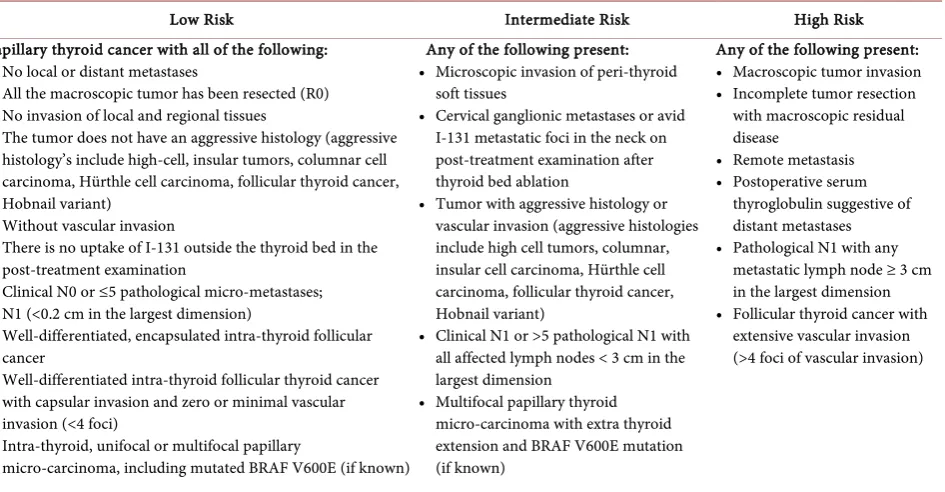
• Minimally invasive follicular thyroid cancer, which demonstrates only the invasion of the tumor capsule without vascular invasion (low-risk tumor ac-cording to the guidelines of the American Thyroid Association [ATA]) (Table 3);
• Encapsulated angioinvasive follicular thyroid cancer, which demonstrates minor vascular invasion (≤4 foci of angioinvasion within the tumor or tumor capsule) with or without capsular invasion (low-risk ATA tumor) (Table 3); • Widely invasive follicular thyroid cancer, which is characterized by:
o Wide invasion of the tumor capsule;
o A multinodular tumor without a well-defined capsule that invades the
nor-mal thyroid surrounding the tumor; and/or
o Extensive vascular invasion (>4 foci of angioinvasion) (high-risk ATA
tu-mor) (Table 3).
Regional metastasis to cervical lymph nodes is somewhat rare in follicular cancer, being present in 5% to 13% of cases in the initial presentation [53] [72]. Distance dissemination is more common in the initial presentation compared to papillary cancer and is observed in 10% to 33% of patients, most often it presents with hematological dissemination to the lungs or bone (lytic lesions) even in those with small primary tumors, although tumors smaller than 2 cm in size have not been associated with metastatic disease [73]. The 10-year survival rates for follicular thyroid cancer are 70% to 95%, slightly worse than those for papil-lary cancer, possibly due to late presentation and the presence of distant metas-tases in the initial diagnosis.
[image:9.595.67.540.470.716.2]Hürthle cell carcinoma (also known as oncocytes, or Askanasy cells), although
Table 3. ATA risk stratification system to estimate the risk of persistent/recurrent disease.
Low Risk Intermediate Risk High Risk
Papillary thyroid cancer with all of the following: • No local or distant metastases
• All the macroscopic tumor has been resected (R0) • No invasion of local and regional tissues
• The tumor does not have an aggressive histology (aggressive histology’s include high-cell, insular tumors, columnar cell carcinoma, Hürthle cell carcinoma, follicular thyroid cancer, Hobnail variant)
• Without vascular invasion
• There is no uptake of I-131 outside the thyroid bed in the post-treatment examination
• Clinical N0 or ≤5 pathological micro-metastases; N1 (<0.2 cm in the largest dimension)
• Well-differentiated, encapsulated intra-thyroid follicular cancer
• Well-differentiated intra-thyroid follicular thyroid cancer with capsular invasion and zero or minimal vascular invasion (<4 foci)
• Intra-thyroid, unifocal or multifocal papillary
micro-carcinoma, including mutated BRAF V600E (if known)
Any of the following present: • Microscopic invasion of peri-thyroid
soft tissues
• Cervical ganglionic metastases or avid I-131 metastatic foci in the neck on post-treatment examination after thyroid bed ablation
• Tumor with aggressive histology or vascular invasion (aggressive histologies include high cell tumors, columnar, insular cell carcinoma, Hürthle cell carcinoma, follicular thyroid cancer, Hobnail variant)
• Clinical N1 or >5 pathological N1 with all affected lymph nodes < 3 cm in the largest dimension
• Multifocal papillary thyroid micro-carcinoma with extra thyroid extension and BRAF V600E mutation (if known)
Any of the following present: • Macroscopic tumor invasion • Incomplete tumor resection
with macroscopic residual disease
• Remote metastasis • Postoperative serum
thyroglobulin suggestive of distant metastases • Pathological N1 with any
metastatic lymph node ≥ 3 cm in the largest dimension • Follicular thyroid cancer with
DOI: 10.4236/ijohns.2019.86024 226 Int. J. Otolaryngology and Head & Neck Surgery
considered a variant of follicular cancer, deserves a separate discussion since it comprises 2% of all thyroid neoplasms and has a biological behavior and a natu-ral history that distinguishes them from follicular cancer [8] [53] [74]. These tumors are formed by sheets of polygonal and hyperchromatic cells that contain abundant mitochondria [74]. Hürthle tumors are characterized by the presence of a cell population of “oncocytes”, mostly eosinophilic oxyphilic cells with ab-undant cytoplasm, very compact mitochondria and round oval nuclei with prom-inent nucleoli [74]. Like follicular cancer, Hürthle carcinoma requires a defini-tive pathological study to identify vascular or capsular invasion [53] [72]. Unlike follicular cancer, Hürthle carcinomas are often multifocal (30%), have regional lymph node metastases (25%), and often fail to concentrate radioactive iodine
[53]. In part, due to these factors, patients with Hürthle carcinoma have higher tumor recurrence rates and lower survival rates compared to patients with pa-pillary or follicular carcinomas [53].
Medullary thyroid carcinoma is a neuroendocrine tumor of the parafollicular cells or C cells of the thyroid gland [75]. Approximately 1.7% of thyroid neop-lasms are medullary carcinomas [8][75]. Although most cases are sporadic, 15% to 25% of cases are part of an autosomal dominant hereditary syndrome [75]. Calcitonin production is a characteristic feature of this tumor [53]. C cells origi-nate in the embryonic neural crest; As a result, medullary carcinomas often have the clinical and histological features of other neuroendocrine tumors such as carcinoid tumors and pancreatic islet cell tumors.
The sporadic form of medullary thyroid cancer typically presents as a unila-teral solitary nodule (75% to 95% of patients) in the fifth decade of life [76] [77]
[78] [79]. Family forms, such as multiple endocrine neoplasia (MEN 2A),
mul-tiple endocrine neoplasia (MEN 2B) and familial spinal cancer, occur in the fourth decade and are typically multifocal [76] [77] [78] [79]. Due to the embryological origin of medullary thyroid cancer (C cells), these tumors are located in the up-per poles of the thyroid gland where these cells reside [53]. It is believed that the presence of C cell hyperplasia is an omen for the development of hereditary spinal cancer [53][76] [77] [78] [79]. These tumors are not encapsulated, nor well defined, and consist of a heterogeneous mixture of fusiform or round cells
[53][76] [77] [78] [79]. The cells are separated by fibrous septa and amyloid, the latter of which helps in the diagnosis of spinal cancer by immunohistochemical staining for calcitonin and carcinoembryonic antigen [53]. Although these tu-mors grow slowly, they have a tendency to metastasize early, usually before the primary tumor has reached 2 cm [53].
Approximately 50% to 70% of patients with medullary thyroid cancer have clinically detectable cervical lymph node involvement at the time of diagnosis
DOI: 10.4236/ijohns.2019.86024 227 Int. J. Otolaryngology and Head & Neck Surgery
and undifferentiated (anaplastic) thyroid cancers. When the disease is limited to the thyroid gland, the 10-year survival rate is 90% compared to patients with dis-tant metastatic disease that has a 10-year survival of only 20% [81].
Anaplastic thyroid tumors are undifferentiated tumors of the thyroid follicu-lar epithelium representing less than 1% of all malignant thyroid tumors [82]. These neoplasms are highly aggressive and are considered one of the most lethal malignancies, with a mortality close to 100% [82] [83]. It is believed that these tumors arise from well differentiated thyroid tumors, but over time they suffer from dedifferentiation [81] [84]. Because activating mutations of the BRAF and RAS genes are observed in both well-differentiated thyroid malignancies and in anaplastic thyroid cancer, it is suspected that these are early events in the path-way of this disease [85]. Late events in disease progression that are most com-monly seen in anaplastic cancer compared to well-differentiated tumors include mutations in the p53 tumor suppressor protein [86] [87] [88] [89], 16p [90], ca-tenin (cadherin-associated protein), beta 1, and PIK3CA [91].
The annual incidence of anaplastic cancer is approximately one to two cases per million people and represents between 0.8% and 9.8% of all thyroid cancers worldwide [82] [92] [93] [94] [95]. Patients with anaplastic cancer are generally older at the time of diagnosis than those with differentiated cancer; The average age at diagnosis is 65 years, and less than 10% of patients are under 50 years [96] [97]. The vast majority of patients with anaplastic thyroid cancer (60% to 70%) are women [96] [97]. About 20% of patients with anaplastic thyroid cancer have a history of differentiated thyroid cancer, and 20% to 30% of patients have syn-chronous differentiated cancer [98] [99] [100] [101][102]. The vast majority of synchronous thyroid tumors are papillary carcinomasbut coexisting follicular tumors have also been identified. Approximately 10% of patients with Hürthle cell thyroid tumors have foci of anaplastic cancer within Hürthle cell cancer
[103].
Patients with anaplastic thyroid carcinoma usually manifest clinically with a rapidly growing tumor and symptoms of dysphagia, dysphonia, or dyspnea sec-ondary to extrinsic compression of the tumor that is often fixed to adjacent structures [53]. However, regional or distant metastases are evident at the time of diagnosis in 90% of cases [101] [103] [104] [105]. Regional extension sites may include peri-thyroid fat and pre-thyroid muscles, lymph nodes, larynx, tra-chea, esophagus, tonsils, large neck vessels, and the mediastinum [101]. Metas-tatic disease at diagnosis is found in 15% to 50% of cases [98] [99] [100][102]. The most common site of distant metastases is the lungs (up to 90% of cases)
[99] [100]. These metastases are usually massive intrapulmonary lesions, but
there may be pleural involvement. About 5% to 15% of patients have bone me-tastases [98] [99] [100] [102]. 5% of patients have brain metastases, and some have metastases in the skin, liver, kidneys, pancreas, heart and adrenal glands
[99] [100][101][106] [107] [108] [109] [110].
en-DOI: 10.4236/ijohns.2019.86024 228 Int. J. Otolaryngology and Head & Neck Surgery
sure diagnosis and rule out possible lymphoma [53]. Cells are characteristically large and multinucleated with nuclear polymorphism and high mitotic activity
[99]. Surgery rarely has a role in this disease; the most common procedures per-formed are isthmusectomy or cytoreduction to alleviate tracheal compression
[81]. In rare cases that anaplastic carcinoma is diagnosed in the intrathyroid stage, without a coexisting well differentiated thyroid cancer component, thyroid lobectomy with wide margins of adjacent soft tissue on the side of the tumor is an appropriate surgical management [98]. If the anaplastic tumor is very small and completely confined to the thyroid, total thyroidectomy with complete tu-mor resection does not prolong survival compared to ipsilateral thyroid lobect-omy and if it is associated with a higher complication rate [100][102]. However, some experts prefer total or near total thyroidectomy with dissection of the cen-tral and lateral lymph nodes of the neck [111]. The reason for this is that diffe-rentiated thyroid cancer and anaplastic thyroid cancer often coexist, and total thyroidectomy offers a greater chance of complete resection [111]. For patients with small intra-thyroid anaplastic tumors associated with a differentiated thy-roid cancer, total thythy-roidectomy is recommended, if complete macroscopic re-section and minimal morbidity can be performed, to facilitate subsequent treat-ment of differentiated cancer [111].
Anaplastic thyroid cancers are extremely aggressive, with a specific mortality close to 100%. The average survival ranges from three to seven months, and the one and five year survival rates, are 20% to 35% percent and 5% to 14%, respec-tively [101] [102] [105] [112] [113] [114], with 90% of patients dying of the dis-ease within 6 months of diagnosis, usually secondary to local progression [81].
Primary thyroid lymphoma is a rare diagnosis, but it should always be consi-dered in the differential diagnosis of patients with thyroid nodules, goiter, and carcinomas, mainly because their prognosis and treatment differ substantially from other disorders. Lymphomas of the thyroid gland typically manifest in the seventh decade of life (the median and median age is between 65 and 75 years), affect women more commonly than men (with a female 4:1 predominance), and are often associated with a history of Hashimoto’s thyroiditis [115]-[120]. They represent less than 2% of all thyroid neoplasms and often present as a rapidly growing tumor with symptoms of dysphagia and dysphonia, possibly confusing the diagnosis with anaplastic thyroid carcinoma [121]. In a Danish epidemio-logical survey, the annual incidence rate was estimated at 2.1 cases per million people [115]. Pre-existing chronic autoimmune thyroiditis (Hashimoto’s disease) is the only known risk factor for primary thyroid lymphoma and is present in approximately half of patients [122]. Among patients with Hashimoto’s thyroi-ditis, the risk of thyroid lymphoma is at least 60 times higher than in patients without thyroiditis [115][119] [120].
Thyroid lymphoma can be primary or secondary, they are almost always non- Hodgkin (B-cells), since thyroid Hodgkin lymphoma is extremely rare [115] [118]
thy-DOI: 10.4236/ijohns.2019.86024 229 Int. J. Otolaryngology and Head & Neck Surgery
roid gland. Occasional cases of T lymphocyte lymphomas have been described, often in endemic areas for adult T-cell leukemia/lymphoma associated with lym-photropic virus-T (HTLV)-I [123] [124]. Sixty percent to 80% of thyroid lym-phomas are diffuse large B-cells of the germinal center type [116] [117] [118]
[125] [126]. The second most common subtype (about 30% of cases) is
lym-phoma of the extra-ganglion marginal marginal zone [32]. Other less common histological subtypes include follicular lymphomas; Small extra lymph node lymphomas have also been described [32]. Extra-lymph node marginal lym-phomas of the type of mucous-associated lymphoid tissue (MALT) are generally associated with Hashimoto’s thyroiditis [127].
Histologically, the cells are monomorphic and stain positively for lymphocyte markers such as CD20 [81]. Tumors of MALT origin generally have a better prognosis and can often be treated with radiation therapy alone, rather than the multimodal therapy necessary to treat lymphomas other than MALT [81]. Sur-vival rates for lymphoma located in the thyroid gland (stage IE) are generally favorable, with a 5-year survival rate of 75% to 85%. However, patients with dis-eases on both sides of the diaphragm (stage IIIE) or disseminated disease (stage IV) have a 5-year survival rate of less than 35% [53].
4. Diagnosis
Thyroid cancer is discovered incidentally in the vast majority of cases during imaging studies (computed tomography, positron emission tomography, mag-netic resonance imaging or ultrasonography) performed for reasons unrelated to the thyroid. The vast majority of patients with thyroid cancer have no specific symptoms and the results of these incidentalomas will trigger a diagnostic evalu-ation. When patients present to a doctor with a specific symptom, it is often with the finding of a new tumor/thyroid nodule, an increase in size of a previously detected nodule, pain secondary to a nodule hemorrhage, or a lymph node palpa-ble cervical [53]. Symptoms of dysphagia, dysphonia, or dyspnea often predict a poor prognosis since these symptoms are the result of a local invasion and are usually due to undifferentiated thyroid cancer, since differentiated tumors rarely invade surrounding structures [5].
ab-DOI: 10.4236/ijohns.2019.86024 230 Int. J. Otolaryngology and Head & Neck Surgery
sence of these findings, the presence of slightly grown lymph nodes (1 to 2 cm) together with a thyroid nodule suggests regional metastases [53] [81]. Palpable lymphadenopathy is most frequently identified along the middle and lower por-tion of the jugular chain. Finally, before any surgical intervenpor-tion, the extent of the disease in the neck should be evaluated in anticipation of surgical position-ing [53].
All patients undergoing thyroid surgery should have a preoperative evaluation of the voice as part of their preoperative physical examination. This should in-clude the description of the patient if he has voice changes, as well as the evalua-tion of the voice doctor (recommendaevalua-tion # 40 of the American Thyroid Associ-ation [ATA]) [34]. The preoperative laryngeal examination should be performed in all patients with voice abnormalities in the preoperative period, a history of cervical or upper thoracic surgery, which puts the recurrent laryngeal or vagus nerve at risk, and in patients with known thyroid cancer with extra posterior thyroid extension or extensive central nodal metastases (ATA recommendation # 41) [34].
The prevalence of palpable thyroid nodules in the general population is ap-proximately 5% to 7% in women and 1% in men living in parts of the world with sufficient iodine [1] [2]. In contrast, high-resolution neck and thyroid ultra-sound can detect thyroid nodules in approximately 19% to 68% of randomly se-lected people, with higher frequencies in women and the elderly [3] [4]. The clinical importance of thyroid nodules lies in the need to rule out thyroid cancer, which occurs between 7% and 15% of cases, varying according to age, sex, radia-tion exposure history, family history, among other factors [5] [6].
If a thyroid nodule larger than 1 cm in any diameter is identified, a serum lev-el of thyroid stimulating hormone (TSH) should be obtained (recommendation 2 ATA) [34]. If the TSH is low, a thyroid scan should be performed (the only in-dication today to perform this study) to document if the thyroid nodule is hyperfunctional (“hot”, that is, the uptake of the marker is greater than the nor-mal thyroid), isofuncionante (“warm”, that is, the uptake of the marker is equal to the surrounding thyroid) or not functioning (“cold”, that is, it has a lower up-take than the thyroid tissue) [128]. Because hyperfunctional thyroid nodules rarely contain malignancy, if one that corresponds to the nodule in question is found, a cytological evaluation is not necessary [34]. High serum levels of TSH, even within high ranges of normality, are associated with an increased risk of malignancy in the thyroid nodule, as well as a more advanced stage of thyroid cancer [129].
During the initial assessment of thyroid nodules, it is not recommended to routinely obtain serum thyroglobulin (Tg) (ATA recommendation 3) [34]. Se-rum levels of Tg may be elevated in the vast majority of thyroid diseases (benign and malignant) and is an insensitive and nonspecific test for thyroid cancer
DOI: 10.4236/ijohns.2019.86024 231 Int. J. Otolaryngology and Head & Neck Surgery
[134] [135], with mixed results, therefore, the ATA cannot recommend either
for or against the measurement Routine serum calcitonin in patients with thy-roid nodules (ATA recommendation 4) [34].
High-resolution neck and thyroid ultrasound should be performed in all pa-tients suspected of having thyroid nodules, nodular goiter, or any radiographic abnormality that suggests a thyroid nodule detected incidentally in another im-aging study (computed tomography or magnetic resonance imim-aging), or 18FDG- PET) (ATA recommendation 6) [34]. Ultrasound of the neck and thyroid should evaluate the following characteristics [34]: the thyroid parenchyma (if homoge-neous or heterogehomoge-neous), the size of the thyroid gland, the size, location, and ul-trasonographic characteristics of any nodule, and finally the presence or absence of suspicious cervical lymph nodes in the central or lateral compartments [34] [53]. Table 4 shows the characteristics that should be assessed in the high-resolution neck and thyroid ultrasound.
The ultrasonographic pattern associated with a thyroid nodule confers a risk of malignancy, and combined with the size of the nodule, guides decision mak-ing (Table 5). The ultrasound pattern of high suspicion of malignancy includes solid, hypoechoic nodules, or nodules with mixed components (solid hypoechoic and partially cystic nodule) with one or more of the following characteristics: ir-regular margins (infiltrative, micro-lobulated), microcalcifications, higher form than wide, calcifications at the edge of the cyst, evidence of extra thyroid exten-sion [136] [137] [138].
[image:15.595.190.540.518.733.2]The most accurate and cost-effective method for evaluating thyroid nodules is fine needle aspiration biopsy (FNAB) (ATA recommendation 7) [34]. Thyroid nodules with a higher probability of obtaining a non-diagnostic cytology (cystic component greater than 25% to 50%) or a sampling error (nodules difficult to palpate or located in the posterior portion of the thyroid lobe), it is preferred to perform a FNAB guided by ultrasound [139] [140]. Figure 1 and Figure 2 provide
Table 4. The characteristics that should be assessed in the ultrasound [233] [234].
• Node size (in three dimensions)
• The location (example—right upper lobe/if anterior or posterior) • Description of the ultrasonographic characteristics of the thyroid nodule:
oComposition of the nodule: Solid, cystic or spongiform oEcogenicity:
Isoechoic, hyperechoic, hypoechoic oMargins:
Regular Irregular:
Defined as infiltrative, microlobed or spiculated oPresence and type of calcifications:
Marcocalcifications or microcalcifications oShape:
If the nodule is taller than wide oVascularity:
DOI: 10.4236/ijohns.2019.86024 232 Int. J. Otolaryngology and Head & Neck Surgery Table 5. Ultrasonographic patterns of thyroid nodules, estimated risk of malignancy, and management guide for thyroid nodules with FNAB [34] [143].
Ultrasonographic
Pattern Ultrasonographic Characteristics Estimated Risk of Malignancy Size to perform FNAB
High Risk
Hypoechoic, solid nodules, or nodules with mixed components (solid and partially cystic hypoechoic nodule) with one or more of the following characteristics: irregular margins (infiltrative, microlobed), microcalcifications, taller than wide, calcifications on the edge of the cyst, evidence of extra thyroid extension
Greater than 70% - 90%
FNAB is recommended if its dimensions are equal to or greater than 1.0 cm
Intermediate Risk Hypoechoic solid nodule with smooth (regular) margins without microcalcifications, no evidence of extra thyroid extension, and the
shape is not taller than wide 10% al 20%
FNAB is recommended if its dimensions are equal to or greater than 1.0 cm
Low Risk Isoechoic or hyperechoic solid nodule, or partially cystic nodule with eccentric solid areas, no microcalcification, no irregular margin, no
evidence of extra thyroid extension, no taller than wide 5% al 10%
FNAB is recommended if its dimensions are equal to or greater than 1.5 cm
Very Low Risk Spongiform or partially cystic nodules without any of the ultrasonographic features described in low, intermediate, or high
suspicion patterns Less than 3%
FNAB can be considered if its dimensions are equal to or greater than 2.0 cm
Observation without BAAF is also reasonable
[image:16.595.61.542.101.537.2]Benign Purely cystic nodules (without solid component) Less than 1% Do not perform FNAB
Figure 1. Algorithm for the initial evaluation and treatment of patients with thyroid nodules according to the ul-trasonographic pattern.
Figure 2. Algorithm for the treatment of patients with thyroid nodules according to the pattern the result of the FNAB [143].
Suspicious Thyroid Nodule Elevated or Normal TSH
Neck Ultrasound
High Risk Ulltrasonographic
Pattern
FNAB in nodules equal or greater than 1 cm
Iintermediate Risk Ulltrasonographic
Pattern
FNAB in nodules equal or greater than 1 cm
Low Risk Ulltrasonographic
Pattern
FNAB in nodules equal or greater than 1.5 cm
Veru Low Risk Ulltrasonographic
Pattern
FNAB in nodules equal or greater than 2.0 cm
Benign Ulltrasonographic
Pattern
No FNAB Requiered
No nodule identified Thyroid nodule does not
meet criteria for FNAB
No FNAB Requiered
Bethesda System Non-Diagnostic Biopsy Repeat FNAB (Ultrasound) Benign No Surgery Requiered Follow-Up
AUS / FLUS
Repeat FNAV, or Molecular Test, or
Lobectomy
Follicular Neoplasm / Suspicious for Follicular Neoplasm
[image:16.595.120.537.578.695.2]DOI: 10.4236/ijohns.2019.86024 233 Int. J. Otolaryngology and Head & Neck Surgery
an algorithm for the initial evaluation and management of patients with thyroid nodules based on their ultrasonographic pattern and the results of the FNAB
[34].
Non-diagnostic or unsatisfactory FNABs (Bethesda 1) are those that do not meet the quantitative or qualitative requirements established to say that the cy-tological assessment is adequate (i.e., the presence of at least six groups of well-visualized follicular cells, each group containing at least 10 well-preserved epithelial cells, preferably in a single lamella) [141] [142] [143]. When a BAAF is performed in a thyroid nodule and the initial cytology result is non-diagnostic, the BAAF should be repeated with the support of ultrasound; and if available, the cytological evaluation should be performed at the time of the FNAB (rec-ommendation 10 of the ATA) [34] [144] [145] [146]. It has been suggested that FNAB should be repeated no earlier than three months after the initial FNAB to avoid a falsely positive interpretation due to biopsy-induced reactive changes
[147]. Two recent studies have questioned the need for a waiting period of three months after the first FNAB because they found no correlation between the di-agnostic performance and accuracy of the second FNAB and the waiting time between procedures [148] [149]. The ATA tells us that a waiting period of three months after a non-diagnostic biopsy is likely not necessary [34]. Thyroid no-dules that have had multiple FNABs that turned out to be non-diagnostic with-out having a highly suspected ultrasonographic pattern may be recommended observation vs. surgical excision to have a definitive histopathological diagnosis (ATA recommendation 10) [34].
In published series of patients classified according to the Bethesda system, non-diagnostic samples constituted 2% to 16% of all FNAB samples, of which 7% to 26% were resected [150] [151] [152]. The frequency of malignancy among all FNABs initially rated as non-diagnostic was 2% to 4% and among the non- diagnostic samples that were finally resected the frequency of malignancy 9% to 32% [150] [151] [152].
If the thyroid nodule turns out to be benign in cytology after a FNAB (Be-thesda 2), no additional diagnostic studies or immediate treatment are required (ATA recommendation 11) [34]. Although prospective studies are lacking, the rates of malignancy in the retrospective series range from 1% to 2% [143] [153] [154] [155].
DOI: 10.4236/ijohns.2019.86024 234 Int. J. Otolaryngology and Head & Neck Surgery
considered as cancer, and 10% to 30% if NIFT is considered as a cancer [143]. For thyroid nodules with atypical cytology of undetermined significance or follicular lesion of undetermined significance after a FNAB, with worrying clin-ical and ultrasonographic characteristics, the assessment can be continued by repeating the BAAF or if you have the technology you can use molecular tests to complement the risk assessment of malignancy instead of proceeding directly with either a surveillance strategy or diagnostic surgery (lobectomy) [143]. Pa-tient preference should be considered in decision making (recommendation 15 of the ATA) [34]. If the FNAB is not repeated, and molecular tests are not per-formed, or both studies proved inconclusive, a diagnostic surgical excision can be performed for thyroid nodules with Bethesda 3 classification, according to clinical risk factors, ultrasound pattern and patient preference (ATA recommen-dation 15) [34].
The diagnostic category of the Bethesda IV, follicular neoplasm/suspected cy-tology of follicular neoplasm is used for cellular aspirates:
• Composed of follicular cells arranged in an altered architectural pattern racterized by cell crowding and/or microfilm formation, lacking nuclear cha-racteristics of papillary carcinoma; or
• Compounds almost exclusively of oncocytic cells (Hurthle) [141][143] [157] [158].
This is an intermediate risk category of malignancy in the Bethesda system, with an estimated risk of malignancy between 10% to 40% if NIFT is not consi-dered as cancer, and between 25% to 40% if NIFT is consiconsi-dered as cancer [143]. This category represents 1% to 25% (average, 10%) of all FNAB samples [34].
Diagnostic surgical excision (lobectomy) is the long-established standard for the treatment of thyroid nodules with Bethesda IV cytology. However, today if the technology is taken into account, after taking into account the clinical as-sessment and ultrasonographic characteristics, molecular tests can be used to complement the assessment of the risk of malignancy rather than proceed di-rectly with surgery (recommendation 16 of the ATA) [34]. Patient preference should be considered in clinical decision making. If molecular tests cannot be performed or are undetermined, surgical removal can be considered for the de-finitive diagnosis of thyroid nodules classified as Bethesda IV (ATA recommen-dation 16) [34].
DOI: 10.4236/ijohns.2019.86024 235 Int. J. Otolaryngology and Head & Neck Surgery
If the FNAB results in a suspicious cytology for papillary thyroid carcinoma, surgical treatment should be very similar to the management of a frankly re-ported FNAB. Factors that we must take into account in offering the definitive treatment with a suspicious cytology for papillary thyroid carcinoma, are the clini-cal risk factors, the ultrasonographic characteristics, the patient’s preference and possibly the results of the molecular tests (BRAF, RAS, RET/PTC, PAX8/PPAR) (ATA recommendation 17) [34].
If the cytological result is a diagnosis of primary thyroid malignancy, Bethesda VI, surgery is generally recommended (ATA recommendation 12) [34]. A diag-nostic cytology of primary thyroid malignancy will almost always lead to thyroid surgery. However, in some parts of the world under active research protocol ac-tive surveillance can be offered as an alternaac-tive to immediate surgery in certain patients who meet some very specific criteria [160] [161]:
• Patients with very low risk tumors (for example, papillary microcarcinomas without clinically evident metastases or local invasion, and without convinc-ing cytological evidence of aggressive disease);
• Patients with high surgical risk due to multiple comorbidities;
• Patients with a relatively short lifespan (for example, severe cardiopulmonary disease, other malignant diseases, very old age);
• Patients with concurrent medical or surgical problems that must be addressed before thyroid surgery.
5. Molecular Studies in the Valuation of Thyroid Nodes
In recent years, advances have been made in the identification of genes related to the origin of thyroid cancer (see Table 1). Studies of the patterns of genetic alte-rations found in thyroid tumors suggest that there are differences in the patho-genesis of different types of thyroid tumors, which probably explains the range of biological behavior observed between different types of thyroid neoplasms
[81]. The genomic panorama of papillary thyroid cancer was recently described as part of The Cancer Genome Atlas (TCGA) project in which a low frequency of somatic mutations was found compared to other carcinomas and there was a dominant role and mutual exclusivity of generating genetic mutations, somatic in the MAPK and PI3K pathways [162]. In approximately 50% to 60% of cases, a constitutive activation of the BRAF kinase, a member of the Ras/MAPK pathway, is present and generally results from a substitution of amino acids V600E [32]
[162]. BRAF normally depends on the activation of Ras to propagate
extracellu-lar signal transduction [30].
DOI: 10.4236/ijohns.2019.86024 236 Int. J. Otolaryngology and Head & Neck Surgery
Somatic mutations have been identified in the Ras oncogene (H-, K-, N-Ras) in benign and malignant thyroid tumors (in 12% of papillary thyroid carcino-mas in TCGA), and therefore appear to be an early event in thyroid tumorigene-sis [162]. Some studies suggest that Ras mutations are more prevalent in follicu-lar thyroid cancers, the follicufollicu-lar variant of papilfollicu-lary thyroid cancer, and in fol-licular adenomas [163]. Ras mutations can result in allelic loss or in chromo-somal rearrangements that lead to increased rates of thyroid follicular cancer formation [163]. There are differences in signaling in papillary thyroid tumors driven by Ras and BRAF V600E; Papillary tumors with BRAF mutations signal primarily through MAPK while papillary tumors with Ras mutations signal through MAPK and PI3K; This may have broad implications for targeted therapies [30] [163].
Chromosomal rearrangements have been observed in the formation of RET/ PTC fusion oncogenes; radiation-induced papillary tumors harbor this alteration
[164]. There are other relatively rare oncogenic fusions described in papillary
thyroid tumors such as BRAF, PAX8/PPARG, ETV6/NTRK3 and RBPMS/NTRK3
[165].
The RET proto-oncogene is a tyrosine kinase receptor that is expressed pri-marily in tumors of neural crest origin, which explains the high incidence of mutations in medullary thyroid cancers that originate in parafollicular cells (C cells) [166]. The RET gene is found on chromosome 10 and germline mutations result in missense activating mutations that are responsible for 95% of hereditary medullary thyroid carcinomas, including those associated with multiple endo-crine neoplasia 2A and 2B [166] [167]. In 80% of cases of medullary thyroid can-cer, the disease is sporadic, without a hereditary etiology, but a somatic mutation is identified in the RET gene in 40% of these sporadic cases [79] [81] [167]. In sporadic cases, mutations are found most often in codon 918 that results in the constitutive activation of the RET tyrosine kinase receptor [75]. Almost all pa-tients with multiple endocrine neoplasia 2A and 2B that are transmitted in an autosomal dominant manner will develop medullary thyroid cancer and the de-tection of germline mutations in the RET gene has been of great value in the early identification of patients who have a genetic basis for your disease [75]. Even in patients with sporadic medullary thyroid carcinoma, 6% to 10% of these patients will have a mutation in the RET proto-oncogene germ line, which re-veals a new family of patients with previously undiagnosed medullary thyroid carcinoma [81]. The discovery of the RET proto-oncogene has had a very im-portant clinical impact, which affects screening and prophylactic treatment of patients who are members of families with multiple endocrine neoplasia and rel-atives of medullary thyroid cancer [81]. The somatic mutation in the Ras gene is observed in approximately 15% of patients with sporadic medullary thyroid car-cinoma [167].
expres-DOI: 10.4236/ijohns.2019.86024 237 Int. J. Otolaryngology and Head & Neck Surgery
sion classifier gene (GEC 167; expression of messenger RNA of 167 genes) [169], and the immunohistochemistry of galectin-3 (in cell blocks) [170]. There is cur-rently no single optimal molecular test that can definitively confirm or rule out a malignant neoplasm in all cases of undetermined cytology, and more studies are needed long-term results that demonstrate clinical utility before the standard becomes, but the future of the evaluation of thyroid nodules and management is going in this direction.
6. Treatment of Thyroid Cancer
The treatment of thyroid tumors, and in some cases, when more tissue is needed to properly diagnose a thyroid nodule, is surgical resection. The goal of thyroid cancer management remains the complete elimination of the disease with mi-nimal morbidity [81]. Adequate surgical treatment will allow careful postopera-tive follow-up, adjuvant therapies if necessary, and minimizes the possibility of disease recurrence.
Surgery for thyroid cancer is a vital element of a multifaceted treatment ap-proach. The recommended operation must be compatible with the general man-agement strategy and the monitoring plan recommended by the multidiscipli-nary team. Experienced surgeons should be referred to patients with high-risk characteristics (clinical disease N1, concern for invasion of the recurrent laryn-geal nerve, or extremely invasive disease), since both the quality of the surgery and the experience of the surgeon may have a significant impact on clinical out-comes and complication rates [171] [172] [173] [174].
Because papillary thyroid cancer has an extremely low mortality rate, recur-rence of the disease has become the main objective of interest when deciding on optimal surgical management for most patients [81]. For patients with papillary thyroid cancer measuring more than 1 cm, the surgery that has historically been recommended is a total thyroidectomy that certainly remains the appropriate operation for well-differentiated high-risk thyroid cancers [34]. The reasons used to consider performing a total thyroidectomy in low-risk thyroid carcino-ma include lesions identified within the contralateral thyroid lobe because papil-lary thyroid cancer foci are found bilaterally in up to 85% of cases and in 5% to 10% of cases of recurrence the focus of recurrence is in the contralateral lobe when a thyroid lobectomy is performed [81]. From the postoperative point of view, the remaining thyroid tissue, if a more conservative resection is performed, makes radioactive iodine ablation of the remaining gland prohibitive. In addi-tion, the measurement of serum thyroglobulin as a marker of persistent or re-current disease after thyroid lobectomy is more difficult to interpret given the remaining thyroid tissue [81]. A total thyroidectomy avoids these difficulties and minimizes re-operative surgery that is associated with an increase in complica-tion rates.
metastas-DOI: 10.4236/ijohns.2019.86024 238 Int. J. Otolaryngology and Head & Neck Surgery
es (cN0), the initial surgical procedure should be a thyroid lobectomy unless there are clear indications to remove the contralateral lobe (ATA recommenda-tion 35) [34]. Thyroid lobectomy is a suitable treatment for small, unifocal in-tra-thyroid carcinomas, in the absence of previous radiation to the head and neck, familial thyroid carcinomas, or clinically detectable cervical lymph node metastases (ATA recommendation 35) [34]. The patient’s preference should al-ways be taken into account during the treatment discussion.
For patients with thyroid cancer greater than 1 cm and less than 4 cm without extra thyroid extension, and without clinical evidence of nodal metastases (cN0), the initial surgical procedure may be a bilateral procedure (almost total or total thyroidectomy) or a unilateral procedure (lobectomy) (ATA recommendation 35)
[34]. Thyroid lobectomy may be the initial treatment for low-risk papillary and follicular carcinomas; however, the team managing the patient can choose total thyroidectomy to allow treatment with radioactive iodine or to facilitate the fol-low-up of these patients (ATA recommendation 35) [34]. The patient’s prefe-rence should always be taken into account during the treatment discussion.
There is controversy over whether it should be performed and the extent of prophylactic dissection of the lymph nodes in order to prevent local recurrence, provide more accurate staging, and increase survival. The distinction between a dissection of the therapeutic versus prophylactic (or elective) central compart-ment is that a therapeutic dissection implies that nodal disease has already oc-curred and has been detected clinically or by preoperative imaging (cN1 disease)
[53][81]. A dissection of the elective or prophylactic central compartment im-plies that there is no clinical or radiographic evidence of nodal metastases [53] [81]. This difference is important because the impact of having clinically detect-able lymph nodes on survival and local recurrence may differ compared to mi-croscopically detected disease. Similarly, a dissection of the central compartment can be ipsilateral (the same side as the dominant tumor) or bilateral (ipsilateral and contralateral) and it is important to document this distinction in the surgical note.
DOI: 10.4236/ijohns.2019.86024 239 Int. J. Otolaryngology and Head & Neck Surgery
in the lateral compartment should be performed in patients with metastatic lat-eral cervical lymphadenopathy proven by biopsy (ATA recommendation 37)
[34]. The isolated removal of the affected lymph nodes, known as “berry pick-ing,” violates the central compartment without adequately addressing the full extent of the disease and may be associated with higher rates of recurrence and morbidity in revision surgery [81].
Usually, the diagnosis of a follicular cell carcinoma or Hürthle is made after the surgical procedure, which is usually a thyroid lobectomy. In these circums-tances, a total thyroidectomy is often performed in high-risk patients when it is anticipated that the patient will require adjuvant treatment with radioactive iodine, since all thyroid tissue must be removed for radioactive iodine to be ef-fective [53][81]. Patients who underwent a thyroid lobectomy should be offered to complete the total thyroidectomy to patients who would have recommended a bilateral thyroidectomy if the diagnosis had been available before the initial sur-gery (ATA recommendation 38) [34]. Therapeutic dissection of the lymph nodes in the central compartment should be included if the lymph nodes are clinically involved (ATA recommendation 38) [34]. Thyroid lobectomy alone can be con-sidered as a sufficient management for low-risk papillary and follicular carcino-mas (ATA recommendation 38) [34]. Ablation with radioactive iodine instead of completing thyroidectomy is not routinely recommended; however, it can be used to burn the remaining lobe in selected cases (ATA recommendation 38)
[35].
Anaplastic carcinoma represents a unique challenge because it is rarely diag-nosed in a timely manner, so surgical management is usually only offered as a palliative option [53][81] [95]. In the rare case in which anaplastic carcinoma has been diagnosed incidentally or at the beginning of its evolution, total thy-roidectomy with central compartment lymphadenectomy and ipsilateral mod-ified radical lymphadenectomy offers the best chance of survival in the excep-tional case that the tumor is intra-thyroid [91] [95] [101]. Given the aggressive nature and limited survival for patients with anaplastic carcinoma, aggressive surgical intervention involving resection of adjacent structures, such as the la-rynx, pharynx or esophagus, is often avoided due to the associated excessive morbidity [101]. Resection of disease that extends beyond the thyroid gland may be appropriate in highly selected individuals as part of a multimodal treatment regimen along with radiation, chemotherapy, and immunotherapy [99].
7. Staging of Thyroid Cancer
DOI: 10.4236/ijohns.2019.86024 240 Int. J. Otolaryngology and Head & Neck Surgery
the medical literature between 1960 and 1970 several articles were published confirming that the cell of origin of thyroid cancer was crucial to discuss the prognosis of these tumors [176] [177]. The Mayo Clinic group reported its re-sults from a population of 859 patients with papillary thyroid cancer treated at their institution between 1940 and 1970. Their results suggested that an ad-vanced age at diagnosis, extra thyroid extension, and metastasis at a distance they were strong predictors of death. These results were replicated by several groups including that of Mazzaferri who reported similar results to those of the Mayo Clinic in a population of 576 patients with papillary thyroid cancer [178] [179].
The Mayo Clinic combined the risk factors of age, histological grade of the tumor, extent of the disease, and the size of the lesion in the AGES system to predict the risk of mortality (low risk or high risk). Subsequently, this system was improved to include resection quality by reporting the system as MACIS
[180]. Cady et al. they reviewed the Lahey clinic database that included more than 800 patients treated over a period of four decades reporting very similar results introducing the AMES system that included age, distant metastasis, extra thyroid extension, and the size of the lesion by classifying patients in high risk or low risk groups for mortality [181]. A similar group of risk factors was reported by the Memorial Sloan Kettering Cancer Center group that resulted in the GAMES system (which included the histological grade) [182]. They separated patients and tumors into two groups, one high risk and the other low risk for mortality [182]. They also introduced an intermediate group for young patients with tumor risk factors of poor prognosis, or for elderly patients without tumor risk factors for poor prognosis [183]. The impact of lymph node metastases on thyroid cancer mortality is very limited, which is why it has not been included as a risk factor in most predictive mortality systems. The first works of Cady et al.
they suggested that lymph node metastases had a protective effect [184], a find-ing that can be explained because their cohort consisted of young patients, and the association of young age with excellent survival and a higher incidence of lymph node metastases. Subsequently Hughes et al. showed that in patients younger than 45 years regional metastases were not associated with a higher mortality. However, in older patients, lymph node metastases had a significant impact on mortality [185]. From the last edition of the AJCC, nodal metastases (N) were included as part of staging in patients older than 45 years [186].
Many similar risk prediction tools have been published focusing on the risk of death from well-differentiated thyroid cancer [187]. Unfortunately, none of the staging systems, including the AJCC system, have been shown to be superior
[175]. The ATA in 2009 and with its recent modifications in 2015 published
DOI: 10.4236/ijohns.2019.86024 241 Int. J. Otolaryngology and Head & Neck Surgery
in individuals who develop thyroid cancer at an early age [175]. Unlike the pre-viously cited staging systems that calculate the risk of death, nodal metastases do have an intermediate risk of recurrence. As almost no well-differentiated thyroid cancer patient is going to die of their disease, a staging system designed to pre-dict the risk of recurrence rather than mortality can prove to be of greater clini-cal utility for modern physicians.
Staging using the AJCC system is recommended for all patients with differen-tiated thyroid cancer, depending on its usefulness in predicting disease mortality and its requirement for cancer registries (ATA recommendation 478) [175]. The 8th edition of the AJCC staging system modified the definitions of the primary tumor and nodal metastases (Tables 6-8) [175]. Age at the time of diagnosis is
Table 6. AJCC staging system for papillary, follicular, poorly differentiated, Hürthle cell, and anaplastic thyroid cancer [175].
Definition of the Primary Tumor (T)
TX—Primary tumor cannot be evaluated
T0—No evidence of primary tumor
T1—Tumor ≥ 2 cm in the largest dimension limited to the thyroid: T1a—Tumor ≤ 1 cm in the largest dimension limited to the thyroid
T1b—Tumor > 1 cm, but ≤ 2 cm in the largest dimension limited to the thyroid
T2—Tumor > 2 cm, but ≤ 4 cm in the largest dimension limited to the thyroid
T3—Tumor > 4 cm limited to the thyroid or extra gross thyroid extension that invades only the pre-thyroid muscles:
T3a—Tumor > 4 cm limited to the thyroid
T3b—Extra macroscopic thyroid extension that invades only pre-thyroid muscles (sternohyoid, sternothyroid, thyroid or omohyoid muscles) of a tumor of any size
T4—Includes extra gross thyroid extension:
T4a—Extra macroscopic thyroid extension that invades subcutaneous soft tissue, larynx, trachea, esophagus, or recurrent laryngeal nerve of a tumor of any size
T4b—Extra macroscopic thyroid extension that invades the prevertebral fascia, or covers the carotid artery, or mediastinal vessels, of a tumor of any size
Definition of regional lymph nodes (N)
NX—Regional lymph nodes cannot be evaluated
N0—There is no evidence of loco-regional lymph node metastasis:
N0a—One or more benign lymph nodes cytologically or histologically confirmed N0b—There is no radiological or clinical evidence of regional crazy lymph node metastases
N1—Metastasis to regional nodes:
N1a—Metastasis to lymph nodes of level VI or VII (pretracheal, paratracheal or prelaringeal/Delphiano or upper mediastinal). This may be a unilateral or bilateral disease. N1b—Metastasis in the lateral, lateral bilateral lymph nodes,
or contralateral (level I, II, III, IV or V), or retropharyngeal lymph nodes Definition of distant metastasis (M)
DOI: 10.4236/ijohns.2019.86024 242 Int. J. Otolaryngology and Head & Neck Surgery Table 7. Prognostic groups based on AJCC staging in well differentiated thyroid cancer [175].
Age at diagnosis T N M Stage
<55 years Any T Any N M0 I
<55 years Any T Any N M1 II
≥55 years T1 N0/NX M0 I
≥55 years T1 N1 M0 II
≥55 years T2 N0/NX M0 I
≥55 years T2 N1 M0 II
≥55 years T3a/T3b Any N M0 II
≥55 years T4a Any N M0 III
≥55 years T4b Any N M0 IVA
≥55 years Any T Any N M1 IVB
Table 8. Prognostic groups based on AJCC staging in anaplastic thyroid cancer [175].
T N M Stage
T1-T3a N0/NX M0 IVA
T1-T3a N1 MO IVB
T3b Any N M0 IVB
T4 Any N M0 IVB
Any T Any N M1 IVC
perhaps one of the most important predictive factors for patients with well-dif- ferentiated thyroid cancer, as evidenced by their inclusion in the AJCC manual, as well as in each of the other staging systems mentioned previously [175] [178]-
[183]. It has also been shown in some studies that the male gender is an
inde-pendent predictor of survival, since in these studies thyroid cancer is more ag-gressive in men [188] [189] [190], although this variable is not specifically in-cluded in any system of staging because. In general, the prognosis of patients with well-differentiated thyroid carcinoma is based on their age, sex, extent of disease and the size of their primary tumor. The issue of lymph node metastases and prognosis is still debated as previously mentioned in the text, since lymph node involvement predicts local recurrence but does not contribute significantly to patient survival [175]. Involvement of lymph nodes affects the classification of AJCC staging only in patients older than 55 years [175].
[image:26.595.209.539.323.425.2]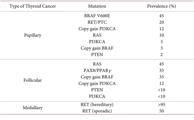
![Table 2. Pathological classification of malignant thyroid tumors [8] [50].](https://thumb-us.123doks.com/thumbv2/123dok_us/8754630.390136/7.595.208.538.80.405/table-pathological-classification-malignant-thyroid-tumors.webp)
![Table 4. The characteristics that should be assessed in the ultrasound [233] [234].](https://thumb-us.123doks.com/thumbv2/123dok_us/8754630.390136/15.595.190.540.518.733/table-characteristics-assessed-ultrasound.webp)
![Figure 2. Algorithm for the treatment of patients with thyroid nodules according to the pattern the result of the FNAB [143]](https://thumb-us.123doks.com/thumbv2/123dok_us/8754630.390136/16.595.62.545.98.534/figure-algorithm-treatment-patients-thyroid-nodules-according-pattern.webp)
![Table 7. Prognostic groups based on AJCC staging in well differentiated thyroid cancer [175]](https://thumb-us.123doks.com/thumbv2/123dok_us/8754630.390136/26.595.209.539.323.425/table-prognostic-groups-based-staging-differentiated-thyroid-cancer.webp)
![Table 9. Relative stage-specific survival for thyroid cancer [175].](https://thumb-us.123doks.com/thumbv2/123dok_us/8754630.390136/27.595.211.539.539.732/table-relative-stage-specific-survival-thyroid-cancer.webp)
![Table 10. ATA modified initial risk stratification system [34].](https://thumb-us.123doks.com/thumbv2/123dok_us/8754630.390136/28.595.204.538.85.455/table-ata-modified-initial-risk-stratification-system.webp)