Functional Properties of Cartilaginous Tissues Engineered From
Infrapatellar Fat Pad Derived Mesenchymal Stem Cells
Conor Timothy Buckley, PhD1#, Tatiana Vinardell, DVM1#, Stephen Desmond Thorpe, BAI, Matthew George Haugh, PhD, Elena Jones2, PhD, Dennis McGonagle, PhD, FRCI2 and Daniel John Kelly, PhD1*
1
Trinity Centre for Bioengineering, School of Engineering, Trinity College Dublin, Ireland.
2
NIHR Leeds Biomedical Research Unit, Leeds Institute of Molecular Medicine, University of Leeds, United Kingdom.
#Both of these authors contributed equally to this study
*Corresponding author
E-mail address: [email protected]
Address: Trinity Centre for Bioengineering
School of Engineering Trinity College Dublin
Ireland
Telephone: +353-1-896-3947
Fax: +353-1-679-5554
Keywords Cartilage repair, agarose hydrogel, infrapatellar fat pad, functional tissue engineering, Mesenchymal Stem Cells.
Word count: 3237 (Excluding Abstract and References)
Abstract
Introduction
Autologous chondrocyte implantation (ACI) is an established technique for the treatment of articular cartilage defects and is now in clinical use (Brittberg et al., 1994; Peterson et al., 2000; Horas et al., 2003; Ruano-Ravina and Jato Diaz, 2006). However, limitations still exist with both first and second generation ACI techniques in relation to the cell source procured. Issues such as dedifferentiation during monolayer expansion of isolated chondrocytes (Benya et al., 1978; Diaz-Romero et al., 2005), age related capacity (Barbero et al., 2004) and poor chondrogenic potential of osteoarthritic (OA) derived cells (Tallheden et al., 2005) have prompted investigations into the therapeutic potential of utilising mesenchymal stem cells (MSCs) as an alternative cell source for cartilage
repair (Wakitani et al., 1994; Bosnakovski et al., 2004; Bosnakovski et al., 2006; Mauck
et al., 2006; Miyanishi et al., 2006; Coleman et al., 2007; Hannouche et al., 2007; Mauck
et al., 2007; Huang et al., 2008; Kisiday et al., 2008).
cartilage repair, from non-cartilaginous knee joint tissues. It has been demonstrated that tissues from this region including the joint infrapatellar fat pad (IFP) (Dragoo et al., 2003; Wickham et al., 2003; English et al., 2007; Khan et al., 2007; Lee et al., 2008), synovium (Nishimura et al., 1999; Sakaguchi et al., 2005; Mochizuki et al., 2006; Shirasawa et al., 2006; Ando et al., 2007; Marsano et al., 2007; Pei et al., 2008a; Pei et al., 2008b) and synovial fluid (Jones et al., 2008) possess significant chondrogenic potential and perhaps provide a more readily available and clinically feasible source of chondroprogenitor cells. Through in vitro experiments it has been demonstrated that 80% of OA derived IFP pellet cultures were more chondrogenic than control bone marrow MSCs, and in addition exhibited similar chondrogenic capacity to cartilage-derived cultures (English et al., 2007).
sources. The objective of this study is to determine the functional properties of cartilaginous tissues engineered from MSCs isolated from the IFP.
Materials and methods
Cell Isolation and expansion
Porcine IFPs were harvested from the knee joint capsule of two 4 month old porcine donors (~50 kg) within 3 hours of sacrifice. IFPs were washed thoroughly in phosphate buffered saline (PBS) and diced followed by overnight incubation under constant rotation at 37°C with high-glucose Dulbecco’s Modified Eagle Medium (hgDMEM, GlutaMAX™) (GIBCO, Biosciences, Ireland) containing collagenase type II
Hydrogel construct fabrication and culturing
IFP MSCs (P3) were encapsulated in agarose (Type VII) at ~40°C, to yield a final gel concentration of 2% and a cell density of 15×106 cells/mL. The agarose-cell suspension was cast in a stainless steel mould to produce cylindrical discs (Ø 5mm×3mm thickness). Agarose hydrogel constructs were maintained in a chemically defined chondrogenic medium (CDM) consisting of DMEM GlutaMAX™ supplemented with penicillin (100 U/mL)-streptomycin (100 μg/mL) (both GIBCO, Biosciences, Ireland), 100 μg/ml sodium pyruvate, 40 μg/ml L-proline, 50 μg/ml L-ascorbic acid-2-phosphate, 1 mg/ml
BSA, 1× insulin–transferrin–selenium, 100 nM dexamethasone (all from Sigma-Aldrich, Ireland) and 10 ng/ml recombinant human transforming growth factor-β3 (TGF-β3; R&D Systems, UK). Hydrogel constructs were cultured in standard 6 well plates (Sarstedt, Ireland) with two-three constructs per well. Each construct was maintained in 2.5mL of complete medium with complete medium exchanges performed every 3-4 days for the total culture duration of 42 days. The surfaces of wells were monitored and changed when outgrowth occurred (~weekly) to avoid excessive nutrient demands from monolayer cells.
Assessment of functional properties
Constructs were mechanically tested in unconfined compression at days 0, 21 and 42 using a standard materials testing machine with a 5N load cell (Zwick Z005, Roell, Germany). A preload of 0.01N was applied to ensure that the surfaces of the gel constructs were in direct contact with the impermeable loading platens. Stress relaxation tests were performed, consisting of a ramp and hold cycle with a ramp displacement of 0.001mm/s until 10% strain was obtained and maintained until equilibrium was reached. Dynamic tests were performed immediately after the stress relaxation cycle. A cyclic strain of 1% was applied at 0.1Hz and 1Hz. After mechanical testing, constructs were weighed wet and the total mass recorded. Constructs were cored using a 3mm biopsy punch and separated from the annulus; the wet mass of both the core and annulus was recorded and frozen for subsequent biochemical analyses.
Quantitative biochemical analysis
assayed using a chloramine-T assay (Kafienah and Sims, 2004) with a hydroxyproline-to-collagen ratio of 1:7.69 (Ignat'eva et al., 2007).
Histology and Immunohistochemistry
Statistical analysis
Statistical analysis was preformed using one way ANOVA with Tukey post-hoc tests to enable comparisons between groups. Tests were performed using SigmaStat® (Version 3.1) software with 3 samples analysed for each group at each time point. Numerical and graphical results are displayed as mean ± standard deviation. Significance was accepted at a level of p < 0.05. The experiment was also replicated with cells pooled from different animals (n=2 donors). Comparable results were observed in both studies, and the results of the first replicate are presented in the manuscript.
Results
Constructs increased in mechanical properties from an equilibrium modulus of 7.0 kPa (± 0.6) at day 0 reaching 26.2 kPa (± 1.1) by day 42 and a final 1 Hz dynamic modulus, of 272.8 kPa (± 46.8) (Fig. 1A).
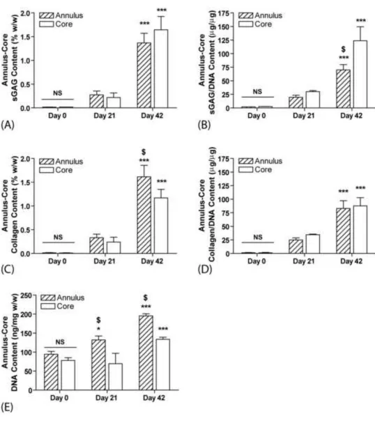
To gain an appreciation of the spatial distribution of biochemical constituents, the annuli and cores of samples were assessed separately (Fig. 2). By day 42, the core regions of constructs contained greater sGAG accumulation compared to the corresponding annular region when expressed in terms of percentage of wet weight (annulus= 1.37 ± 0.20 %w/w and core= 1.64 ± 0.28 %w/w) (Fig 2A), although this was not found to be statistically significant (p > 0.05). Similar differences were also observed in core-annulus sGAG accumulation when normalised by DNA content (Fig. 2B), and were found to be statistically significant (p < 0.01). Interestingly, collagen content was higher in the annular region of IFP constructs compared to the core region when normalised by wet weight (annulus= 1.61 ± 0.24 %w/w, core= 1.17 ± 0.18 %w/w, p < 0.05), although no differences were observed when normalised by DNA content (annulus = 82.88 ± 14.32 µg/µg, core = 87.73 ± 15.00 µg/µg, p > 0.05), (Fig. 2 C&D).
The DNA content increased with time in the annular regions from 94.36 ng/mg w/w (± 7.63) at day 0 to 195.31 ng/mg w/w (± 5.40) at day 42 (~2.1 fold increase) indicating extensive proliferation. The IFP core region DNA content also increased from 77.92 ng/mg w/w (± 7.42) at day 0 to 133.59 ng/mg w/w (± 5.68) at day 42 (~1.7 fold increase), (Fig. 2E).
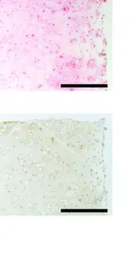
Histological evaluation revealed that constructs stained positively for sulphated proteoglycan and collagen accumulation (Fig. 3 A&B). Immunohistochemical results revealed that constructs stained weakly for collagen I, with more pronounced staining observed for collagen type II after 42 days of culture (Fig. 3 C&D).
constructs after 42 days (Fig. 4). No statistically significant increases occurred in any mechanical or biochemical measurement from day 21 to day 42 without the continued supplementation of TGF-β3 (p > 0.05). Both the equilibrium (TGF+ = 26.26 ± 1.10 kPa, TGF- = 12.30 ± 0.74 kPa, p < 0.001) and 1Hz dynamic moduli (TGF+ = 272.85 ± 46.81 kPa, TGF- = 74.35 ± 10.14 kPa, p < 0.001) were higher for continued supplementation with TGF-β3 from day 21 to 42 compared to withdrawal of TGF-β3 (Fig 4A). Significantly higher levels of sGAG accumulation were observed after 42 days for constructs with continued supplementation of TGF-β3 (TGF+ = 1.47 ± 0.22 %w/w; TGF- = 0.42 ± 0.08 %w/w, p < 0.01), with similar differences for collagen accumulation (TGF+ = 1.44 ± 0.22 %w/w, TGF- = 0.58 ± 0.06 %w/w, p < 0.01) (Fig 4B). These results were also reflected when normalised by DNA content (Fig 4C). For DNA content (Fig 4D), continued supplementation of TGF-β3 also enhanced cellular proliferation (TGF+ = 171.65 ± 3.17 ng/mg w/w; 129.93 ± 11.82 ng/mg, p < 0.01).
Discussion
Alternative cell sources for articular cartilage repair strategies have received significant
attention with the identification of chondro-progenitor populations within bone marrow
and in joint tissues such as synovium, infrapatellar fat pad and within the synovial fluid
itself. In this study we have demonstrated that IFP derived MSCs encapsulated in agarose
synovium-derived cells than to subcutaneous fat-derived cells (Mochizuki et al., 2006). Synovial-membrane derived MSCs have already been shown to possess significant promise as a cell source for cartilage tissue engineering (Sakaguchi et al., 2005; Yokoyama et al., 2005; Ando et al., 2007; Ando et al., 2008; Pei et al., 2008a; Pei et al., 2008b). The findings, in the present study, therefore extend observations in relationship to MSCs isolated from within the joint space (Mochizuki et al., 2006; Yoshimura et al., 2007) and suggest that IFP derived MSCs proliferate and undergo robust chondrogenesis within agarose hydrogels.
The spatial distribution of sGAG within the agarose hydrogels was generally different regionally, with greater amounts of sGAG accumulation in the core of constructs compared to the annulus. Higher levels of core sGAG content has previously been reported by our laboratory for agarose encapsulated porcine bone marrow derived MSCs (Thorpe et al., 2008). This may simply be due to diffusion of sGAG from the periphery of the hydrogel into the media, or alternatively spatial gradients in the concentration of nutrients, growth factors, oxygen etc. could be leading to the development of a more chondrogenic environment within the core of the constructs. Interestingly, greater proliferation and collagen accumulation was observed in the annulus, suggesting an environment in this region that promotes a more proliferative phenotype compared to the construct core.
et al., 2008). However differences due to age, species and culture conditions mean that future studies comparing donor matched chondrocytes and IFP MSCs will be critical. In addition, longer term studies will be required to assess if the mechanical properties of cartilaginous tissue generated using IFP MSCs plateau like that observed with bone marrow MSCs after extended culture periods (Mauck et al., 2006).
The motivation behind the removal of TGF-β3 from culture was based upon the published findings of Byers et al. (2008) for chondrocyte seeded agarose hydrogels. The authors demonstrated that two week transient exposure to TGF-β3 in a chemically defined medium elicited a superior response to that observed after continuous growth factor supplementation, with certain mechanical and biochemical levels achieved comparable to those of native cartilage. In the present study, the removal of TGF-β3 from culture after 21 days did not significantly enhance either the mechanical properties or biochemical content of constructs at 42 days compared to constructs exposed to continued TGF-β3 supplementation. Removal of TGF- β has also been shown to be sub-optimal for bone marrow derived MSCs in agarose hydrogels seeded at a similar density (Huang et al., in press), although a pronounced positive effect on the equilibrium modulus and sGAG accumulation was observed in that study for higher seeding densities (60 million/ml). Further studies to determine the optimal cell seeding density in order to engineer more functional cartilaginous tissues from IFP derived MSCs are required.
has been suggested that they may provide a useful animal model system to evaluate tissue engineering strategies (Ringe et al., 2002). Regardless it is unclear if the results presented here would be comparable when using IFP MSCs derived from aged or diseased human tissue, although studies investigating the chondrogenic potential of such cells are promising 32,56.
Given its anatomical location and ease of accessibility for harvesting biopsies of tissue, the IFP may provide an attractive source of cells for articular cartilage repair. In
the present work we add to this emerging concept by showing good in vitro functionality
of cartilaginous tissues engineered using IFP derived MSCs. A critical next step is to
assess in vivo the ability of cartilaginous tissues engineered using IFP MSC-agarose
constructs to repair large chondral or osteochondral defects. Clinical studies have already
been undertaken by other groups investigating agarose-alginate hydrogels seeded with
chondrocytes for cartilage defect repair (Selmi et al., 2007; Selmi et al., 2008). In this
clinical context, the IFP may also possess other benefits as a source of cells for cartilage
repair. Although this work utilised expanded cells, future work will also investigate the
feasibility and practicalities of utilising unexpanded cells from freshly digested IFP
tissue, since the yield of putative MSCs may be higher in such tissues. If successful, such
advantages would significantly accelerate translation into a clinical setting by avoiding
the time and cost hindrances associated with large scale cell expansion. The present
porcine studies therefore provide a rationale for the exploration of IFP in man as a source
Acknowledgements
Figure Legends
Fig 1: Mechanical and total biochemical properties of IFP constructs at days 0, 21 and 42. (A) Equilibrium modulus and 1Hz dynamic modulus (B) TotalDNA content (ng/mg w/w) (C) Total sGAG and collagen content (% w/w) (D) Total sGAG and collagen content normalised to DNA (µg/µg). (NS) represents no significance, (*) p < 0.05, (***) p < 0.001 represents significance compared to earlier time point(s), n=3 for each group.
Fig 2: Annulus-core biochemical analysis of IFP constructs at days 0, 21 and 42. (A)
sGAG content (% w/w) (B) sGAG/DNA content (µg/µg) (C) Collagen content (% w/w)
(D) Collagen/DNA content (µg/µg) and (E) DNA content (ng/mg w/w). (NS) represents no significance, (*) p < 0.05, (***) p < 0.001 represents significance compared to earlier time point(s), ($) p < 0.01 compared to corresponding core at the same time point, n=3 for each group.
Fig 3: Histological evaluation of constructs after 42 days of culture (A) Alcian blue staining for sGAG (B) Picrosirius Red staining for collagen, (C) Immunohistochemical staining for type I collagen (D) Immunohistochemical staining for type II collagen. Scale bar is 1mm.
Fig 4: The effect of removal of TGF-β3 from culture after 21 days. Data represents mechanical properties and total biochemical analysis of constructs at day 42. (A)
Equilibrium modulus and 1Hz dynamic modulus (B) Total sGAG and collagen content (% w/w) (C) Total sGAG and collagen content normalised to DNA (µg/µg) and (D)
References
Ando, W., Tateishi, K., Hart, D. A., Katakai, D., Tanaka, Y., Nakata, K., Hashimoto, J., Fujie, H., Shino, K., Yoshikawa, H., Nakamura, N., 2007. Cartilage repair using an in vitro generated scaffold-free tissue-engineered construct derived from porcine synovial mesenchymal stem cells. Biomaterials 28, 5462-5470.
Ando, W., Tateishi, K., Katakai, D., Hart, D. A., Higuchi, C., Nakata, K., Hashimoto, J., Fujie, H., Shino, K., Yoshikawa, H., Nakamura, N., 2008. In vitro generation of a scaffold-free tissue-engineered construct (TEC) derived from human synovial mesenchymal stem cells: biological and mechanical properties and further chondrogenic potential. Tissue Eng Part A 14, 2041-2049.
Barbero, A., Grogan, S., Schäfer, D., Heberer, M., Mainil-Varlet, P., Martin, I., 2004. Age related changes in human articular chondrocyte yield, proliferation and post-expansion chondrogenic capacity. Osteoarthritis and Cartilage 12, 476-484.
Barry, F., Boynton, R. E., Liu, B., Murphy, J. M., 2001. Chondrogenic differentiation of mesenchymal stem cells from bone marrow: differentiation-dependent gene expression of matrix components. Exp Cell Res 268, 189-200.
Benya, P. D., Padilla, S. R., Nimni, M. E., 1978. Independent regulation of collagen types by chondrocytes during the loss of differentiated function in culture. Cell 15, 1313-1321.
Bosnakovski, D., Mizuno, M., Kim, G., Ishiguro, T., Okumura, M., Iwanaga, T., Kadosawa, T., Fujinaga, T., 2004. Chondrogenic differentiation of bovine bone marrow mesenchymal stem cells in pellet cultural system. Exp Hematol 32, 502-509.
Bosnakovski, D., Mizuno, M., Kim, G., Takagi, S., Okumura, M., Fujinaga, T., 2006. Chondrogenic differentiation of bovine bone marrow mesenchymal stem cells (MSCs) in different hydrogels: influence of collagen type II extracellular matrix on MSC chondrogenesis. Biotechnol Bioeng 93, 1152-1163.
Brittberg, M., Lindahl, A., Nilsson, A., Ohlsson, C., Isaksson, O., Peterson, L., 1994. Treatment of deep cartilage defects in the knee with autologous chondrocyte transplantation. New England Journal of Medicine 331, 889-895.
Bruder, S. P., Jaiswal, N., Haynesworth, S. E., 1997. Growth kinetics, self-renewal, and the osteogenic potential of purified human mesenchymal stem cells during extensive subcultivation and following cryopreservation. Journal of Cellular Biochemistry 64, 278-294.
biomechanical and biochemical maturation of tissue-engineered cartilage. Tissue Eng Part A 14, 1821-1834.
Caplan, A. I., 1991. Mesenchymal stem cells. Journal of Orthopaedic Research 9, 641-650.
Coleman, R. M., Case, N. D., Guldberg, R. E., 2007. Hydrogel effects on bone marrow stromal cell response to chondrogenic growth factors. Biomaterials 28, 2077-2086.
Diaz-Romero, J., Gaillard, J. P., Grogan, S. P., Nesic, D., Trub, T., Mainil-Varlet, P., 2005. Immunophenotypic analysis of human articular chondrocytes: changes in surface markers associated with cell expansion in monolayer culture. J Cell Physiol 202, 731-742.
Dragoo, J. L., Samimi, B., Zhu, M., Hame, S. L., Thomas, B. J., Lieberman, J. R., Hedrick, M. H., Benhaim, P., 2003. Tissue-engineered cartilage and bone using stem cells from human infrapatellar fat pads. J Bone Joint Surg Br 85, 740-747.
Egrise, D., Martin, D., Vienne, A., Neve, P., Schoutens, A., 1992. The number of fibroblastic colonies formed from bone marrow is decreased and the in vitro proliferation rate of trabecular bone cells increased in aged rats. Bone 13, 355-361.
English, A., Jones, E. A., Corscadden, D., Henshaw, K., Chapman, T., Emery, P., McGonagle, D., 2007. A comparative assessment of cartilage and joint fat pad as a potential source of cells for autologous therapy development in knee osteoarthritis. Rheumatology 46, 1676-1683.
Erickson, I. E., Huang, A. H., Chung, C., Li, R. T., Burdick, J. A., Mauck, R. L., 2009. Differential maturation and structure-function relationships in mesenchymal stem cell- and chondrocyte-seeded hydrogels. Tissue Eng Part A 15, 1041-1052.
Hannouche, D., Terai, H., Fuchs, J. R., Terada, S., Zand, S., Nasseri, B. A., Petite, H., Sedel, L., Vacanti, J. P., 2007. Engineering of implantable cartilaginous structures from bone marrow-derived mesenchymal stem cells. Tissue Eng 13, 87-99.
Horas, U., Pelinkovic, D., Herr, G., Aigner, T., Schnettler, R., 2003. Autologous chondrocyte implantation and osteochondral cylinder transplantation in cartilage repair of the knee joint. A prospective, comparative trial. Journal of Bone and Joint Surgery - Series A 85, 185-192.
Huang, A. H., Yeger-McKeever, M., Stein, A., Mauck, R. L., 2008. Tensile properties of engineered cartilage formed from chondrocyte- and MSC-laden hydrogels. Osteoarthritis and Cartilage 16, 1074-1082.
Ignat'eva, N. Y., Danilov, N. A., Averkiev, S. V., Obrezkova, M. V., Lunin, V. V., Sobol, E. N., 2007. Determination of hydroxyproline in tissues and the evaluation of the collagen content of the tissues. J Anal Chem 62, 51-57.
Johnstone, B., Hering, T. M., Caplan, A. I., Goldberg, V. M., Yoo, J. U., 1998. In vitro chondrogenesis of bone marrow-derived mesenchymal progenitor cells. Experimental Cell Research 238, 265-272.
Jones, E. A., Crawford, A., English, A., Henshaw, K., Mundy, J., Corscadden, D., Chapman, T., Emery, P., Hatton, P., McGonagle, D., 2008. Synovial fluid mesenchymal stem cells in health and early osteoarthritis: Detection and functional evaluation at the single-cell level. Arthritis and Rheumatism 58, 1731-1740.
Jones, E. A., Kinsey, S. E., English, A., Jones, R. A., Straszynski, L., Meredith, D. M., Markham, A. F., Jack, A., Emery, P., McGonagle, D., 2002. Isolation and characterization of bone marrow multipotential mesenchymal progenitor cells. Arthritis Rheum 46, 3349-3360.
Kadiyala, S., Young, R. G., Thiede, M. A., Bruder, S. P., 1997. Culture expanded canine mesenchymal stem cells possess osteochondrogenic potential in vivo and in vitro. Cell Transplantation 6, 125-134.
Kafienah, W., Sims, T. J., 2004. Biochemical methods for the analysis of tissue-engineered cartilage. Methods Mol Biol 238, 217-230.
Khan, W. S., Adesida, A. B., Hardingham, T. E., 2007. Hypoxic conditions increase hypoxia-inducible transcription factor 2alpha and enhance chondrogenesis in stem cells from the infrapatellar fat pad of osteoarthritis patients. Arthritis Res Ther 9, R55.
Kisiday, J. D., Kopesky, P. W., Evans, C. H., Grodzinsky, A. J., McIlwraith, C. W., Frisbie, D. D., 2008. Evaluation of adult equine bone marrow- and adipose-derived progenitor cell chondrogenesis in hydrogel cultures. J Orthop Res 26, 322-331.
Lee, S. Y., Nakagawa, T., Reddi, A. H., 2008. Induction of chondrogenesis and expression of superficial zone protein (SZP)/lubricin by mesenchymal progenitors in the infrapatellar fat pad of the knee joint treated with TGF-beta1 and BMP-7. Biochem Biophys Res Commun 376, 148-153.
Maniatopoulos, C., Sodek, J., Melcher, A. H., 1988. Bone formation in vitro by stromal cells obtained from bone marrow of young adult rats. Cell and Tissue Research 254, 317-330.
Marsano, A., Millward-Sadler, S. J., Salter, D. M., Adesida, A., Hardingham, T., Tognana, E., Kon, E., Chiari-Grisar, C., Nehrer, S., Jakob, M., Martin, I., 2007. Differential cartilaginous tissue formation by human synovial membrane, fat pad, meniscus cells and articular chondrocytes. Osteoarthritis Cartilage 15, 48-58.
Mauck, R. L., Byers, B. A., Yuan, X., Tuan, R. S., 2007. Regulation of cartilaginous ECM gene transcription by chondrocytes and MSCs in 3D culture in response to dynamic loading. Biomech Model Mechanobiol 6, 113-125.
Mauck, R. L., Yuan, X., Tuan, R. S., 2006. Chondrogenic differentiation and functional maturation of bovine mesenchymal stem cells in long-term agarose culture. Osteoarthritis Cartilage 14, 179-189.
Miyanishi, K., Trindade, M. C., Lindsey, D. P., Beaupre, G. S., Carter, D. R., Goodman, S. B., Schurman, D. J., Smith, R. L., 2006. Effects of hydrostatic pressure and transforming growth factor-beta 3 on adult human mesenchymal stem cell chondrogenesis in vitro. Tissue Eng 12, 1419-1428.
Mochizuki, T., Muneta, T., Sakaguchi, Y., Nimura, A., Yokoyama, A., Koga, H., Sekiya, I., 2006. Higher chondrogenic potential of fibrous synovium- and adipose synovium-derived cells compared with subcutaneous fat-derived cells: distinguishing properties of mesenchymal stem cells in humans. Arthritis Rheum 54, 843-853.
Nishimura, K., Solchaga, L. A., Caplan, A. I., Yoo, J. U., Goldberg, V. M., Johnstone, B., 1999. Chondroprogenitor cells of synovial tissue. Arthritis and Rheumatism 42, 2631-2637.
Pei, M., He, F., Boyce, B. M., Kish, V. L., 2008a. Repair of full-thickness femoral condyle cartilage defects using allogeneic synovial cell-engineered tissue constructs. Osteoarthritis Cartilage.
Peterson, L., Minas, T., Brittberg, M., Nilsson, A., Sjo?gren-Jansson, E., Lindahl, A., 2000. Two-to 9-year outcome after autologous chondrocyte transplantation of the knee. Clinical Orthopaedics and Related Research, 212-234.
Pittenger, M. F., Mackay, A. M., Beck, S. C., Jaiswal, R. K., Douglas, R., Mosca, J. D., Moorman, M. A., Simonetti, D. W., Craig, S., Marshak, D. R., 1999. Multilineage potential of adult human mesenchymal stem cells. Science 284, 143-147.
Ringe, J., Kaps, C., Schmitt, B., Buscher, K., Bartel, J., Smolian, H., Schultz, O., Burmester, G. R., Haupl, T., Sittinger, M., 2002. Porcine mesenchymal stem cells. Induction of distinct mesenchymal cell lineages. Cell Tissue Res 307, 321-327.
Ruano-Ravina, A., Jato Diaz, M., 2006. Autologous chondrocyte implantation: a systematic review. Osteoarthritis Cartilage 14, 47-51.
Sakaguchi, Y., Sekiya, I., Yagishita, K., Muneta, T., 2005. Comparison of human stem cells derived from various mesenchymal tissues: superiority of synovium as a cell source. Arthritis Rheum 52, 2521-2529.
Selmi, T. A. S., Neyret, P., Verdonk, P. C. M., Barnouin, L., 2007. Autologous chondrocyte transplantation in combination with an alginate-agarose based hydrogel (Cartipatch). Techniques in Knee Surgery 6, 253-258.
Selmi, T. A. S., Verdonk, P., Chambat, P., Dubrana, F., Potel, J. F., Barnouin, L., Neyret, P., 2008. Autologous chondrocyte implantation in a novel alginate-agarose hydrogel: Outcome at two years. Journal of Bone and Joint Surgery - Series B 90, 597-604.
Shirasawa, S., Sekiya, I., Sakaguchi, Y., Yagishita, K., Ichinose, S., Muneta, T., 2006. In vitro chondrogenesis of human synovium-derived mesenchymal stem cells: optimal condition and comparison with bone marrow-derived cells. J Cell Biochem 97, 84-97.
Tallheden, T., Bengtsson, C., Brantsing, C., Sjo?gren-Jansson, E., Carlsson, L., Peterson, L., Brittberg, M., Lindahl, A., 2005. Proliferation and differentiation potential of chondrocytes from osteoarthritic patients. Arthritis research & therapy. 7, R560-568.
Thorpe, S. D., Buckley, C. T., Vinardell, T., O'Brien, F. J., Campbell, V. A., Kelly, D. J., 2008. Dynamic compression can inhibit chondrogenesis of mesenchymal stem cells. Biochem Biophys Res Commun 377, 458-462.
Vacanti, V., Kong, E., Suzuki, G., Sato, K., Canty, J. M., Lee, T., 2005. Phenotypic changes of adult porcine mesenchymal stem cells induced by prolonged passaging in culture. J Cell Physiol 205, 194-201.
Wakitani, S., Goto, T., Pineda, S. J., Young, R. G., Mansour, J. M., Caplan, A. I., Goldberg, V. M., 1994. Mesenchymal cell-based repair of large, full-thickness defects of articular cartilage. J Bone Joint Surg Am 76, 579-592.
Wakitani, S., Nawata, M., Tensho, K., Okabe, T., Machida, H., Ohgushi, H., 2007. Repair of articular cartilage defects in the patello-femoral joint with autologous bone marrow mesenchymal cell transplantation: three case reports involving nine defects in five knees. Journal of tissue engineering and regenerative medicine 1, 74-79.
Wickham, M. Q., Erickson, G. R., Gimble, J. M., Vail, T. P., Guilak, F., 2003. Multipotent stromal cells derived from the infrapatellar fat pad of the knee. Clin Orthop Relat Res, 196-212.
Worster, A. A., Brower-Toland, B. D., Fortier, L. A., Bent, S. J., Williams, J., Nixon, A. J., 2001. Chondrocytic differentiation of mesenchymal stem cells sequentially exposed to transforming growth factor-beta1 in monolayer and insulin-like growth factor-I in a three-dimensional matrix. J Orthop Res 19, 738-749.
Yokoyama, A., Sekiya, I., Miyazaki, K., Ichinose, S., Hata, Y., Muneta, T., 2005. In vitro cartilage formation of composites of synovium-derived mesenchymal stem cells with collagen gel. Cell Tissue Res 322, 289-298.
Yoo, J. U., Barthel, T. S., Nishimura, K., Solchaga, L., Caplan, A. I., Goldberg, V. M., Johnstone, B., 1998. The chondrogenic potential of human bone-marrow-derived mesenchymal progenitor cells. Journal of Bone and Joint Surgery - Series A 80, 1745-1757.
Figure 1
Figure 2
Figure 3
Figure 4
Conflict of interest statement
The authors have nothing to disclose.