Original Article
A nonalcoholic fatty liver disease cirrhosis model in
gerbil: the dynamic relationship between hepatic
lipid metabolism and cirrhosis
Wei Li1,2, Zheng Guan3, Jean C Brisset6, Qiaojuan Shi2, Qi Lou2, Yue Ma2, Su Suriguga4, Huazhong Ying2,
Xiaoying Sa2, Zhenwen Chen5, Wim J Quax3, Xiaofeng Chu2
1Laboratory Animal Center, Zhejiang University, Hangzhou, Zhejiang, China; 2Laboratory Animal Center, Zhejiang Academy of Medical Sciences, Hangzhou, Zhejiang, China; Departments of 3Chemical and Pharmaceutical Biol-ogy, 4Pharmaceutical Technology and Biopharmacy, GUIDE, University of Groningen, Groningen, The Netherlands; 5Department of Laboratory Animal Science, School of Basic Medical Science, Capital Medical University, Beijing, China; 6Department of Radiology, New York University, New York, State of New York, The United States
Received November 2, 2017; Accepted November 21, 2017; Epub January 1, 2018; Published January 15, 2018
Abstract: Nonalcoholic fatty liver disease (NAFLD) usually takes decades to develop into cirrhosis, which limits the longitudinal study of NAFLD. This work aims at developing a NAFLD-caused cirrhosis model in gerbil and examining the dynamic relationship between hepatic lipid metabolism and cirrhosis. We fed gerbil a high-fat and high-cho-lesterol diet (HFHCD) for 24 weeks, and recorded the gerbil’s phenotype at 3, 6, 9, 12, 15, 18, 21, 24 weeks. The model’s pathological process, lipid metabolism, oxidative stress, liver collagen deposition and presence of relevant cytokines were tested and evaluated during the full-time frame of disease onset. The gerbil model can induce non-alcoholic steatohepatitis (NASH) within 9 weeks, and can develop cirrhosis after 21 weeks induction. The model’s lipids metabolism disorder is accompanied with the liver damage development. During the NAFLD progression, triglycerides (TG) and free fatty acids (FFA) have presented distinct rise and fall tendency, and the turning points are at the fibrosis stage. Besides that, the ratios of total cholesterol (CHO) to high-density lipoprotein cholesterol (HDL-C) exhibited constant growth tendency, and have a good linear relationship with hepatic stellate cells (HSC) (R2 = 0.802, P < 0.001). The gerbil NAFLD cirrhosis model has been developed and possesses positive correlation
between lipids metabolism and cirrhosis. The compelling rise and fall tendency of TG and FFA indicated that the fibrosis progression can lead to impairment in lipoprotein synthesis and engender decreased TG level. CHO/HDL-C ratios can imply the fibrosis progress and be used as a blood indicator for disease prediction and prevention.
Keywords: Cirrhosis, fibrosis, hepatic lipid metabolism, nonalcoholic fatty liver disease (NAFLD), gerbil
Introduction
Cirrhosis is a severe stage of liver dysfunction and is most commonly caused by alcohol, viral hepatitis, and NAFLD. Since NAFLD has become a leading health problem worldwide, especially for those people who are living with obesity [1], the lack of a suitable animal model for NAFLD-caused cirrhosis has aroused more and more attention. It has been suggested that 20% of the adult population in the world now has NAFLD [2], with an increase of 90% morbidity in obese populations [3]. The whole natural histo-ry of NAFLD has four stages: simple steatosis, NASH, fibrosis and cirrhosis. Fibrosis can lead
to an increased morbidity rate from cirrhosis, hepatic failure, metabolic syndrome associated cardiovascular and cerebrovascular diseases, and hepatic carcinoma [4]. The clinical pro- gression from simple steatosis to cirrhosis of NAFLD usually takes decades, strongly impact-ing the NAFLD-caused cirrhosis mechanism research and related drug development. For this reason, finding a suitable animal model for NAFLD-caused cirrhosis study becomes crucial.
Nonalcoholic fatty liver disease cirrhosis model in gerbil
model. Most commonly used genetic models are related to lipid metabolism genes, such as ob/ob mouse, db/db mouse, (fa/fa) Zucker rats, PPARα-/- mouse, and PNPLA3 transgenic mouse. Whereas, spontaneous mutations are very rare in the clinic, and normally those cases are based on multiple gene mutations, which causes differences between clinic and the genetic models. For those drug and/or diet-induced models, the typical diets used to in- duce NAFLD are methionine choline deficient (MCD) diet [5, 6], high-fat diet (HFD, including high-fat and high-fat, high-cholesterol diets), and high-carbohydrate diet (HCD) [7]. Com- pared with the diet of human the MCD diet is abnormal, but HFD and HCD are similar. Additionally, comparing with the other animal models, our literature searches show that the rat HFD model is the more widely used model to investigate the etiology and pathogenesis of NAFLD [8-10]. However, this type of model cannot induce cirrhosis [11, 12]. The induced animal can reach NASH stage and fibrosis stage, but the fibrosis is mild and unstable. So in the past decade, a great emphasis has be- en placed on the animal models for NAFLD study.
The Mongolian gerbil (Merionesunguiculatus) is a fat-sensitive animal [13-18]. Its plasma lipid response to dietary fatty acids is more sensi-tive than humans, even without dietary cho- lesterol [13-18]. Due to its reported lipemic responses to dietary fat and cholesterol [13-20], the gerbil has been chosen to establish NAFLD animal model. Moreover, previous re- search findings have pointed out that NASH is associated with metabolic syndrome [21-25], and gerbil has the tendency to acquire diabe- tes spontaneously [26]. We attempt to estab-lish a stable animal model, which shares the major clinical pathogenic factors: HFD, male, and lipid metabolism disorder [2]. Since we have clearly observed fibrosis stage and veri-fied the stability of a gerbil NASH model in our former work [27, 28], the gerbil NASH model was used for developing a NAFLD-caused cir-rhosis model and studying the mechanism. This is the first study to undertake a longitudinal observation of the whole natural history of NAFLD in gerbil and to investigate the dynamic relationship between hepatic lipid metabolism and cirrhosis. This is also the first comprehen-sive description of the new established
NAFLD-caused cirrhosis model. The model’s patho- logical process, lipid metabolism, oxidative str- ess, liver collagen deposition and related cyto-kines were examined and evaluated in this research.
Methods
Animals
A total of 72 male littermate Mongolian ger- bils (Merionesunguiculatus) were obtained fr- om Zhejiang Academy of Medical Sciences, Zhejiang Center of Laboratory Animals (Hang- zhou, China). All experiments were performed in accordance to the guidelines for animal care and use of Zhejiang province and Zhejiang Academy of Medical Sciences, and approved by the Ethics Committee of Zhejiang Academy of Medical Sciences (No. SCXK (Zhe) 2008-0033, and No. SYXK (Zhe) 2008-0113). HFHCD for induction of NAFLD and cirrhosis
Gerbils were divided randomly into nine gr- oups: one control group (n = 8) and eight time point model groups (each group, n = 8). The control group animals were fed a normal diet for 24 weeks, while the model group animals were fed an HFHCD. Based on former reports [29], we developed a new diet recipe (China Patent No. CN 102106476 A) to establish the model, which is similar to the western diet and containing the following components: 10% egg yolk powder (Zhejiang Changxing Ags Biologi- cal Products Co., Ltd.), 7% lard oil (purchas- ed from local supermarket), 2.5% cholesterol (Huadong Pharmaceutical Co., Ltd) and 0.2% cholate (Huadong Pharmaceutical Co., Ltd). The nutritional differences of the diets are shown in Supplementary Table 1.
Experimental design
choles-terol (LDL-C) and glucose (GLU). Liver sam- ples were obtained from the central part of the largest liver lobe. Liver homogenate was prepared to determine the content of FFA; and the following fibrosis indexes: type I collagen, type III collagen, transforming growth factor β1 (TGF-β1), and platelet-derived growth factor (PDGF), which were examined by western blot-ting (see Supplementary). Another part of the liver sample was fixed by 10% formalin and embedded in paraffin for histological study, and then were stained with hematoxylin and eosin (HE) and with Masson trichrome (for collagen). Besides, liver biopsy specimens we- re also immunohistochemically examined for Kupffer cells by assessing the expression of the receptor CD68, and HSC by evaluating the activation of the marker of HSC: alpha-smooth muscle actin (αSMA). As a double check, type I collagen and type III collagen were appraised as well as Kupffer cells and HSC by immunohis-tochemical examination. Meanwhile, for lipid accumulation assessment, electron micro-scope was employed to observe the liver tissue.
Biochemical analysis
The levels of serum ALT, AST, CHO, TG, GLU, LDL-C, and HDL-C were determined by an auto-matic chemical analyzer (HITACHI 7100, Ja- pan). Commercial kits (Huifeng Science & Te- chnology Co. Ltd., Shanghai, China; Jiancheng Biotech. Sci. Inc., Nanjing, China) were used for these analysis. All the procedures were fol-lowed as those stated in the protocols of the detection kits. Likewise, the levels of FFA in liver were examined by a Nonesterified free fatty acids assay kit (Jiancheng Biotech. Sci. Inc., Nanjing, China).
Histopathology, electron microscope observa-tion and immunohistochemical analysis
For histopathological study, the liver sections were stained with HE and with Masson’s tri-chrome. Histological steatosis, inflammation, and fibrosis were assessed semiquantitatively by a single-blinded pathologist, according to the scoring system proposed by Kleiner et al. [31]. During the assessment, a Leica micro-scope (Leica DM2500, Germany) was used for the observation. In each liver section, images of 10 random fields were taken (10 × 2.5
origi-nal magnification; 0.22 mm2 total area per
image) for the judgment.
For transmission electron microscope obser- vation, one animal was selected randomly from each group. After the liver was washed by using 0.9% physiological saline, a piece of the hepatic tissue was cut off from the central part of the largest liver lobe (about 1 × 1 × 1 mm). And then, the tissue was fixed in 2.5% glutaraldehyde for 4 hours at 4°C. Later, the samples were sent to electron microscopic laboratory (Virus Research Institute, Chinese Academy of Sciences), to gain the transmission electron microscope result.
The Kupffer cell, hepatic stellate cell, type I collagen, and type III collagen were detected using the common immunohistochemical me- thod (see Supplementary). In order to show the fibrosis progression during the HFHCD induc-tion clearly, an improved German immuno- histochemical scoring (GIS) system [32] was employed to access the immunohistochemical slices. The percentage of positive cells was graded as follows: 0, negative; 1, up to 10% positive cells; 2, 11% to 20%; 3, 21% to 30%; 4, 31% to 40%; 5, 41% to 50%; 6, 51% to 60%; 7, 61% to 70%; 8, 71% to 80%; and 9, > 80%. The intensity of marker expression was graded as follows: 0, negative; 1, weakly positive; 2, mod-erately positive; 3, strongly positive. The final immunoreactive score equals to the product of the percentage of positive cells multiply the highest staining intensity.
Statistical analysis
Nonalcoholic fatty liver disease cirrhosis model in gerbil
Results
Histological and biochemical assessment of HFHCD induced NAFLD and cirrhosis
In the morphological observations, the control group gerbils have shown soft livers with nor-mal color and smooth surfaces, whereas the model group gerbils presented different de- grees of pathological liver over time. The liver of model group animals became swollen and hard and turned pale yellow, the edge of the pathological liver developed into a blunt shape, and the liver surface changed into an irregular pattern with whitish micronodules and mac-ronodules (Figure 1F), which indicated the liv- er damage changes over the HFHCD induction time.
The comparison of histopathological study of the model group and control group has also shown a similar tendency as morphology ob- servation. As exhibited in Figure 1A, no dis-cernible histological alterations could be identi-fied in control group. They had complete cyto-plasm, sinusoidal spaces, distinct nucleus, nucleolus and central vein. In contrast, the mo- del group presented a full blown cirrhosis fea-ture accompanied with steatosis (Figure 1E),
and a significantly higher fibrosis score (Sup- plementary Table 2). These results provided microscopic evidence that indicated the sig- nificant liver damage changes over the HFH- CD induction time. The independent pathologi-cal judgment [31] (Supplementary Table 2) show the obvious steatosis stage (Figure 1B) between 3 to 6 weeks, the NASH stage (Figure 1C, 1D) between 9 to 18 weeks, and liver fi- brosis stage (Figure 1D) after 12 weeks HFHCD induction. Finally, the cirrhosis model (Figure 1E, 1F) can be seen to have been established after 21 weeks HFHCD induction (Figure 2). Supplementary Tables 3 and 4 presented the results of raised ALT, AST, GLU, TG, CHO and body weight of model animals (0-9 weeks). These results exhibited the well repeated ger- bil NASH model as we have reported before [27, 28].
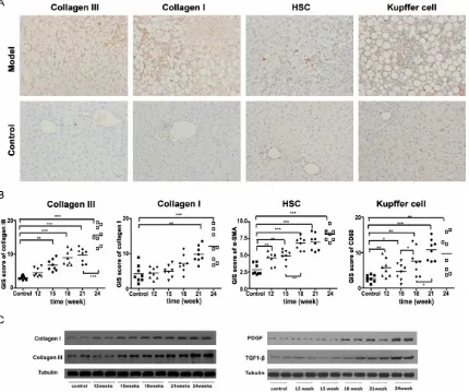
In accord with the pathological assessment and transaminases results, in comparison to the control group, the immunohistochemical analysis and western blot results also show a compelling increase of type I (P < 0.05, after 21 weeks) and type III collagens (P < 0.01, after 15 weeks) in the model group (Figure 3). As shown in Figure 3B, After 15 weeks of HFHCD
induction, collagen III was increasing consis-tently (P < 0.001); on the other hand, collagen I was significantly increased again at week 24 (P < 0.01). Both of the type I and type III collagens reached the biochemical peak at week 24, which strengthen the assurance of the model. Activation of Kupffer cell and HSC
Kupffer cells and hepatic stellate cells are gen-erally regarded as keys of the fibrosis and cir-rhosis mechanisms. In this study, Kupffer cells and hepatic stellate cells were evaluated by assessing the expression of the receptor CD- 68, and αSMA, the marker of activated HSC. As expected, the control group animals only have few Kupffer cells, and no activated HSC has been observed (Figure 3). Whereas, the model groups show many Kupffer cells and HSC (Figure 3) after 12 weeks of HFHCD in- duction. Compare with the control group, the increase of Kupffer cells after 12 weeks HF- HCD induction is significant: 12 weeks, P < 0.01; 15 weeks, P < 0.05; 18 weeks, P < 0.01; 21 weeks, P < 0.001; and 24 weeks, P < 0.01. The comparison of the activated Kupffer cells between 18 weeks and 21 weeks model gr- oups also shows compelling difference (P < 0.05). The HSC changes of model groups over the HFHCD induction time are significant too, as well as the tendency of Kupffer cells. HSC appeared together with the ballooning hepato-cytes. Compared with the control group, the increase of HSC numbers after 12 weeks HF- HCD induction is notable: 12 weeks, P < 0.01; 15 weeks, P < 0.01; 18 weeks, P < 0.001; 21
correlation coefficient between the other vari-ables and HSC measurements were calculated to be 0.68 for TGF-β1, 0.41 for PDGF and 0.62 for Kupffer cell (Figure 4B). The strong positive correlation between those chronic inflamma-tion factors and HSC implies that the HFHCD induced cirrhosis may be partly caused by the chronic inflammation factors [11, 33], which directly induced the activation of HSC. Besides that, partial correlation analysis shows that TGF-β1 has a strong positivecorrelation with HSC, its partial correlation coefficient is 0.65, which is similar to their correlation coefficient mentionedabove (0.68, TGF-β1 and HSC). This result suggests that TGF-β1 and HSC has a strong and direct positive correlation. The fol-lowed-up statistical analysis also indicates TGF-β1 is prior to HSC and is the Granger-cau- se of HSC (P < 0.05, optimal lagorder is deter-mined by Akaike information criteria) [34, 35]. Since TGF-β1 is the Granger-cause of HSC, along with HFHCD feeding time, we calculated their linear regression formula: HSC = 2.467 + 1.279e-5 × TGF-β1 + 1.831e-1 × Time (R2 =
0.764, TGF-β1 is based on the gray scale val-ues of western blotting result [36]) (Figure 4B), and their coefficients are significant (P < 0.001). Its good linear relation offers TGF-β1 as a new supplement indicator for fibrosis diag-nosis and prediction.
Dynamic relationship between hepatic lipid metabolism and cirrhosis
[image:5.612.94.368.81.238.2]The lipid accumulation in liver was observed by transmission electron microscopy. The ul-
Figure 2. The four stages of the NAFLD-cirrhosis gerbil model and the tested indexes in this study.
weeks, P < 0.001; and 24 weeks, P < 0.001.
Hepatic expression of fibro -genesis mediators implicated in the fibrogenic process
Nonalcoholic fatty liver disease cirrhosis model in gerbil
trastructure of hepatic cells was normal in con-trol group: the membrane was well defined; chromatin was uniformly distributed. No fat droplet could be found in mitochondria, endo-cytoplasmic reticulum or liver cell. The cell nucleus was overall in shape (Figure 5A). In contrast, the model group animals have many fat droplets and formation of vacuoles in liver cells. After 3 weeks HFHCD induction, the small lipid droplets appeared everywhere in the cell (Figure 5B, 5C). And then, during the HFHCD induction process, the fat droplets accumulat-ed into some big lipid droplets (Figure 5E, 5F). The observed lipid accumulation changes in- dicated the severity degree of hepatic lipid metabolism disorder, which also complies with the NAFLD progression. In addition, total CHO,
[image:6.612.95.525.76.435.2]LDL-C, FFA and HDL-C were increased dramati-cally in relation to the HFHCD induction time (Figures 5D and 4A). To illustrate the dynamic relationship between hepatic lipid metabolism and cirrhosis, a focused principal components analysis (FPCA) has been performed [37]. Sin- ce TG and FFA have shown distinct rise and fall tendency, and the turning point is at 18 weeks and 12 weeks (fibrosis stage). Accordingly, FPCA also reflected the differences. The analy-sis has shown significant positive correlation between HSC and all the tested factors (P < 0.05), except FFA and TG (Figure 4B). FPCA revealed that HDL-C, LDL-C, and CHO belong to the main factors in this experiment, and their increment can promote the fibrosis progression (Figure 4B).
Discussion
Our novel data shows that in the first 9 weeks HFHCD induction, the model’s body weight, GLU, ALT, AST, CHO, and TG levels were signifi-cantly increased (Supplementary Tables 3 and 4). The histological scoring system also shows the NASH features. Therefore, the gerbil NASH model has been repeated well as our former studies [27, 28]. During the whole stu- dy period, a number of features in common with human disease were found: (i) the exces-sive accumulation of FFA, (ii) blood lipid me- tabolism disorder, (iii) cellular ballooning, (iv) immunohistochemistry changes, and (v) liver morphology modifications [38-40]. Progres-
[image:7.612.91.491.72.451.2]sion of the cirrhosis from NAFLD was clearly subdivided into stages (Figure 2) allowing the study of the progression of the disease and the correlation with the HFHCD induction time. The result indicates a strong positive correla-tion between lipids metabolism and cirrhosis, which mimics the clinical disease. Therefore, the gerbil NAFLD cirrhosis model has been established and can be used to study NAFLD progression, especially the cirrhosis stage, for a better understanding of the disease patho-physiology and for medicine development. Besides, during the longitudinal study we have observed a distinct rise and fall tendency of TG and FFA levels, and their turning points are
Nonalcoholic fatty liver disease cirrhosis model in gerbil
in the fibrosis stage. The decreased TG level seems rare based on our knowledge. Coin- cidently, a Harvard group has also reported the similar tendency recently [41]. Their clinical study statistically analyzed 11947 cases, and pointed out that lower triglyceride levels may indicate more advanced liver disease. The fi- brosis progression caused liver dysfunction can possibly lead to resultant impairment in lipoprotein synthesis and result in decreased triglyceride export. The accompanied FFA level changes are the support of the inference. Since the gerbil NAFLD cirrhosis model has been well developed, which not only possess the similar clinical features but also can be clearly divided into stages, we tried FPCA to find out the closest indexes of HSC for the dynamic monitoring and disease progression prognosis. The FPCA [37] result indicated that HDL-C, LDL-C, and CHO are closely related with HSC and other fibrosis indicators in the NA- FLD-cirrhosis progression. The similar increas-ing tendency of Kupffer cells, HSC, TGF-β1, and PDGF also supports previous research into this area which indicated that the Kupffer cells can secrete TGF-β1, and PDGF to stimulate fibrosis development through the mitogenic
stimuli of them on HSC [42]. Surprisingly, the growth of HDL-C was found abnormal during the HFHCD induction time, which has aroused our attention. In this study, HDL-C not only has strong positive correlation with HSC (r = 0.556, P < 0.05), but also has a good linear relation-ship with HSC along with the HFHCD induction time (Figure 4B, HSC = 1.047 + 0.756 × HDL-C + 0.178 × Time, R2 = 0.823, P < 0.001). A few
NAFLD studies [43, 44] have also reported the same HDL-C increase tendency, which may suggest the special mechanism during the NAFLD-caused fibrosis progression. So we lo- oked into the CHO/HDL-C ratio to further in- spect the model. It is interesting to note that the CHO/HDL-C ratios show constant growth tendency during the HFHCD induction, and have good linear relationship with HSC (HSC = 3.542 - 0.655 × CHO/HDL-C + 0.237 × Time, R2 = 0.802, P < 0.001). These results are
[image:8.612.93.526.70.289.2]dramatically consistent with the clinic reports [45, 46] and may imply the special status of HDL-C in the NAFLD progression. Considering the possible HFD or HFHCD differences, envi-ronment differences, and animal differences in other labs, we suggest using CHO/HDL-C ratio as a convenient blood indicator for the standard gerbil model assessment. For this
purpose, the blood sample can be collected from the tail vein or the cheek vein (300 μL blood is enough for detecting more than two indexes), and the regression equation might be used to determine the NAFLD stage and suit-able experiment time.
As we presented in the result, the pros and cons of this gerbil NAFLD cirrhosis model can easily be listed. First of all, this gerbil model can induce NASH within 9 weeks, and can develop stable cirrhosis after 21 weeks HFH- CD induction (Supplementary Table 2). This is in contrast to, as Robert Schierwagen reported, most of the existing HFD induced NAFLD mod-els that need a minimum of 15 weeks induc- tion and that hardly show stable fibrosis [23, 47-49]. Secondly, the model has the same fibrosis progression (eg. HSC activation, colla-gen III and collacolla-gen I deposition, bridging fibro-sis, and cirrhosis) as in the clinic [38-40], and leads to a stable cirrhosis stage. It can thus be suggested that the gerbil model can cover the whole NAFLD disease spectrum to make up the NAFLD model deficiency [11, 12]. Having the same features and etiologies as the clini- cal process combined with its stable cirrhosis stage, is key advantage of this gerbil model for the NAFLD and cirrhosis investigations. As still remaining limitations of the gerbil model we can mention that the additional diet cho- lesterol may cause some metabolism differ-ences. Further studies are required to address whether alterations in diet composition can lead to a refined model, which can completely reproduce the human disease mechanism. Be- sides that, the 21 weeks cirrhosis induction time is still not very convenient. If the research purpose is only to study the cirrhosis stage, it would be meaningful to further improve the HFHCD recipe for the gerbil model.
In summary, we have firstly established a ger- bil NAFLD cirrhosis model, which has stable fibrosis stage and can be used for NAFLD longi-tudinal study. The study shows for the first time that the lipids metabolism disorder is accom- panied with liver damage during the whole NAFLD progression, and they have a positive correlation along with the HFHCD induction time. The dynamic observation also initially de- tected the noticeable rise and fall tendency of TG and FFA levels during the NAFLD progres-sion. Moreover, the good linear relationship between CHO/HDL-C and HSC pioneered that
the CHO/HDL-C ratio can be a convenient bio-marker for the NAFLD progression judgment, especially for the fibrosis progression diagnosis and prediction.
Acknowledgements
The work is supported by National Sci-Tech Support Plan of China (2015BAI09B01-02). W.L. is supported by the Zhejiang Natural Science Foundation (LQ16H030003). Z.G. is supported by the China Scholarship Council (201408330157). We thank Honggang Guo and Lingqun Lu from Laboratory Animal Center, Zhejiang Academy of Medical Sciences, for their skilled technical assistance. We also want to thank Yafeng Song from Department of Chemical and Pharmaceutical Biology, Uni- versity of Groningen for the proof reading. Disclosure of conflict of interest
None.
Address correspondence to: Dr. Zhenwen Chen, Department of Laboratory Animal Science, School of Basic Medical Science, Capital Medical Univer- sity, Beijing 100069, China. Tel: +86 10 83911495; E-mail: czwen@ccmu.edu.cn; Dr. Wim J Quax, Department of Chemical and Pharmaceutical Biology, GUIDE, University of Groningen, Groningen, 9713 AV, The Netherlands. Tel: +31 503632558; Fax: +31 503633000; E-mail: w.j.quax@rug.nl; Dr. Xiaofeng Chu, Laboratory Animal Center, Zhejiang Academy of Medical Sciences, Hangzhou 310013, Zhejiang, China. Tel: +86 571 86952350; Fax: +86 571 88208070; E-mail: sydw@zjinfo.gov.cn
References
[1] Henao-Mejia J, Elinav E, Jin C, Hao L, Mehal WZ, Strowig T, Thaiss CA, Kau AL, Eisenbarth SC, Jurczak MJ, Camporez JP, Shulman GI, Gor-don JI, Hoffman HM and Flavell RA. Inflamma-some-mediated dysbiosis regulates progres-sion of NAFLD and obesity. Nature 2012; 482: 179-185.
[2] Chalasani N, Younossi Z, Lavine JE, Diehl AM, Brunt EM, Cusi K, Charlton M and Sanyal AJ. The diagnosis and management of non-alco-holic fatty liver disease: practice guideline by the American association for the study of liver diseases, American college of gastroenterolo-gy, and the American gastroenterological asso-ciation. Hepatology 2012; 55: 2005-2023. [3] Torres DM and Harrison SA. Diagnosis and
Nonalcoholic fatty liver disease cirrhosis model in gerbil
[4] Younossi ZM, Otgonsuren M, Henry L, Venkate-san C, Mishra A, Erario M and Hunt S. Associa-tion of nonalcoholic fatty liver disease (NAFLD) with hepatocellular carcinoma (HCC) in the United States from 2004 to 2009. Hepatology 2015; 62: 1723-1730.
[5] Chae MK, Park SG, Song SO, Kang ES, Cha BS, Lee HC and Lee BW. Pentoxifylline attenuates methionine- and choline-deficient-diet-induced steatohepatitis by suppressing TNF-alpha ex-pression and endoplasmic reticulum stress. Exp Diabetes Res 2012; 2012: 762565. [6] Loyer X, Paradis V, Hénique C, Vion AC, Colnot
N, Guerin CL, Devue C, On S, Scetbun J, Ro-main M, Paul JL, Rothenberg ME, Marcellin P, Durand F, Bedossa P, Prip-Buus C, Baugé E, Staels B, Boulanger CM, Tedgui A, Rautou PE. Liver microRNA-21 is overexpressed in non-al-coholic steatohepatitis and contributes to the disease in experimental models by inhibiting PPARα expression. Gut 2015; 65: 1882-1894. [7] Feldstein AE, Canbay A, Guicciardi ME, Higuchi
H, Bronk SF and Gores GJ. Diet associated he-patic steatosis sensitizes to Fas mediated liver injury in mice. J Hepatol 2003; 39: 978-983. [8] Pasarin M, Abraldes JG, Rodriguez-Vilarrupla
A, La Mura V, Garcia-Pagan JC and Bosch J. In-sulin resistance and liver microcirculation in a rat model of early NAFLD. J Hepatol 2011; 55: 1095-1102.
[9] Thomsen KL, Gronbaek H, Glavind E, Hebbard L, Jessen N, Clouston A, George J and Vilstrup H. Experimental nonalcoholic steatohepatitis compromises ureagenesis, an essential he-patic metabolic function. Am J Physiol Gastro-intest Liver Physiol 2014; 307: G295-301. [10] Thomsen KL, Hebbard L, Glavind E, Clouston
A, Vilstrup H, George J and Gronbaek H. Non-alcoholic steatohepatitis weakens the acute phase response to endotoxin in rats. Liver Int 2014; 34: 1584-1592.
[11] Kucera O and Cervinkova Z. Experimental models of non-alcoholic fatty liver disease in rats. World J Gastroenterol 2014; 20: 8364-8376.
[12] Hebbard L and George J. Animal models of nonalcoholic fatty liver disease. Nat Rev Gas-troenterol Hepatol 2011; 8: 35-44.
[13] Hegsted DM and Gallagher A. Dietary fat and cholesterol and serum cholesterol in the ger-bil. J Lipid Res 1967; 8: 210-214.
[14] Mercer NJ and Holub BJ. Response of free and esterified plasma cholesterol levels in the Mongolian gerbil to the fatty acid composition of dietary lipid. Lipids 1979; 14: 1009-1014. [15] Nicolosi RJ, Marlett JA, Morello AM, Flanagan
SA and Hegsted DM. Influence of dietary un-saturated and un-saturated fat on the plasma
li-poproteins of Mongolian gerbils. Atherosclero-sis 1981; 38: 359-371.
[16] Andersen DB and Holub BJ. Effects of dietary cholesterol level and type of dietary carbohy-drate on hepatic and plasma sterols in the ger-bil. Can J Physiol Pharmacol 1982; 60: 885-892.
[17] Dictenberg JB, Pronczuk A and Hayes KC. Hy-perlipidemic effects of trans fatty acids are ac-centuated by dietary cholesterol in gerbils. J Nutr Biochem 1995; 6: 353-361.
[18] Wijendran V, Pronczuk A, Bertoli C and Hayes KC. Dietary trans-18:1 raises plasma triglycer-ides and VLDL cholesterol when replacing ei-ther 16:0 or 18:0 in gerbils. J Nutr Biochem 2003; 14: 584-590.
[19] Forsythe WA 3rd. Comparison of dietary casein or soy protein effects on plasma lipids and hor-mone concentrations in the gerbil (Meriones unguiculatus). J Nutr 1986; 116: 1165-1171. [20] Tracy E, Tasker and Potter SM. Effects of
di-etary protein source on plasma lipids, HMG CoA reductase activity, and hepatic glutathi-one levels in gerbils. J Nutr Biochem 1993; 4: 458-462.
[21] Marchesini G, Brizi M, Bianchi G, Tomassetti S, Bugianesi E, Lenzi M, McCullough AJ, Natale S, Forlani G and Melchionda N. Nonalcoholic fatty liver disease: a feature of the metabolic syn-drome. Diabetes 2001; 50: 1844-1850. [22] Sanyal AJ, Campbell-Sargent C, Mirshahi F,
Rizzo WB, Contos MJ, Sterling RK, Luketic VA, Shiffman ML and Clore JN. Nonalcoholic ste-atohepatitis: association of insulin resistance and mitochondrial abnormalities. Gastroenter-ology 2001; 120: 1183-1192.
[23] Schierwagen R, Maybuchen L, Zimmer S, Hit-tatiya K, Back C, Klein S, Uschner FE, Reul W, Boor P, Nickenig G, Strassburg CP, Trautwein C, Plat J, Lutjohann D, Sauerbruch T, Tacke F and Trebicka J. Seven weeks of Western diet in apolipoprotein-E-deficient mice induce meta-bolic syndrome and non-alcoholic steatohepa-titis with liver fibrosis. Sci Rep 2015; 5: 12931. [24] Meex RC, Hoy AJ, Morris A, Brown RD, Lo JC,
Burke M, Goode RJ, Kingwell BA, Kraakman MJ, Febbraio MA, Greve JW, Rensen SS, Molloy MP, Lancaster GI, Bruce CR and Watt MJ. Fe-tuin B is a secreted hepatocyte factor linking steatosis to impaired glucose metabolism. Cell Metab 2015; 22: 1078-1089.
[25] Pais R, Barritt AS 4th, Calmus Y, Scatton O, Runge T, Lebray P, Poynard T, Ratziu V and Con-ti F. NAFLD and liver transplantaCon-tion: Current burden and expected challenges. J Hepatol 2016; 65: 1245-1257.
[27] Li W, Shi QJ, Guo HG, Lou Q, Lu LQ and Sa XY. Dynamic analysis of the pathogenesis and bio-chemistry of nonalcoholic fatty liver disease in gerbils. Chinese Journal of Laboratory Animal Science 2011; 21: 44-48.
[28] Ying HZ, Liu YH, Yu B, Wang ZY, Zang JN and Yu CH. Dietary quercetin ameliorates nonalcohol-ic steatohepatitis induced by a high-fat diet in gerbils. Food Chem Toxicol 2013; 52: 53-60. [29] Ioannou GN, Morrow OB, Connole ML and Lee
SP. Association between dietary nutrient com-position and the incidence of cirrhosis or liver cancer in the United States population. Hepa-tology 2009; 50: 175-184.
[30] Panchal SK, Poudyal H, Arumugam TV and Brown L. Rutin attenuates metabolic changes, nonalcoholic steatohepatitis, and cardiovascu-lar remodeling in high-carbohydrate, high-fat diet-fed rats. J Nutr 2011; 141: 1062-1069. [31] Kleiner DE, Brunt EM, Van Natta M, Behling C,
Contos MJ, Cummings OW, Ferrell LD, Liu YC, Torbenson MS, Unalp-Arida A, Yeh M, Mc-Cullough AJ and Sanyal AJ. Design and valida-tion of a histological scoring system for nonal-coholic fatty liver disease. Hepatology 2005; 41: 1313-1321.
[32] Remmele W and Schicketanz KH. Immunohis-tochemical determination of estrogen and pro-gesterone receptor content in human breast cancer. Computer-assisted image analysis (QIC score) vs. subjective grading (IRS). Pathol Res Pract 1993; 189: 862-866.
[33] Marra F, Gastaldelli A, Svegliati Baroni G, Tell G and Tiribelli C. Molecular basis and mecha-nisms of progression of non-alcoholic steato-hepatitis. Trends Mol Med 2008; 14: 72-81. [34] Zeileis A and Hothorn T. Diagnostic checking in
regression relationships. R News 2002; 2: 7-10.
[35] Sherman KE, Guedj J, Shata MT, Blackard JT, Rouster SD, Castro M, Feinberg J, Sterling RK, Goodman Z, Aronow BJ and Perelson AS. Mod-ulation of HCV replication after combination antiretroviral therapy in HCV/HIV co-infected patients. Sci Transl Med 2014; 6: 246ra298. [36] Yang Q, Pan Q, Li C, Xu Y, Wen C and Sun F.
NRAGE is involved in homologous recombina-tion repair to resist the DNA-damaging chemo-therapy and composes a ternary complex with RNF8-BARD1 to promote cell survival in squa-mous esophageal tumorigenesis. Cell Death Differ 2016; 23: 1406-1416.
[37] Bayen E, Pradat-Diehl P, Jourdan C, Ghout I, Bosserelle V, Azerad S, Weiss JJ, Joel ME, Ae-gerter P, Azouvi P; Steering Committee of the PariS-TBI study. Predictors of informal care burden 1 year after a severe traumatic brain injury: results from the PariS-TBI study. J Head Trauma Rehabil 2013; 28: 408-418.
[38] Garcia-Monzon C, Lo Iacono O, Mayoral R, Gon-zalez-Rodriguez A, Miquilena-Colina ME, Loza-no-Rodriguez T, Garcia-Pozo L, Vargas-Castril-lon J, Casado M, Bosca L, Valverde AM and Martin-Sanz P. Hepatic insulin resistance is associated with increased apoptosis and fibro-genesis in nonalcoholic steatohepatitis and chronic hepatitis C. J Hepatol 2011; 54: 142-152.
[39] Pirhonen J, Arola J, Sadevirta S, Luukkonen P, Karppinen SM, Pihlajaniemi T, Isomaki A, Huk-kanen M, Yki-Jarvinen H and Ikonen E. Contin-uous grading of early fibrosis in NAFLD using label-free imaging: a proof-of-concept study. PLoS One 2016; 11: e0147804.
[40] Vespasiani-Gentilucci U, Carotti S, Perrone G, Mazzarelli C, Galati G, Onetti-Muda A, Picardi A and Morini S. Hepatic toll-like receptor 4 ex-pression is associated with portal inflamma-tion and fibrosis in patients with NAFLD. Liver Int 2015; 35: 569-581.
[41] Jiang ZG, Tsugawa Y, Tapper EB, Lai M, Afdhal N, Robson SC and Mukamal KJ. Low-fasting triglyceride levels are associated with non-in-vasive markers of advanced liver fibrosis among adults in the United States. Aliment Pharmacol Ther 2015; 42: 106-116.
[42] Arrese M, Cabrera D, Kalergis AM and Feld-stein AE. Innate immunity and inflammation in NAFLD/NASH. Dig Dis Sci 2016; 61: 1294-1303.
[43] Aller R, Izaola O, Ruiz-Rebollo L, Pacheco D and de Luis DA. Predictive factors of non-alco-holic steatohepatitis: relationship with meta-bolic syndrome. Nutr Hosp 2015; 31: 2496-2502.
[44] Zhang L, Zhang Z, Li Y, Liao S, Wu X, Chang Q and Liang B. Cholesterol induces lipoprotein li-pase expression in a tree shrew (Tupaia be-langeri chinensis) model of non-alcoholic fatty liver disease. Sci Rep 2015; 5: 15970. [45] Wu KT, Kuo PL, Su SB, Chen YY, Yeh ML, Huang
CI, Yang JF, Lin CI, Hsieh MH, Hsieh MY, Huang CF, Lin WY, Yu ML, Dai CY and Wang HY. Nonal-coholic fatty liver disease severity is associat-ed with the ratios of total cholesterol and triglycerides to high-density lipoprotein choles-terol. J Clin Lipidol 2016; 10: 420-425, e421. [46] Sookoian S, Castano GO, Scian R, Mallardi P,
Fernandez Gianotti T, Burgueno AL, San Marti-no J and Pirola CJ. Genetic variation in trans-membrane 6 superfamily member 2 and the risk of nonalcoholic fatty liver disease and his-tological disease severity. Hepatology 2015; 61: 515-525.
Nonalcoholic fatty liver disease cirrhosis model in gerbil
[48] Ito M, Suzuki J, Tsujioka S, Sasaki M, Gomori A, Shirakura T, Hirose H, Ito M, Ishihara A, Iwaasa H and Kanatani A. Longitudinal analysis of mu-rine steatohepatitis model induced by chronic exposure to high-fat diet. Hepatol Res 2007; 37: 50-57.
Supplementary materials & methods: western blot, immunohistochemical method
Western blot: type I collagen, type III collagen, TGF-β1, and PDGF
Hepatic cells were lysed in RIPA lysis buffer (Beyotime Biotechnology, Jiangsu, China) with 1 mM phenyl-methylsulphonyl fluoride (PMSF) (Amresco, Solon, OH, USA). After the samples were centrifuged at 10000 g for 5 minutes, protein levels in the supernatants were measured by a BCA assay kit (Beyotime Biotechnology, Jiangsu, China). For each sample, 50 μg of protein was separated on 10% polyacryl-amide electrophoresis (160 V, 2 h), and transferred to a polyvinylidenedifluoride membrane (400 mA, 2 h at 4°C). The membranes were preblocked (1 h at room temperature) with 5% skimmed milk in Tris-buffered saline-Tween-20. Subsequently, membranes were incubated overnight at 4°C with the primary antibodies: anti-Collagen I, anti-Collagen III, anti-TGFβ1, and anti-PDGF (Abcam, Cambridge, MA, USA). After wash three times, the membranes were incubated with secondary antibodies HRP-labeled goat anti-rabbit IgG (H + L) (Beyotime Biotechnology, Jiangsu, China) (1:5000) for 1 hour and followed by another three washes for preparing the visualization. Finally, the results were visualized with enhanced chemiluminescence (ECL, Millipore, Billerica, MA), and were quantitative analyzed by using a BioshineChemiQ 4600 mini chemiluminescence imaging system (Bioshine, Shanghai, China).
Immunohistochemical method: Kupffer cell, hepatic stellate cell, type I collagen, and type III collagen
After the liver sections were dewaxed in xylene and graded alcohols, the sections were hydrated and washed in phosphate-buffered saline. Afterwards, the peroxidase in the sample was inhibited by 3% H2O2 for 10 minutes, and then the sections were pretreated in a pressure cooker in sodium citrate buf-fer, followed by an incubation step with 5% bull serum albumin for 30 minutes to block the slides. The primary antibodies used were: α-SMA (product code: ab5694, Abcam, Cambridge, MA, USA), anti-CD68 (product code: ab53444, Abcam, Cambridge, MA, USA), anti-collagen I (product code: ab34710, Abcam, Cambridge, MA, USA), and anti-collagen III (product code: ab7778, Abcam, Cambridge, MA, USA). These primary antibodies were applied overnight in a refrigerator at 4°C. After washing, a second-ary antibody, anti-rabbit mouse immunoglobulins (clone MR12/53, DakoDiagnostika, Hamburg, Germany) was added inside and incubated at 37°C for 30 minutes. Reaction products were visualized by incubation with diaminobenzidine (Liquid DAB+, DakoDiagnostika, Hamburg, Germany) and counter-stained with hematoxylin. And then, a leica microscope (Leica DM2500, Germany) was used to measure the optical density values. We scanned 10 images for each slice (10 × 2.5 original magnification; 0.22 mm2 total area per image).
Supplementary Table 1. Nutritional differences between standard diet and high-fat, high-cholesterol diet
Standard diet High-fat high-cholesterol diet Total calorific value 16.55 MJ/kg 19.19 MJ/kg
Carbohydrates 44.70% 36.40%
Crude protein 22.10% 19.30%
Fat (cholesterol) Minor* 2.74%
Other fat 3.00% 13.26%
Crude fiber 3.30% 3.50%
Crude ash 7.80% 3.20%
Nonalcoholic fatty liver disease cirrhosis model in gerbil
Supplementary Table 2. NAFLD clinical research network scoring system definitions and scores
Item Definition 3 Time (week)
6 9 12 15 18 21 24
Steatosis Grade < 33% 8 0 1 0 0 0 0 0
33%-66% 0 8 7 6 3 0 0 0
> 66% 0 0 0 2 5 8 8 8
Microvesicular steatosis Not present 8 8 2 0 1 0 0 0
Present 0 0 6 8 7 8 8 8
Fibrosis Stage None 8 8 8 4 0 2 1 0
Perisinusoidal or periportal 0 0 0 4 0 0 0 0
Mild, zone 3, perisinusoidal 0 0 0 0 2 1 0 0
Moderate, zone 3, perisinusoidal 0 0 0 0 5 2 2 0
Portal/periportal 0 0 0 0 1 3 3 3
Bridging fibrosis 0 0 0 0 0 0 1 2
Cirrhosis 0 0 0 0 0 0 1 3
Inflammation Lobular inflammation No foci 8 5 7 4 1 0 1 0
< foci per 200 × field 0 3 1 4 7 8 7 8
> 2 foci per 200 × field 0 0 0 0 0 0 0 0
Portal inflammation None to minimal 8 8 3 4 2 3 5 4
Greater than minimal 0 0 5 4 6 5 3 4
Liver cell injury Ballooning None 8 3 0 0 0 0 0 0
Few balloon cells 0 5 1 0 0 0 0 0
Many cells/prominent ballooning 0 0 7 8 8 8 8 8
Result Steatosis NASH
without fibrosis
NASH with
fibrosis Advanced fibrosis/ cirrhosis
(n = 8, score means the number of animals with described symptom at each time point. NAFLD: nonalcoholic fatty liver disease; NASH: nonalco-holicsteatohepatitis).
Supplementary Table 3. Biochemical characteristics of control group and model groups during the first 9 weeks HFHCD induction
Control 3 weeks 6 weeks 9 weeks
ALT (U/L) 70.44 ± 4.03 73.80 ± 9.93 88.00 ± 15.80** 77.71 ± 21.26 AST (U/L) 164.38 ± 17.85 192.67 ± 35.42* 179.25 ± 29.26 193.86 ± 31.36* GLU (mmol/L) 4.55 ± 0.40 6.28 ± 1.13** 6.56 ± 0.56** 7.48 ± 1.55** TG (mmol/L) 1.11 ± 0.43 1.95 ± 0.40** 1.51 ± 0.43* 0.48 ± 0.14** CHO (mmol/L) 2.66 ± 0.70 10.42 ± 2.47** 7.89 ± 2.89** 4.75 ± 0.87** *P < 0.05, **P < 0.01.