Original Article
Roles of lncRNA H19 and MALAT1 as biomarkers in
patients with white-coat hypertension
Huiyuan Ma, Peng Su, Nan Wang
Department of Cardiology, Gansu Provincial Hospital, Lanzhou, Gansu, People’s Republic of China
Received December 9, 2016; Accepted January 21, 2017;Epub March 1, 2017; Published March 15, 2017
Abstract: Background and aim: White-coat hypertension (WCH), characterized by elevated clinic blood pressure (BP) but normal home or ambulatory BP, is a clinical observation that requires recognition and timely diagnostic follow-up. Until now, the role of long non-coding RNAs (lncRNAs) in WCH has been unclear. In this study, we have identified deregulated lncRNAs in patients with WCH and assessed their diagnostic value. Methods: The expression levels of lncRNAs in subjects with WCH (n=35) as well as those with hypertension (HT; n=35) and normotension (NT) were acquired by microarray analysis and validated by quantitative PCR. The relationship between the lncRNA expression and clinical BP or 24-hour ambulatory blood pressure monitoring (ABPM) was analyzed using the Pear-son correlation test. Receiver operating characteristic analysis was performed to assess the diagnostic value of the selected lncRNAs. Results: Microarray data revealed 29 aberrantly expressed lncRNAs between the groups. Among these, H19 and MALAT1 were markedly upregulated in the WCH group compared to those in the NT and HT groups (P<0.01). The expression of circulating H19 and MALAT1 was negatively associated with ABPM, whereas there was no rectilinear correlation between their expression levels and clinical BP. The values for area under the curve (AUC) (95% CI) for H19 and MALAT1 were 0.76 (0.65-0.86) and 0.80 (0.69-0.89), respectively, for distinguishing WCH from HT. Conclusion: The expression levels of H19 and MALAT1 were significantly increased in subjects with WCH compared to those with NT and HT, suggesting their potential role as novel noninvasive biomarkers.
Keywords: White-coat hypertension, diagnosis, lncRNAs
Introduction
Hypertension (HT) is the most important risk
factor for death and disability worldwide, affect-ing more than one billion individuals and caus-ing an estimated 9.4 million deaths every year [1]. Chronic exposure to increased blood pres-sure (BP) induces several changes in the struc-ture and function of tissues and organ systems [2], and under these circumstances, medical treatment should be provided without delay [3].
Hypertension is defined as systolic blood pres
-sure (SBP) ≥140 mmHg and/or diastolic blood pressure (DBP) ≥90 mmHg, which are conven
-tionally measured at the doctor’s office. On the
other hand, guidelines recommend ruling out white-coat hypertension (WCH) before starting
antihypertensive treatment [4]. WCH is defined
by the occurrence of elevated BP at the
doc-tor’s office and low ambulatory or home BP, and is widely regarded as a condition reflecting a
pronounced pressor response to BP
measure-ment in the clinic in normotensive (NT) individu
-als [5]. However, it remains controversial as to whether WCH has the same cardiovascular risk
as sustained HT [6-8]. Since the use of 24-hour
ambulatory blood pressure monitoring (ABPM) is limited in the clinic environment, it would be
useful to find new noninvasive biomarkers that could distinguish WCH from sustained HT.
[16] and myocardial infarction [17]. However, whether circulating lncRNAs could become promising biomarkers for the diagnosis of WCH
or essential HT remains elusive.
This study indicates the feasibility of lncRNA expression quantification in the peripheral -blood mononuclear cells (PBMCs) of patients with elevated clinical BP, and shows that the lncRNAs H19 and MALAT1 could serve as novel noninvasive diagnostic biomarkers for
differen-tiating WCH from HT. Aberrantly expressed
lncRNAs in the PBMCs of subjects were deter-mined using microarray analysis and were sub-sequently validated by quantitative PCR (qPCR). LncRNAs, H19 and MALAT1, which were previ-ously demonstrated to have cardiac disease involvement, were explored as candidate circu-lating biomarkers for the diagnosis of WCH by performing receiver operating characteristic
(ROC) analysis.
Materials and methods
Study population
From 2013 to 2015, 35 untreated patients with WCH (23 males, 12 females; Age 44.2±8.4 years) and 35 newly diagnosed and untreated
patients with HT (25 males, 10 females; Age 45.3±5.6 years) as well as 35 NT volunteers
(25 males, 10 females; Age 46.1±4.7 years) were recruited from Gansu Provincial Hospital. Subjects were instructed to sit in a quiet room for at least 5 min prior to checking their blood pressure, which was taken using a mercury sphygmomanometer on three occasions within
a span of five days. Home or office BP measure -ments were taken twice in the morning and
twice in the evening for five to seven consecu
-tive days [18]. The aveage of the three mea -surements was calculated as the SBP and DBP.
ABPM (TM-2430, A&D Medical, Japan) was
then performed on the left arm. Daytime BP was recorded at 20-min intervals from 8:00 AM to 10:00 PM and was considered for analysis. WCH was diagnosed by the following critria:
Clinical systolic BP≥140 mmHg and diastolic BP≥90 mmHg; Elevated office BP≥140/90
mmHg and 24-h ABPM<130/80 mmHg; Daytime AB-PM<135/85 mmHg; nighttime ABPM<120/70 mmHg [19]. Exclusion criteria were as follows: (a) Patients with malignant tumors and other severe systemic diseases (such as renal failure or hepatic disease); (b)
Unavailable baseline information on conven-tional BP, ABPM, and cardiovascular disease risk factors; (c) Antihy pertensive drug treat-ment at baseline; and (d) A history of stent or
surgery. The Research Ethics Committee of the
Gansu Provincial Hospital approved this study, and informed consent was obtained from each patient. Whole peripheral venous blood sam-ples were drawn into commonly used test tubes
containing EDTA and stored at -80°C within one hour.
RNA extraction and complementary DNA syn-thesis
One milliliter (mL) of peripheral blood was care -fully transferred to a centrifuge tube containing equal amounts of lymphocyte separation
medi-um (Haoyang Biological Manufacture CO. LTD, Tianjin, China) followed by centrifugation at
1500×g for 15 min. PMBCs were then
trans-ferred to a new tube for RNA extraction. Total RNA was extracted from PMBCs using TRIzol
reagent (Invitrogen, Carlsbad, CA),
strictlyac-cording to the manufacturer’s protocol. The reaction mixture (20 μL) containing 2 μg of total
RNA was reversely transcribed to
complemen-tary DNA (cDNA) using a Reverse Transcription Kit (Takara, Dalian, China). Reverse transcrip -tion was performed at 65°C for 1 min, 30°C for 5 min, 37°C for 15 min, and 90°C for 5 min, and the cDNA obtained was stored at -20°C. Microarray analysis and data analysis
To screen the differential lncRNA expression in WCH and HT patients, we performed lncRNA expression profiling in PMBC samples from the WCH and HT groups, and 35 healthy donors
using the Arraystar lncRNA Array (V2.0) system. Samples with an RNA integrity number >8 were processed for hybridization. After isolation, RNA samples were labeled using the Quick Amp Labeling kit (Arraystar, Rockville, MD) and hybridized on a Human LncRNA Array (version 2.0) station. Scanned images were imported into an Axon GenePix 4000B microarray scan-ner (KangChen Bio-tech, Shanghai, China). Quantile normalization and data processing were performed using the GeneSpringGX v. 11.0 software package (KangChen Bio-tech)
for data analysis. The threshold value for signifi
Real-time PCR
To confirm the findings obtained by analyzing the lncRNA profiles, quantitative realtime PCR
was performed using the Kapa SYBR fast qPCR kit (Kapa Biosystems, MA, USA), according to the manufacturer’s instructions. GAPDH was used as an endogenous control for data nor-malization. Sequences of primers used are as follows: H19 forward,
5’-ATCGGCTCTGGAAGGT-GAAG-3’; H19 reverse,
5’-TGGTGGCTGGTGGTC-AAC-3’; MALAT1 forward, 5’-CAGCAGTTCGT-GGTGAAGATAG-3’; MALAT1 reverse, 5’-GCCT-CCTCCGTGTGGTTG-3’; β-actin forward, 5’-AGT-TGCGTTACACCCTTTCTTG-3’; and β-actin reve-
rse, 5’-CACCTTCACCGTTCCAGTTTT-3’.
Quantit-ative PCR was performed at 95°C for 2 min, folowed by 40 cycles at 95°C for 15 s and 60°C for 1 min, using the Corbett Research 6000 Detection System (Applied Biosystems, CA). All
samples were analyzed in triplicate. The fold
change for lncRNA expression was obtained
using the 2-ΔΔCt method and results are pre -sented as the mean ± standard error of samples.
Statistical analysis
The data were analyzed using SPSS software
version 20.0 (SPSS Inc., Chicago, IL). A P-value
of <0.05 was considered statistically signifi
-cant. Data for continuous variables are pre-sented as mean ± standard deviation. Categorical variables were expressed as
per-centages. The Student’s t-test or
non-paramet-ric ANOVA (Mann-Whitney test) were used to
compare the differences in lncRNA levels between the groups. Associations between lncRNA expression and AMPB levels were determined using the Pearson correlation test.
The Spearman correlation test was used for nominal data. To evaluate the diagnostic accu -racy of lncRNA for separate WCH from
sus-tained HT or healthy individuals, we built ROC curves. The AUC was used to assess the diag -nostic values of lncRNAs.
Results
Clinical characteristics of study subjects
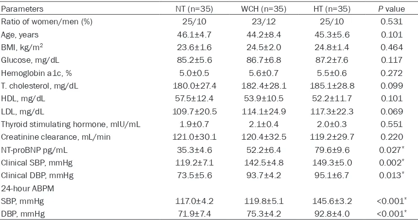
As shown in Table 1, among several clinical parameters compared between the groups, except for BP, N-terminal pro brain natriuretic
peptide (NT-proBNP) was expressed at higher levels in the WCH group than in the NT group (P=0.017) and NT-proBNP was significantly upregulated in the HT group compared to that in the WCH group (P=0.004). This phenomenon was in accordance with a previous finding that NT-proBNP in plasma is a new biomarker that
differentiates WCH from sustained hyperten-Table 1. Demographic characteristics, clinical blood pressure, and abpm of study groups
Parameters NT (n=35) WCH (n=35) HT (n=35) P value
Ratio of women/men (%) 25/10 23/12 25/10 0.531
Age, years 46.1±4.7 44.2±8.4 45.3±5.6 0.101
BMI, kg/m2 23.6±1.6 24.5±2.0 24.8±1.4 0.464
Glucose, mg/dL 85.2±5.6 86.7±6.8 87.2±7.6 0.117
Hemoglobin a1c, % 5.0±0.5 5.6±0.7 5.5±0.6 0.272
T. cholesterol, mg/dL 180.0±27.4 182.4±28.1 185.1±28.8 0.099
HDL, mg/dL 57.5±12.4 53.9±10.5 52.2±11.7 0.101
LDL, mg/dL 109.7±20.5 114.1±24.9 117.3±22.3 0.069
Thyroid stimulating hormone, mIU/mL 1.9±0.7 2.1±0.4 2.0±0.3 0.551
Creatinine clearance, mL/min 121.0±30.1 120.4±32.5 119.2±29.7 0.220
NT-proBNP pg/mL 35.3±4.6 52.2±6.4 79.6±9.6 0.027*
Clinical SBP, mmHg 119.2±7.1 142.5±4.8 149.3±5.0 0.002*
Clinical DBP, mmHg 73.5±5.6 93.7±4.2 95.1±6.7 0.013*
24-hour ABPM
SBP, mmHg 117.0±4.2 119.8±5.1 145.6±3.2 <0.001*
DBP, mmHg 71.9±7.4 75.3±4.2 92.8±4.0 <0.001*
Note: ABPM = ambulatory blood pressure monitoring, BMI = body mass index, HDL = High-density lipoprotein, LDL =
Low-den-sity lipoprotein, NT-proBNP = N-terminal pro brain natriuretic peptide, HT = hypertension, WCH = white coat hypertension, NT =
[image:3.612.92.522.83.309.2]sion [20]. However, there were no statistically
significant differences detected in other select
-ed clinical indexes among the groups. The clini -cal BP of each subject is listed in Table 1. The
results of 24-hour ABPM showed that subjects
in the NT and WCH groups had lower BP than patients in the HT group (P<0.01 for all). LncRNA expression profiles in PMBC of pa -tients with WCH and HT
To obtain an lncRNA signature from the PMBCs of patients with WCH and HT, we performed
microarray analyses of healthy volunteers and
patients with WCH and HT. Interestingly, micro -array data revealed 29 aberrantly expressed lncRNAs among the three study groups. Figure 1 shows seven lncRNAs were downregulated in
the HT group compared with those in other
groups, and 20 lncRNAs were upregulated in
patients with HT compared with those mea -sured in patients with WCH as well as healthy
volunteers (fold change, ≥1.5, P<0.05).
However, only lncRNA H19 and MALAT1 showed high expression in the WCH group and low
expression in the HT and NT groups, thus
prompting us to explore their potential for
dif-ferentiating WCH from HT. Expression levels of
lncRNA H19 and MALAT1 were determined in the PMBCs of 70 patients with elevated clinical BP and 35 healthy individuals by using quanti-tative real-time PCR. In agreement with the microarray data, the relative expression of H19 and MALAT1 was significantly higher in the WCH group than in the NT and HT groups
(P<0.01 for all; Figure 2). The expression level
of H19 was higher in the HT group than in the NT group (P<0.01; Figure 2A), but MALAT1 did
[image:4.612.97.523.68.470.2]not exhibit a significant difference between the HT and NT groups (Fig-ure 2B).
Figure 2. Relative expression levels of H19 (A), and MALAT1 (B) in the PBMC samples of healthy individuals (NT), patients with white-coat hypertension (WCH), and patients with essential hypertension (HT). *P<0.05 versus NT group, **P<0.05 versus HT group, ***P<0.05 NT group versus HT group.
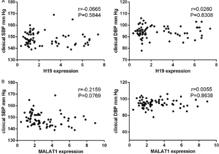
Figure 3. Pearson correlation test demonstrating the relationship between lncRNA H19 (A) and MALAT1 (B) relative expression levels with clinical systolic blood pressure (SBP) and clinical diastolic blood pressure (DBP).
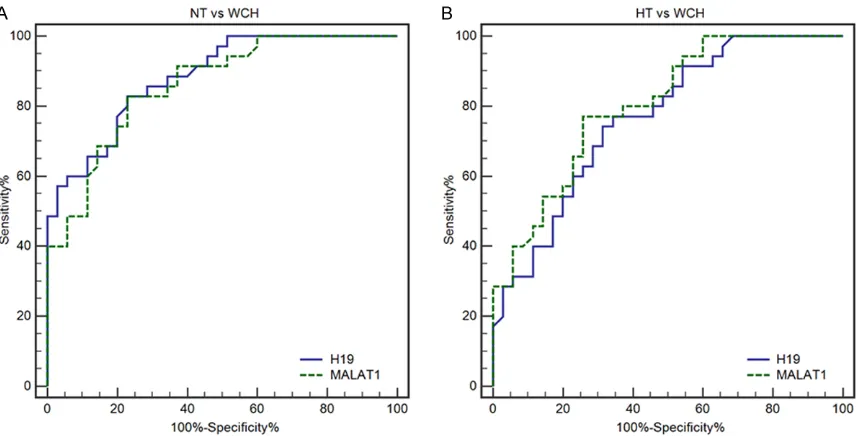
The value of lncRNAs for distinguishing WCH from NT and HT
To explore the diagnostic value of lncRNAs in differentiating patients with WCH from HT or NT
[image:5.612.90.520.322.625.2]Figure 4. Pearson correlation test demonstrating the relationship between lncRNA H19 (A) and MALAT1 (B) rela -tive expression levels with ambulatory 24-hour systolic blood pressure (SBP) and diastolic blood pressure (DBP). P<0.001 for all, r = coefficient of product-moment correlation.
Table 2. Sensitivity, specificity, AUC, cut-off, and asymptomatic signifi
-cance of ROC analysis of lncRNAs
AUC (95% CI) Sensitivity (%) Specificity (%) Cut-off Youden P value
NT vs WCH
H19 0.88 (0.78-0.95) 82.7 77.2 3.00 0.61 <0.001
MALAT1 0.85 (0.76-0.93) 83.0 77.1 2.95 0.60 <0.001 WCH vs HT
H19 0.76 (0.65-0.86) 74.29 68.57 3.52 0.43 <0.001
MALAT1 0.80 (0.69-0.89) 77.14 74.29 3.13 0.52 <0.001
ROC = receiver operating characteristic, AUC = area under the curve, CI = confidence interval, HT = hypertension, NT = normotensive, WCH = white coat hypertension.
SBP (Figure 3B) or DBP (Figure 3B). The ambu -latory 24-hour SBP and DBP were negatively correlated with H19 expression levels (r=-0.7231, P<0.01 and r=-0.5489, P<0.01, respectively; Figure 4A) and with MALAT1 expression levels (r=-0.6084, P<0.01 and r=-0.4376, P<0.01, respectively; Figure 4B).
ROC curves were then constructed and AUC values were generated to assess the power of the lncRNA H19 and MALAT1 for distinguishing
0.86) and 0.80 (0.69-0.89), respectively (Figure 5B). Detailed information on the ability of lncRNA H19 and MALAT1 to differentiate
between patients with or without real HT is pre -sented in Table 2.
Discussion
In this study, we investigated lncRNA expres-sion signature in the PBMCs of patients with
between the NT, WCH and HT groups. As illus -trated in Table 2, lncRNA H19 and MALAT1 had AUC values of 0.88 (0.78-0.95) and 0.85 (0.76-0.93), respectively, which
demonstrates their suffi -ciency in distinguishing
WCH from NT individuals
(Figure 5A). When differ-entiating patients with
WCH from HT, lncRNA
(0.65-sustained hypertension and white-coat
hyper-tension, compared with healthy subjects. The
data revealed that the expression levels of H19 and MALAT1 were significantly increased in subjects with WCH compared to those with NT and HT and further exploration find these two
lncRNAs showed good performance to
distin-guish WCH from HT individuals. To our knowl
-edge, we are the first to report a link between
lncRNA and WCH.
Emerging evidence suggests that lncRNA sig-natures of normal cancer tissues and metasta-ses are used to classify different cancer types, indicating the potential of these lncRNAs as biomarkers for diagnosis, prognosis, and thera-py [21-23]. Recent studies also revealed that lncRNAs are potential biomarkers and thera-peutic targets in cardiovascular disease [24].
The H19 gene, discovered 25 years ago, pro-duces a non-coding RNA, which is abundantly expressed during embryonic development and is down-regulated after birth [25]. A recent study suggested that the physiological role of H19 is to limit growth of the placenta prior to birth by regulating the Insulin-like growth factor 1 receptor through miR-675 [26]. Emerging evi-dence had highlighted the important roles of H19 during the complex process of tumorigen-esis [27]. Meanwhile, it might predict progres-sion and metastasis in cancers, such that the
expression of H19 was associated with
histo-logical grade TNM and tumor invasion depth in
gastric cancer [28]. Furthermore, studies are gradually uncovering the function of H19 in the heart. Liu et al detected the RNA level of H19 and its encoded miR-675 expression level in both normal and diseased hearts with
patho-logical cardiac hypertrophy in mice, and verified
their upregulation in pathological cardiac hyper-trophy and heart failure. Further experiments revealed a novel function of H19-miR-675 axis that targets CaMKIId, a multifunctional serine/ threonine protein kinase mainly found in the heart, as a negative regulator of cardiomyocyte hypertrophy [29]. Greco et al found lncRNA H19
was significantly upregulated in the left ventric -ular biopsies of 18 non-end-stage ischemic dilated cardiomyopathy patients compared
with 17 control subjects, and confirmed its
upregulation in a mouse model of cardiac hypertrophy [30]. In the present study, we determined the upregulation of lncRNA H19 in the PBMCs of patients with increased clinical
BP, and ROC analysis suggested it had enough power to distinguish WCH from HT individuals.
[image:7.612.93.522.70.288.2]Metastasis associated lung adenocarcinoma transcript 1 (MALAT1), which was first associ -ated with metastasis of lung tumors [31], is a typical multifunctional gene that is important in a wide array of cancers [32, 33], and MALAT1 silencing may be an effective therapeutic
Figure 5. Receiver operating characteristic curve analysis demonstrating the diagnostic performance of lncRNA
approach against tumors [34]. A recent study has revealed that MALAT1 deregulation is impli-cated in the pathogenesis of diabetic retinopa-thy and its knockdown could regulate retinal endothelial cell proliferation, migration, and tube formation and ameliorate diabetic retinop-athy in vivo [35]. In addition, Katharina et al elu-cidated that genetic ablation or pharmacologi-cal inhibition of MALAT1 inhibited proliferation of endothelial cells and reduces vascular growth in mice [36]. Furthermore, MALAT1 was
significantly upregulated in human umbilical
vein endothelial cells subjected to transforming growth factor-1 treatment [37]. However, MALAT1 lncRNA has not yet been studied in HT
or WCH. We revealed that circulating MALAT1 expression was higher in patients with WCH
than in NT or HT subjects, and identified its
diagnostic value for differentiating WCH from
NT and HT. To our knowledge, we are the first to
demonstrate deregulated expression of lncRNA H19 and MALAT1 in the PMBCs of patients with elevated BP, and to establish them as novel potential noninvasive biomarkers for
differenti-ating between WCH and HT. This is important
as misdiagnosis of WCH can contribute to inap-propriate anti-hypertensive treatment and adverse events. Furthermore, similar observa-tions have been made for WCH, suggesting a
complementary role of out-of-office and office
BP values in the determination of patients’ prognoses [38]. At present, ABPM, which may accurately grade the severity of hypertension and predict the cardiovascular risk of patients,
is the gold standard in the identification of
WCH. However, the disadvantages of ABPM are its limited availability and discomfort as well as the reluctance of some patients to use it. In summary, our data suggest that lncRNA H19 and MALAT1 could serve as novel noninvasive
biomarkers for distinguishing WCH from HT
individuals, and provide crucial insights into the diagnosis of WCH and could extend the under-standing of the role of lncRNAs in cardiovascu-lar disease. However, this study has limitations. We explored the diagnostic value of lncRNA H19 and MALAT1 in a small cohort and did not
confirm our findings in another group with a
larger number of subjects. In addition, we did not investigate the expression levels of these lncRNAs in samples after anti-hypertensive treatment and compare the differences, which may help to discover more differential lncRNAs
during this process. Thus, further studies are
required to understand the exact mechanisms
underlying these observations in WCH and HT.
Acknowledgements
This study was supported by Gansu
provin-ce natural scienprovin-ce foundation project (No: 1208RJZA110).
Disclosure of conflict of interest
None.
Address correspondence to: Nan Wang, Department of Cardiology, Gansu Provincial Hospital, 204 Dongg- angxilu, Lanzhou 730000, Gansu Province, People’s Republic of China. E-mail: wangnan_gs@126.com
References
[1] Ettehad D, Emdin CA, Kiran A, Anderson SG, Callender T, Emberson J, Chalmers J, Rodgers A and Rahimi K. Blood pressure lowering for prevention of cardiovascular disease and death: A systematic review and meta-analysis. Lancet 2016; 387: 957-967.
[2] Perlini S and Grassi G. Hypertension-related target organ damage: Is it a continuum? J Hy-pertens 2013; 31: 1083-1085.
[3] Harbaoui B, Courand PY, Defforges A, Khettab F, Milon H, Girerd N and Lantelme P. Cumula-tive effects of several target organ damages in risk assessment in hypertension. Am J Hyper-tens 2016; 29: 234-244.
[4] Blacher J, Halimi JM, Hanon O, Mourad JJ, Pathak A, Schnebert B, Girerd X; Société fran-çaise d’hypertension artérielle. [Management of arterial hypertension in adults: 2013 guide-lines of the French Society of Arterial Hyperten-sion]. Presse Med 2013; 42: 819-825. [5] Mancia G, Bombelli M, Seravalle G and Grassi
G. Diagnosis and management of patients with white-coat and masked hypertension. Nat Rev Cardiol 2011; 8: 686-693.
[6] Yavuzer S, Yavuzer H, Cengiz M, Erman H, Al-tiparmak MR, Korkmazer B, Balci H, Simsek G, Yaldiran AL, Karter Y and Uzun H. Endothelial damage in white coat hypertension: Role of lectin-like oxidized low-density lipoprotein-1. J Hum Hypertens 2015; 29: 92-98.
car-diovascular outcome. Hypertension 2014; 63: 675-682.
[8] Mancia G. Clinical significance of white-coat hypertension. J Hypertens 2016; 34: 623-626. [9] Chen YA and Aravin AA. Non-coding RNAs in
transcriptional regulation: the review for cur-rent molecular biology reports. Curr Mol Biol Rep 2015; 1: 10-18.
[10] Fatica A and Bozzoni I. Long non-coding RNAs: New players in cell differentiation and develop-ment. Nat Rev Genet 2014; 15: 7-21.
[11] Bouckenheimer J, Assou S, Riquier S, Hou C, Philippe N, Sansac C, Lavabre-Bertrand T, Commes T, Lemaitre JM, Boureux A and De Vos J. Long non-coding RNAs in human early em-bryonic development and their potential in ART. Hum Reprod Update 2016; 23: 19-40. [12] Ulitsky I and Bartel DP. lincRNAs: Genomics,
evolution, and mechanisms. Cell 2013; 154: 26-46.
[13] Jiang X and Ning Q. The emerging roles of long noncoding RNAs in common cardiovascular diseases. Hypertens Res 2015; 38: 375-379. [14] Visel A, Zhu Y, May D, Afzal V, Gong E, Attanasio
C, Blow MJ, Cohen JC, Rubin EM and Pennac-chio LA. Targeted deletion of the 9p21 non-coding coronary artery disease risk interval in mice. Nature 2010; 464: 409-412.
[15] Matkovich SJ, Edwards JR, Grossenheider TC, de Guzman Strong C and Dorn GW 2nd. Epi-genetic coordination of embryonic heart tran-scription by dynamically regulated long non-coding RNAs. Proc Natl Acad Sci U S A 2014; 111: 12264-12269.
[16] Kumarswamy R, Bauters C, Volkmann I, Maury F, Fetisch J, Holzmann A, Lemesle G, de Groote P, Pinet F and Thum T. Circulating long noncod -ing RNA, LIPCAR, predicts survival in patients with heart failure. Circ Res 2014; 114: 1569-1575.
[17] Yan Y, Zhang B, Liu N, Qi C, Xiao Y, Tian X, Li T and Liu B. Circulating long noncoding RNA UCA1 as a novel biomarker of acute myocardial infarction. Biomed Res Int 2016; 2016: 8079372.
[18] Shimbo D, Abdalla M, Falzon L, Townsend RR and Muntner P. Role of ambulatory and home blood pressure monitoring in clinical practice: a narrative review. Ann Intern Med 2015; 163: 691-700.
[19] Parati G, Stergiou G, O’Brien E, Asmar R, Beilin L, Bilo G, Clement D, de la Sierra A, de Leeuw P, Dolan E, Fagard R, Graves J, Head GA, Imai Y, Kario K, Lurbe E, Mallion JM, Mancia G, Meng-den T, Myers M, Ogedegbe G, Ohkubo T, Om -boni S, Palatini P, Redon J, Ruilope LM, Shen-nan A, Staessen JA, vanMontfrans G, Verdecchia P, Waeber B, Wang J, Zanchetti A, Zhang Y; European Society of Hypertension
Working Group on Blood Pressure Monitoring and Cardiovascular Variability. European Soci-ety of Hypertension practice guidelines for am-bulatory blood pressure monitoring. J Hyper-tens 2014; 32: 1359-1366.
[20] Courand PY, Harbaoui B, Serraille M, Berge C and Lantelme P. Ruling out white coat hyper-tension with NT-proBNP: A new paradigm away from blood pressure assessment. Int J Cardiol 2016; 207: 57-58.
[21] Meng J, Li P, Zhang Q, Yang Z and Fu S. A four-long non-coding RNA signature in predicting breast cancer survival. J Exp Clin Cancer Res 2014; 33: 84.
[22] Chen H, Xu J, Hong J, Tang R, Zhang X and Fang JY. Long noncoding RNA profiles identify five distinct molecular subtypes of colorectal can-cer with clinical relevance. Mol Oncol 2014; 8: 1393-1403.
[23] Zhou M, Zhong L, Xu W, Sun Y, Zhang Z, Zhao H, Yang L and Sun J. Discovery of potential prognostic long non-coding RNA biomarkers for predicting the risk of tumor recurrence of breast cancer patients. Sci Rep 2016; 6: 31038.
[24] Bar C, Chatterjee S and Thum T. Long noncod -ing RNAs in cardiovascular pathology, diagno-sis, and therapy. Circulation 2016; 134: 1484-1499.
[25] Gabory A, Jammes H and Dandolo L. The H19 locus: Role of an imprinted non-coding RNA in growth and development. Bioessays 2010; 32: 473-480.
[26] Keniry A, Oxley D, Monnier P, Kyba M, Dandolo L, Smits G and Reik W. The H19 lincRNA is a developmental reservoir of miR-675 that sup-presses growth and Igf1r. Nat Cell Biol 2012; 14: 659-665.
[27] Raveh E, Matouk IJ, Gilon M and Hochberg A. The H19 long non-coding RNA in cancer initia -tion, progression and metastasis-a proposed unifying theory. Mol Cancer 2015; 14: 184. [28] Jing W, Zhu M, Zhang XW, Pan ZY, Gao SS,
Zhou H, Qiu SL, Liang CZ and Tu JC. The signifi -cance of long noncoding RNA H19 in predict-ing progression and metastasis of cancers: a meta-analysis. Biomed Res Int 2016; 2016: 5902678.
[29] Liu L, An X, Li Z, Song Y, Li L, Zuo S, Liu N, Yang G, Wang H, Cheng X, Zhang Y, Yang X and Wang J. The H19 long noncoding RNA is a novel neg -ative regulator of cardiomyocyte hypertrophy. Cardiovasc Res 2016; 111: 56-65.
[31] Ji P, Diederichs S, Wang W, Boing S, Metzger R, Schneider PM, Tidow N, Brandt B, Buerger H, Bulk E, Thomas M, Berdel WE, Serve H and Muller-Tidow C. MALAT-1, a novel noncoding RNA, and thymosin beta4 predict metastasis and survival in early-stage non-small cell lung cancer. Oncogene 2003; 22: 8031-8041. [32] Wu XS, Wang XA, Wu WG, Hu YP, Li ML, Ding Q,
Weng H, Shu YJ, Liu TY, Jiang L, Cao Y, Bao RF, Mu JS, Tan ZJ, Tao F and Liu YB. MALAT1 pro -motes the proliferation and metastasis of gall-bladder cancer cells by activating the ERK/ MAPK pathway. Cancer Biol Ther 2014; 15: 806-814.
[33] Ying L, Chen Q, Wang Y, Zhou Z, Huang Y and Qiu F. Upregulated MALAT-1 contributes to bladder cancer cell migration by inducing epi-thelial-to-mesenchymal transition. Mol Biosyst 2012; 8: 2289-2294.
[34] Ren D, Li H, Li R, Sun J, Guo P, Han H, Yang Y and Li J. Novel insight into MALAT-1 in cancer: Therapeutic targets and clinical applications. Oncol Lett 2016; 11: 1621-1630.
[35] Liu JY, Yao J, Li XM, Song YC, Wang XQ, Li YJ, Yan B and Jiang Q. Pathogenic role of lncRNA-MALAT1 in endothelial cell dysfunction in dia -betes mellitus. Cell Death Dis 2014; 5: e1506. [36] Michalik KM, You X, Manavski Y, Doddaballa-pur A, Zornig M, Braun T, John D, Ponomareva Y, Chen W, Uchida S, Boon RA and Dimmeler S. Long noncoding RNA MALAT1 regulates endo -thelial cell function and vessel growth. Circ Res 2014; 114: 1389-1397.
[37] Singh KK, Matkar PN, Quan A, Mantella LE, Teoh H, Al-Omran M and Verma S. Investigation of TGFbeta1-Induced long noncoding RNAs in endothelial cells. Int J Vasc Med 2016; 2016: 2459687.