Original Article
Characterization of the N-terminally clipped histone H3
(∆H3) from old chicken and rat liver
Neetika Singh1, Jogeswar S Purohit1,2, S Shanti1, Abhilasha Singh1, Anil K Panigrahi1,3, Madan M Chaturvedi1
1Laboratory for Chromatin Biology, Department of Zoology, University of Delhi, North Campus, Delhi 110007,
India; 2Cluster Innovation Centre, G. C. Narang Marg, University of Delhi, North Campus, Delhi 110007, India; 3Present Address: Department of Pediatric Hematology/Oncology, Texas Children’s Cancer Center, Baylor College
of Medicine, Houston, TX 77030, USA
Received December 27, 2016; Accepted February 14, 2017; Epub May 1, 2017; Published May 15, 2017
Abstract: Site-specific proteolysis of the N- or C- terminus of histone tails has recently emerged as a novel form of irreversible post-translational modifications of histones. A proteolytically cleaved H3 product (named as ∆H3) has recently been reported in the liver of old chicken. In the present study, a comparison of the N-terminal amino acid sequences of the H3 and the ∆H3 revealed that the ∆H3 lacked 23 amino acids from the N-terminus. The proteoly
-sis was observed only in histone H3 in the liver of old chicken, and it was absent in brain and erythrocytes. A ∆H3 like product was also observed in the liver of old rats. Further, the ∆H3 could be probed with H3 specific N-terminal pan acetylated antibody, suggesting that the chromatin domains containing the ∆H3 would be transcriptionally repressive. However, the ∆H3 could be probed with anti-H3K36me2 antibody (broadly a mark of transcriptionally permissive chromatin), suggesting that the ∆H3 though lacked major portion of the N-terminal region, still be impor
-tant in determining the transcriptional status of the chromatin. Therefore, a comparison of the existing epigenetic marks on H3 and ∆H3 is also tabulated in the present study. To ascertain whether the cleavage is dependent on post-translational modifications of histone H3, bacterially expressed recombinant H3 was used as substrate for in vitro cleavage reaction. The analyses of the products revealed that the major cleaved product was different from that of the ∆H3, suggesting that the in vivo generation of ∆H3 requires histone H3 to be chromatin-bound.
Keywords: Histone H3, histone H3 proteolysis, ∆H3, epigenetic marks
Introduction
The N- and the C- terminal tails of core histones are subjected to a diverse array of reversible post-translational modifications. Some of the widely-studied modifications are methylation, acetylation, phosphorylation, ubiquitylation and ADP-ribosylation etc [1]. In addition to these reversible modifications, histones also undergo irreversible modifications by proteoly -sis of the N- or the C- terminal of histone tails, which also have been correlated to epigenetic regulation [2-9].
Proteolysis of the N- or the C- terminal histone tails have been reported in Tetrahymena [2], mammalian kidney cells (BHK cells) infected with Foot and Mouth Disease Virus (FMDV) [10, 11], cycad pollen [12], rat uterus [13], yeast
[14, 15], mouse embryonic stem cells [16], human embryonic stem cells [17], calf thymus [18], monocytes [19] and chicken liver [20]. A recent attempt has been made to divide his-tone proteolysis into hishis-tone degradation and epigenetically inferential tail clipping [21].
Site specific histone H3 proteolysis
above cleavage sites revealed that the mapped cleavage site (i.e., R26/K27) did not coincide with any of the in vitro cleavage sites by GDH [22]. Hence, it was essential to determine the exact site of cleavage on histone H3, and also to explore a possible fate of the cleaved prod -uct. In the present study, by comparing the N-terminal sequences of the H3 and the ∆H3 we identify that the ∆H3 lacks 23 amino acid residues from N-terminal region. Further, a ∆H3 like product was also observed in the liver of old rat while, it was absent in the erythrocytes and brain of the old chicken. To understand whether the cleavage would occur in a modifi -cation free histone H3, bacterially expressed histone was used to assess the cleavage in his-tones devoid of any post-translational modifica -tions. The results suggested that the in vivo
generation of the ∆H3 would require histone H3 to be chromatin-bound.
Materials & methods
Biological materials
White leg horn chicken, Wistar strain albino rats and Parks strain mice were used for the experiments. Rats and mice were of the age groups of ~18 weeks (termed as young) and ~108 weeks (termed as old). Tissues from young (~4 weeks) and old (~24 weeks) chicken were procured from slaughter house. Tissues from these organisms were collected following the guidelines of the departmental animal ethi -cal committee and stored following standard methods [22], until used.
Isolation of nuclei
The nuclei from liver and brain tissues of chick -en and liver of rat and mouse were isolated as previously described [25] with minor modifica -tions [26] and from chicken erythrocytes follow -ing the standard methods [22]. The purity of the nuclei was verified under a phase contrast microscope at each step of nuclei isolation. Nuclei were estimated following already estab -lished methods [27].
Isolation of histones
Histones were extracted from the purified nuclei by acid extraction method [28]. Extracted histones were quantified as previously des-cribed [27]. The histones were analysed on a high resolution SDS-18% PAGE as described [29] with minor modifications [30].
Western blotting
Western Blotting was performed following the standard protocol [31]. Anti-H3 (against full length H3), anti-acetylated anti-H3 (corre -sponding to N-terminal 20 amino acids with Kac, kind gifts from Dr. Sharon Y. Roth, M.D. An-derson Cancer Centre, USA) and H3K36-me2 (Abcam, USA) were used as primary anti-bodies. Signals of the western blots were devel -oped following standard methods [31].
N-terminal sequencing of H3 and ∆H3
Chicken liver total histones were separated on SDS-18% PAGE, transferred onto a PVDF mem -brane in modified transfer buffer (10% metha -nol, 95 mM glycine, 12.5 mM Tris base), and, stained with 0.5% Ponceau S. The H3 and the ∆H3 bands were excised out and sequenced commercially from Department of Chemistry, IIT Mumbai, India. The procedure used a gas phase automated protein sequencer from Shimadzu model PPSQ10. The first 6 amino acids sequences from the N-terminus for both H3 and ∆H3 were aligned manually to identify the cleavage site in H3.
Histone isolation from H1 depleted soluble chromatin of rat liver
H1 depleted soluble chromatin was prepared from nuclei of liver of old rat by standard meth -od [32]. Histones were isolated from the solu -ble chromatin as reported earlier [32].
Purification of histone H3 expressed in bacte -ria
A recombinant Xenopus histone H3 cloned in pET 3a was received as a kind gift from Dr. Tony Kouzarides, University of Cambridge, UK. This plasmid was transformed and histone H3 was expressed in E. coli BL21 (DE3) pLys S strain following standard methods [22]. The histone H3 expressed in bacteria was purified following standard methods [22].
H3 cleavage assay
SDS-18% PAGE and staining with coomassie brilliant blue R-250 [22].
Results
The ∆H3 is an N-terminally 23 amino acids
cleaved product of H3
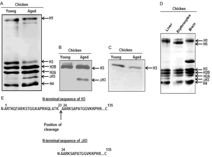
Total histones isolated from liver of young and old chicken were analysed on SDS-PAGE. A faster migrating extra band was observed in the histones isolated from liver of old chicken, with a corresponding reduction in H3 (Figure 1A). The extra band was immunoreactive to anti-H3 antibody (Figure 1B); however, it was not recognized by anti-acetylated H3 antibody (Figure 1C). All these findings suggested that the extra band was indeed the ∆H3, which has
observed that the ∆H3 was absent in the his -tone preparations from brain and erythrocytes of old chicken (Figure 1D). The first 6 amino acids from N-terminus for the H3 and the ∆H3 were identified as ‘ARTKQT’ and ‘AARKSA’, respectively. Both the sequences were aligned manually to the available sequence of H3 to deuce the cleavage site. It revealed that the ∆H3 lacked 23 amino acids from the N-terminus, indicating that the cleavage site was at K23/ A24 of H3 (Figure 1E).
A ∆H3 like product is also observed in liver of
old rat
Histone preparations from liver of young as well as old rat were analysed to explore the
[image:3.612.91.523.71.390.2]Site specific histone H3 proteolysis
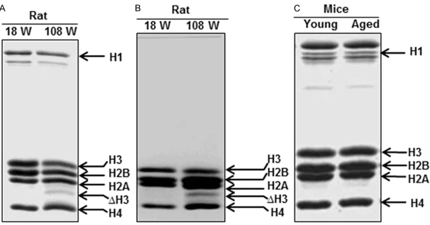
Figure 2. A ∆H3 like product is also generated in liver of old rat. A. Total histones were isolated from the nuclei of
young (18 weeks) and old (108 weeks) rat liver and resolved on SDS-PAGE and stained with 0.1% coomassie blue.
B. Histones prepared from H1 depleted soluble chromatin from the nuclei of liver of young and old rat were resolved on SDS-PAGE and stained as above. C. Total histones from liver of young and old mice were resolved on SDS-PAGE
and stained as above.
Figure 3. The ∆H3 immunoreacts to anti-H3K36me2 antibody similar to H3. (A) Total histones isolated from nuclei of liver of old chicken (CLH), erythrocytes of old chicken (CEH) and liver of old mice (MLH) were analysed by SDS-PAGE and stained with 0.1% coomassie blue. (B) Resolved histones as described in (A) were transferred on to a nitrocellulose membrane and probed with anti-H3antibody and processed by enhanced chemiluminescence. (C)
Resolved histones as described in (A) were transferred on to a nitrocellulose membrane and probed with an anti-H3 antibody specific for dimethylated Lys 36 (K36me2)antibody.
Table 1. Status of various global histone modifications on H3 and ∆H3 Mod
Histone K4me K4me2 K9me K9me2 K9ac K14ac K23ac K27ac K27me2 K56ac
H3 + + + + + + + + + +
∆H3 - - - + +
[image:4.612.93.522.488.635.2]∆H3 like product was also found in the histones isolated from liver of old rat, while it was absent in that of the young (Figure 2A). It was next sought to investigate, whether the ∆H3 like
[image:5.612.92.515.68.262.2]product was chromatin bound in the liver of old rat. Soluble chromatin was prepared from nuclei of liver of young and old rat. Histones were isolated from these soluble chromatin Figure 4. Assessment of cleavage in the post-translational modification free histone H3. The H3 cleavage assay was performed by incubating bacterially expressed histone H3 with GDH in the standard assay condition already developed and subsequently analysed on SDS-PAGE. A. 2 µg of bacterially expressed histones were incubated with 1 µg of chicken liver mitochondrial GDH for different time duration (5, 15, 30, 45, 60, 90, 120 and 180 min). B. 2 µg of bacterially expressed histones were incubated with different amount of GDH (0.1, 0.5, 1 and 2 µg) for 1 h. C. N-terminal sequences of the cleaved products of modification free H3.
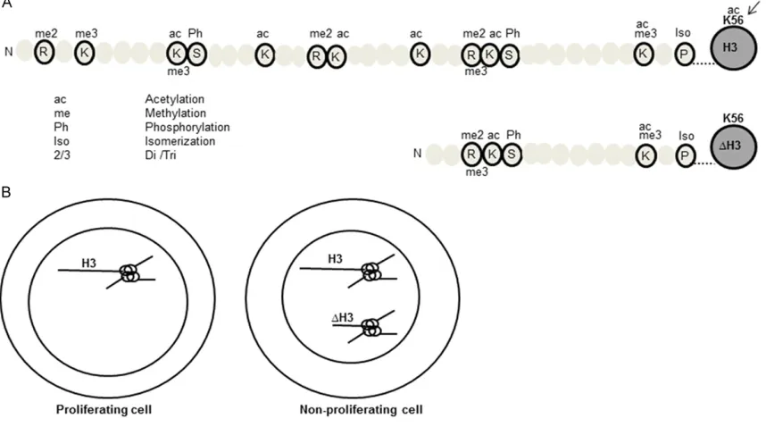
Figure 5. A plausible model to depict the distribution of ∆H3. A. Post-translational modification map of histone H3 and ∆H3. Different modifications are represented by corresponding abbreviations. In general, the activation marks are on the upper side of the peptide and the repressive marks are on the lower side of the peptide. ‘Arrow’ indicates selective presence of K56ac mark in H3. B. A plausible model representing the distribution of H3 and ∆H3 in the
[image:5.612.92.521.356.595.2]Site specific histone H3 proteolysis
fractions and were analysed on SDS-PAGE. A ∆H3 like product was also observed in the his -tones isolated from these soluble chromatin fractions (Figure 2B). This result further con -firmed that the ∆H3 like product was a part of the chromatin and was not produced due to some cytoplasmic contamination during isola-tion. Surprisingly, a ∆H3 like product was absent in histones isolated from liver of old mice (Figure 2C). It would be interesting to investigate histone H3 proteolysis in other organisms, to understand its physiological role.
Evaluation of epigenetic marks on the ∆H3
There have been evidences supporting the role of N-terminal tail clipping of H3 in epigenetic reprogramming [21]. The downstream effect of the proteolysis can be broadly categorized as either formation of transcriptionally permissive or repressive chromatin domains. Since the ∆H3 lacked 23 amino acids from N-terminus, it was intended to evaluate the differences in the epigenetic marks on H3 and ∆H3. A summary of the comparative list of existing modifications on H3 and the ∆H3 is represented in Table 1. From the table, it is evident that the ∆H3 was immunoreactive to anti-H3K27ac antibody, whi-ch is a mark of transcriptionally active whi-chroma -tin [33]. It was thus suggested that the ∆H3 might not be a part of repressive chromatin. Therefore, presence of any other epigenetic mark(s) for transcriptionally permissive chro -matin (such as H3K36me2) was explored in ∆H3. Since the chicken erythrocyte is classi -cally known to possess transcriptionally repre- ssed chromatin [34], chicken erythrocyte tones were used in the present study as his-tones possessing repressive marks. Total his -tones from liver and erythrocytes of old chicken and liver of old mice were isolated and resolved on SDS-PAGE (Figure 3A) and probed with anti-H3 (Figure 3B) and anti-anti-H3K36me2 antibodies (Figure 3C). The ∆H3 was immunoreactive to anti-H3K36me2 antibody, suggesting that it might form a part of transcriptionally permis -sive chromatin. This observation was in confor -mity with the other permissive modification, such as H3K27ac on ∆H3 (Table 1). Though it was difficult to establish a precursor product relation by comparing the anti-H3K36me2 sig -nals of H3 and ∆H3; an increased signal of the same in ∆H3 compared to the H3 of chicken erythrocytes suggested that ∆H3 might be a part of transcriptionally permissive chromatin domains.
Assessment of cleavage in post-translational
modification free histone H3
It was intended to evaluate whether post-trans-lational modifications in H3 were essential for its cleavage. As a bacterially expressed histone H3 would be devoid of any post-translational modification; it was used as a substrate to study the H3 cleavage. Bacterially expressed histone H3 was incubated with chicken liver mitochondrial GDH (demonstrated earlier to have H3 specific protease activity) for increas -ing time durations (Figure 4A) or with increas-ing amount of GDH (Figure 4B). The results indi -cated that the major cleavage of the bacterially expressed histone H3 occurred at the 8th amino acid residue (Figure 4C), and which was differ -ent from the chromatin bound H3, where, the cleavage site was only specific to 23rd amino acid residue.
Discussion
While there are many reports on specific prote -olysis of histone H3 [6, 21]; Cathepsin L and GDH are the only two H3 specific proteases characterized till date [16, 23]. In the in vitro
reaction, chicken liver GDH cleaves histone H3 at multiple sites spanning in the N-terminal region. The results of the present study indi -cate that the identified in vivo cleavage site in histone H3 (K23/A24), coincided with one of such in vitro cleavage sites by GDH. It proposes a strong argument for the in vivo role of GDH in clipping H3 and generating the ∆H3. The above hypothesis is also supported by the observa-tion that in old chicken, there in increased his-tone H3 cleavage and also increased nuclear localization of GDH [35]. On the contrary, the increased nuclear localization was not observed in chicken brain tissue where the ∆H3 was absent [35]. Further, GDH is reported to be excluded from the nuclei and localize in the cytoplasm and mitochondria of Drosophila
[36]. We also failed to detect a ∆H3 like product in the histones from Drosophila (Tiwari and Purohit, unpublished observation). All these observations support the above hypothesized role of GDH in H3 clipping in vivo. However, the confirmation of the same anticipates further investigations, which are underway.
observed in rat liver (present study) and Japanese quail [37]. On the other hand, a ∆H3 like product was not found in the liver of old mice (present study), and in bovine and Calottes
liver, and HeLa cells (data not shown).
Evaluation of epigenetic signatures on the ∆H3 revealed that it lacked most of the acetylation sites in comparison to H3. However, the ∆H3 possessed both activation marks (K27ac, K36me2) and repression marks (K27me2). The above proposed chromatin dynamicity is also well supported by the predominant role of H3K36me2 in transcriptional activation and minor role in repression in various organisms [38-42]. Also, from the present study, an in-creased K36me2 signal in the ∆H3 as com -pared to chicken erythrocyte histone H3, sug -gests that the ∆H3 containing chromatin do-mains might not exist in a transcriptionally repressed state. Furthermore, it has been re- ported that the ∆H3 could also be probed in comparable intensities in S1, S2 and P frac -tions in MNase digested nuclei from liver of old chicken, suggesting that the ∆H3 might form a part of transcriptionally active chromatin [35]. In summary, we suggest that similar to mouse embryonic stem cell differentiation and yeast models [14, 16], the ∆H3 generation also leads to formation of a more dynamic chromatin domain.
A careful comparison of the acetylation marks on H3 and ∆H3 (Table 1) indicates that in con-trast to H3, the ∆H3 lacked K56ac mark (which peaks at S-phase) [43]. The absence of K56ac mark in ∆H3 was probably due to the lack of N-terminal binding site for Rtt 109 (a H3K56 acetyl transferase) [35]. The absence of H3-K56ac mark has been reported to direct chro-matin in an assembled state [44]. In addition, it limits the entry of the cell into S phase. Hence, we propose that such an epigenetically con-noted tail clipping might lead to selective local-ization of the ∆H3 (Figure 5).
A comparison of the in vivo H3 cleavage and the in vitro H3 cleavage reveal that the predom -inant H3 product in the in vitro reaction lacked 8 amino acids from the N-terminus due to cleavage at 8th amino acid residue. On the con-trary, in case of ∆H3, there was a cleavage at 23rd amino acid residue of H3, produced in vivo. These findings suggested that the specific cleavage to produce ∆H3 was probably
fav-oured when H3 remained in a chromatin bound state.
Collectively, the results presented here demon-strate that the ∆H3 is 23 amino acids clipped product of H3. The ∆H3 was found to be pres -ent in the histone preparations from liver of old chicken and it was absent in brain and erythro-cytes. A ∆H3 like product was also observed in liver of old rat. However, detailed investigations need to be done in various organisms to under-stand the physiological role of histone H3 pro -teolysis. We hypothesize that the selective epi-genetic marks on H3 and ∆H3 might be responsible for differential regulation of chro -matin domains. Also, the conformation of the N-terminal tail in chromatin bound histone H3 might determine its specific cleavage.
Acknowledgements
We thank Dr. Sharon Y. Roth-Dent, M.D. An-derson Cancer Center, USA for her generous gifts in the form of anti-H3 and anti-acetylated H3 antibodies, which were used for western blotting. We would also like to thank Dr. Tony Kouzarides for the kind gift of recombinant his -tone H3 clone. Dr. R.S. Tomar is also duly acknowledged for the initial experiments during his Ph.D. tenure. This research work was sup -ported by direct grant and purse grant from Department of Science and Technology (DST), Government of India, New Delhi to MMC; R&D grants from University of Delhi to JSP and MMC. Research fellowships from Council of Scientific and Industrial Research (CSIR) and University Grants Commission (UGC) are also duly acknowledged.
Disclosure of conflict of interest
None.
Address correspondence to: Dr. Madan M Chatur-
vedi, Laboratory for Chromatin Biology, Department of Zoology, North Campus, Delhi University, Room
No. 317, 2nd Floor, Delhi 110007, India. Tel: +91-11-27666051; Fax: +91-11-+91-11-27666051; E-mail: mcha- turvedi@zoology.du.ac.in
References
[1] Bannister AJ and Kouzarides T. Regulation of chromatin by histone modifications. Cell Res
2011; 21: 381-395.
[2] Allis CD, Bowen JK, Abraham GN, Glover CV
-Site specific histone H3 proteolysis
tone H3 in chromatin: a physiologically regu
-lated event in Tetrahymena micronuclei. Cell
1980; 20: 55-64.
[3] Nathan D, Sterner DE and Berger SL. Histone modifications: Now summoning sumoylation.
Proc Natl Acad Sci U S A 2003; 100: 13118-13120.
[4] Kouzarides T. Chromatin modifications and their function. Cell 2007; 128: 693-705.
[5] Zhang K, Chen Y, Zhang Z and Zhao Y. Identifi
-cation and verifi-cation of lysine propionylation
and butyrylation in yeast core histones using
PTMap software. J Proteome Res 2009; 8:
900-906.
[6] Purohit JS, Chaturvedi MM, Panda P. Histone proteases: the tale of tail clippers. Internation
-al Journ-al of Integrative Sciences, Innovation and Technology 2012; 1.
[7] Zhou P, Wu E, Alam HB and Li Y. Histone cleav
-age as a mechanism for epigenetic regulation:
current insights and perspectives. Curr Mol Med 2014; 14: 1164-1172.
[8] Sadakierska-Chudy A and Filip M. A
compre-hensive view of the epigenetic landscape. Part II: Histone post-translational modification, nu -cleosome level, and chromatin regulation by ncRNAs. Neurotox Res 2015; 27: 172-197. [9] Purohit JS and Chaturvedi MM. Chromatin and
aging topics in biomedical gerontology Spring-er press Singapore 2017; 205-241.
[10] Grigera PR and Tisminetzky SG. Histone H3 modification in BHK cells infected with
foot-and-mouth disease virus. Virology 1984; 136: 10-19.
[11] Tesar M and Marquardt O. Foot-and-mouth dis -ease virus prot-ease 3C inhibits cellular
tran-scription and mediates cleavage of histone H3. Virology 1990; 174: 364-374.
[12] Brandt WF, Bohm L and Von Holt C. Proteolytic degradation of histones and site of cleavage in
histone F2al and F3. FEBS Lett 1975; 51: 88-93.
[13] Levitz M, Katz J, Krone P, Prochoroff NN and Troll W. Hormonal influences on histones and
template activity in the rat uterus. Endocrinol-ogy 1974; 94: 633-640.
[14] Santos-Rosa H, Kirmizis A, Nelson C, Bartke T, Saksouk N, Cote J and Kouzarides T. Histone H3 tail clipping regulates gene expression. Nat
Struct Mol Biol 2009; 16: 17-22.
[15] Azad GK and Tomar RS. Partial purification of histone H3 proteolytic activity from the bud
-ding yeast Saccharomyces cerevisiae. Yeast
2016; 33: 217-226.
[16] Duncan EM, Muratore-Schroeder TL, Cook RG, Garcia BA, Shabanowitz J, Hunt DF and Allis
CD. Cathepsin L proteolytically processes
his-tone H3 during mouse embryonic stem cell dif
-ferentiation. Cell 2008; 135: 284-294.
[17] Vossaert L, Meert P, Scheerlinck E, Glibert P,
Van Roy N, Heindryckx B, De Sutter P, Dhae
-nens M and Deforce D. Identification of histone H3 clipping activity in human embryonic stem
cells. Stem Cell Res 2014; 13: 123-134. [18] Eickbush TH, Watson DK and Moudrianakis
EN. A chromatin-bound proteolytic activity with
unique specificity for histone H2A. Cell 1976;
9: 785-792.
[19] Minami J, Takada K, Aoki K, Shimada Y, Okawa Y, Usui N and Ohkawa K. Purification and char
-acterization of C-terminal truncated forms of histone H2A in monocytic THP-1 cells. Int J Bio -chem Cell Biol 2007; 39: 171-180.
[20] Panda P, Chaturvedi MM, Panda AK, Suar M
and Purohit JS. Purification and characteriza
-tion of a novel histone H2A specific protease (H2Asp) from chicken liver nuclear extract.
Gene 2013; 512: 47-54.
[21] Dhaenens M, Glibert P, Meert P, Vossaert L
and Deforce D. Histone proteolysis: a proposal for categorization into ‘clipping’ and ‘degrada
-tion’. Bioessays 2015; 37: 70-79.
[22] Purohit JS, Tomar RS, Panigrahi AK, Pandey
SM, Singh D and Chaturvedi MM. Chicken liver
glutamate dehydrogenase (GDH) demon
-strates a histone H3 specific protease (H3ase)
activity in vitro. Biochimie 2013; 95: 1999-2009.
[23] Mandal P, Verma N, Chauhan S and Tomar RS. Unexpected histone H3 tail-clipping activity of
glutamate dehydrogenase. J Biol Chem 2013; 288: 18743-18757.
[24] Mandal P, Azad GK and Tomar RS. Identifica
-tion of a novel histone H3 specific protease activity in nuclei of chicken liver. Biochem Bio -phys Res Commun 2012; 421: 261-267. [25] Hewish DR and Burgoyne LA. Chromatin
sub-structure. The digestion of chromatin DNA at
regularly spaced sites by a nuclear deoxyribo-nuclease. Biochem Biophys Res Commun 1973; 52: 504-510.
[26] Panda P, Suar M, Singh D, Pandey SM, Chaturvedi MM and Purohit JS.
Characteriza-tion of nuclear glutamate dehydrogenase of
chicken liver and brain. Protein Pept Lett 2011; 18: 1194-1203.
[27] Sollner-Webb B, Camerini-Otero RD and
Felsenfeld G. Chromatin structure as probed by nucleases and proteases: evidence for the central role of histones H3 and H4. Cell 1976;
9: 179-193.
[28] Hoffmann P and Chalkley R. Procedures for minimizing protease activity during isolation of
nuclei, chromatin, and the histones. Methods Cell Biol 1978; 17: 1-12.
[29] Laemmli UK. Cleavage of structural proteins during the assembly of the head of bacterio
[30] Thomas JO and Kornberg RD. The study of
histone--histone associations by chemical cross-linking. Methods Cell Biol 1978; 18: 429-440.
[31] Edmondson DG, Smith MM, Roth SY. Repres
-sion domain of the yeast global repressor Tup1 interacts directly with histones H3 and H4.
Genes Dev 1996; 10: 1247-59.
[32] Panigrahi AK, Tomar RS and Chaturvedi MM. A SWI/SNF-like factor from chicken liver that dis
-rupts nucleosomes and transfers histone oc -tamers in cis and trans. Arch Biochem Biophys 2003; 414: 24-33.
[33] Creyghton MP, Cheng AW, Welstead GG,
Koois-tra T, Carey BW, Steine EJ, Hanna J, Lodato MA, Frampton GM, Sharp PA, Boyer LA, Young RA and Jaenisch R. Histone H3K27ac separates active from poised enhancers and predicts de -velopmental state. Proc Natl Acad Sci U S A 2010; 107: 21931-21936.
[34] Jahan S, Xu W, He S, Gonzalez C, Delcuve GP and Davie JR. The chicken erythrocyte epig -enome. Epigenetics Chromatin 2016; 9: 19. [35] Mandal P, Chauhan S and Tomar RS. H3 clip
-ping activity of glutamate dehydrogenase is regulated by stefin B and chromatin structure.
FEBS J 2014; 281: 5292-5308.
[36] Tiwari AK, Panda P and Purohit JS. Evaluation of sub-cellular distribution of glutamate dehy
-drogenase (GDH) in Drosophila melanogaster larvae. Acta Histochem 2014; 116: 297-303.
[37] Mahendra G and Kanungo MS. Age-related
and steroid induced changes in the histones of
the quail liver. Arch Gerontol Geriatr 2000; 30: 109-114.
[38] Bannister AJ, Schneider R, Myers FA, Thorne AW, Crane-Robinson C and Kouzarides T. Spa
-tial distribution of di- and tri-methyl lysine 36 of histone H3 at active genes. J Biol Chem 2005;
280: 17732-17736.
[39] Rao B, Shibata Y, Strahl BD and Lieb JD. Di
-methylation of histone H3 at lysine 36 demar -cates regulatory and nonregulatory chromatin genome-wide. Mol Cell Biol 2005; 25: 9447-9459.
[40] Karmodiya K, Pradhan SJ, Joshi B, Jangid R, Reddy PC and Galande S. A comprehensive
epigenome map of Plasmodium falciparum re
-veals unique mechanisms of transcriptional regulation and identifies H3K36me2 as a glob
-al mark of gene suppression. Epigenetics
Chromatin 2015; 8: 32.
[41] Li B, Jackson J, Simon MD, Fleharty B, Gogol
M, Seidel C, Workman JL and Shilatifard A. His
-tone H3 lysine 36 dimethylation (H3K36me2) is sufficient to recruit the Rpd3s histone
deacetylase complex and to repress spurious transcription. J Biol Chem 2009; 284: 7970-7976.
[42] Suzuki S, Kato H, Suzuki Y, Chikashige Y, Hira
-oka Y, Kimura H, Nagao K, Obuse C, Takahata S and Murakami Y. Histone H3K36 trimethyl
-ation is essential for multiple silencing mecha
-nisms in fission yeast. Nucleic Acids Res 2016;
44: 4147-4162.
[43] Masumoto H, Hawke D, Kobayashi R and Ver
-reault A. A role for cell-cycle-regulated histone H3 lysine 56 acetylation in the DNA damage
response. Nature 2005; 436: 294-298. [44] Williams SK, Truong D and Tyler JK. Acetylation
in the globular core of histone H3 on lysine-56