Original Article
Inhibitory effect of midkine-binding peptide on tumor
proliferation and migration
Hui-Lian Huang1, Jian-Fen Shen2, Li-Shan Min1, Jin-Liang Ping3, Yong-Liang Lu4, Li-Cheng Dai1
1Huzhou Key Laboratory of Molecular Medicine, Huzhou Central Hospital, Huzhou 313000, China; 2The First Hospital of Jiaxing, Jiaxing 314000, China; 3Department of Pathology, Huzhou Central Hospital, Huzhou 313000, Zhejiang Province, China; 4Huzhou Teacher’s Collage, Huzhou 31300, Zhejiang Province, China
Received February 8, 2015; Accepted April 10, 2015; Epub May 1, 2015; Published May 15, 2015
Abstract: Background: To investigate the inhibitory effect of midkine-binding peptides on human umbilical vein en-dothelial cells (HUVECs) proliferation and angiogenesis of xenograft tumor. Methods: The midkine-binding peptides were panned by Ph.D.-7™ Phage Display Peptide Library Kit, and the specific binding activities of positive clones to target protein were examined by phage ELISA. The effect of midkine-binding peptides on proliferation of HUVECs was confirmed by MTT test. The xenograft tumor model was formed in BALB/c mice with the murine hepatocarci-noma cells H22 (H22). Microvessel density (MVD) was analyzed by immunohistochemistry of factor VIII staining. Results: Midkine-binding peptides have the inhibitory effects on tumor angiogenesis, a proliferation assay using human umbilical vein endothelial cells (HUVECs) indicated that particular midkine-binding peptides significantly inhibited the proliferation of the HUVECs. Midkine-binding peptides were also observed to efficiently suppress an-giogenesis induced by murine hepatocarcinoma H22 cells in BALB/c nude mice. Conclusion: The midkine-binding peptides can inhibit solid tumor growth by retarding the formation of new blood vessels. The results indicate that midkine-binding peptides may represent potent anti-angiogenesis agents in vivo.
Keywords: Midkine, angiogenesis, peptide
Introduction
The realization that angiogenesis is an essen-tial step for tumor growth and initiation of
metastasis [1] has led to significant interest in
the discovery of angiogenic factors [2, 3] and the development of drugs that target these molecules. Several inhibitors of angiogenesis
have been identified, including angiostatin [4]
and endostatin [5]. These agents are able to inhibit tumor growth by suppressing angiogen-esis in xenograft models.
Midkine (MK) is a secreted heparin-binding
growth factor identified as a product of a reti -noic acid response gene [6, 7]. The pathophysi-ological effects of midkine include enhanced plasminogen activator activity [8], oncogenic
transformation of fibroblasts [9], antiapoptotic
activity [10] and angiogenic activity [11]. MK is overexpressed in a variety of cancers such as esophageal [12], gastric [13], colon [14], pan-creatic [15], lung [16] and breast cancers [17], whereas its expression is usually low or
unde-tectable in normal adult tissues. Additionally, MK is expressed in bladder cancer and overex-pression correlates with a poor outcome in patients with invasive cancers [18].
There is evidence that MK plays an important role in angiogenesis, which is a crucial event for tumor development and progression. It has been reported that enhanced tumor growth in MCF-7 breast carcinoma cells due to high levels of MK expression correlate with an increase in vascular density and endothelial proliferation. Moreover, midkine was found to induce a strong angiogenic response in a rabbit corneal assay [19]. An anti-midkine antibody was shown to inhibit the growth of Wilm’s tumor cells in vitro [20], and antisense oligo DNA to MK inhibits the growth of colon carcinoma cells in vivo [21]. Consequently, MK represents a suitable molec-ular target of tumor therapy.
We have confirmed that antisense oligo DNA to MK can significantly inhibit tumor growth in a
tissues [23]. In contrast to large molecules such as antibodies and antisense oligo DNA, peptides are known to exhibit less toxicity and posses better pharmacokinetic properties [24]. In this study, inhibition of tumor growth and suppression of angiogenesis in a murine hepa-tocellular carcinoma model using peptides that bound to MK was examined.
Materials and methods
Cell lines, animals, antibodies and other reagents
Human umbilical vein endothelial cells (HU- VECs) and murine hepatocarcinoma cells H22 (H22) were purchased from American Type Culture Collector (ATCC). All cells were cultured in DMEM medium (Gibco) supplemented with 10% (v/v) heat-inactivated fetal bovine serum (Gibco). Six-week-old female BALB/c nude mi- ce were purchased from Shanghai Bikai Experimental Animal Center and kept in the local central animal facility. The mice were housed under standard conditions and had free access to water and food. Animal proce-dures were performed in accordance to approved protocols and followed recommenda-tions for proper care and use of laboratory ani-mals. Recombinant human midkine (rh-mid-kine) was purchased from BioVendor Laboratory Medicine Inc. Anti-M13 phage antibody was purchased from New England Biolabs Inc. (Beverly, MA, USA). Anti-factor VIII antibody was purchased from DAKO (Denmark). All of the other reagents used were analytical grade and commercially available.
Phage peptide library and bacterial strain
The Ph.D.-7TM Phage Display Peptide Library Kit (2×10¹³ pfu/ml), Ph.D.-C7C™ Phage Display Peptide Library Kit (2×10¹³ pfu/ml) and the host bacterial strain E. coli ER2738 we-
Three rounds of biopanning were performed.
Briefly, 100 µl of rh-midkine (100 µg/ml in 0.1
mol/L NaHCO3, pH 8.6) was coated onto 96-well microtiter plates and incubated overnight at
4°C with gentle agitation in a humidified con -tainer. Following blocking with an appropriate buffer (TBS, pH 7.2, containing 5% BSA), each
well was filled with the phage library (100 µl,
approximately 2.0×10¹⁰ pfu), which was 10-fold diluted with TBST (TBS, 0.05% Tween-20) and incubated at room temperature for 1 h. To
remove non-specific phage binding, the wells
were washed 10 times with TBST (TBS, 0.1%
Tween-20) during the first round of biopanning
and the concentration of Tween-20 was raised to 0.5% in the second and third rounds of bio-panning. The bound phage were eluted with
100 µl of 0.1 mol/L glycine (pH 2.2) at room
temperature for 15 min and neutralized with 15
µl of 1 mol/L Tris-HCl (pH 9.1). The titer of the
phage-solution was determined by infecting E. coli ER2738, followed by counting surviving colonies on LB/IPTG/X-gal agar plates after incubation at 37°C overnight. The rest of the
solution was used for amplification. The ampli
-fied phage particles were puri-fied using PEG/
NaCl.
Peptide-protein binding assays
The binding activity of monoclonal phages for rh-midkine was detected by ELISA, which was essentially performed according to the
manu-facturer’s instructions. Briefly, 100 µl of rh-mid
-kine (100 µg/ml in 0.1 mol/L NaHCO3, pH 8.6) was coated onto 96-well microtiter plates and incubated overnight at 4°C with gentle
[image:2.612.90.418.85.174.2](TBS, 0.1% Tween-20) five times and the
amount of bound phages was detected with horseradish peroxidase (HRP)-conjugated anti-M13 phage antibody (1:5000, Pharmacia #27-9411-01). After the addition of the substrate,
the antibody reaction was analyzed in a microti-ter plate reader at 415 nm. The original phage library without selection was used as the nega-tive control.
Peptides sequencing
Single-stranded DNA (ssDNA) was prepared
from the identified phage clones as described
[image:3.612.91.520.71.229.2]in the kit guidelines and sequencing was carried out by Invitrogen. The primer used for sequencing was: 5’-CCCTCATGTTAGCGTAA- CG-3’. The corresponding amino acid sequenc-es were deduced from the DNA sequencsequenc-es and multiple sequence alignments were performed using the BLAST software package to deter-mine which peptides were related by sequence.
[image:3.612.90.523.311.511.2]Figure 1. Specific binding of selected phage clones to the anti-M13 antibody. 29 clones bound to the target protein; there were 14 cyclic peptides (No. 1-14) (A) and 15 linear peptides (No. 15-29) (B). The original phage peptide li-brary without selection, represented as No. 0, the original lili-brary without selection was used as negative control. All of the treatments were performed in triplicate.
Table 2. Amino acid sequences deduced from the DNA sequences
Groups Clones Sequences Frequency
Cyclic peptides Phage 1, phage 2, phage 3, phage 4, phage 10, phage 11 ACVIHWDFIC 6
Phage 5, phage 12 ACTWLHWWAC 2
Phage 9, phage 14 ACTSTAMDNC 2
Phage 8 ACNSHTDTNC 1
Phage 13 ACMPKVGLNC 1
Phage 6 ACKNTFANVC 1
Phage 7 ACTIPRMPYC 1
Linear peptides Phage 17, phage 19, phage 21, phage 23, phage 24 LLWYDEI 5 Phage 15, phage 18, phage 27, phage 29 HAIYPRH 4
Phage 25, phage 28 PVPRSRP 2
Phage 16 PISSYQR 1
Phage 20 RPPGYIP 1
Phage 22 VPFYSHS 1
Phage 26 RPPGYI P 1
Table 3. Name and sequence of the midkine-binding peptides
Name Amino acid sequence
MK-P1 CVIHWDFIC
MK-P2 CTWLHWWAC
MK-P3 CTSTAMDNC
MK-P4 LLWYDEI
MK-P5 HAIYPRH
[image:3.612.91.288.558.652.2]Peptide synthesis
Peptides were synthesized chemically by the
standard N-(9-fluorenyl-methoxycarbonyl) (Fm-oc) protocol and purified by reversed-phase
high-performance liquid chromatography (RH- HPLC) (Zhongtai, Hangzhou, China). The purity of all products was > 95%.
Cell proliferation assay
HUVEC cells were seeded into 96-well plates
(3000 cells/well) in 100 µl DMEM medium. The
cells were treated with or without the peptides
ed into the subcutaneous on the right side of the back of each mouse. The mice were ran-domly divided into eight groups (12 mice per group). The peptides and 5-FU were injected daily into the peritoneal cavum for the seven treated groups following inoculation. The same volume of a PBS solution was injected into the mice of the negative control group. After twen-ty-one days of treatment, the mice were
sacri-ficed and tumors were removed and weighted.
The percentage of tumor growth inhibition was calculated as: Inhibitory rate (%) = (Wcontrol -Wtreat)/Wcontrol×100.
Immunohistochemistry analysis for microves-sel formation
Tumors were fixed and frozen in Tissue
Freez-ing Medium (Triangle Biomedical Sciences,
Durham, NC). 5-µm-thick sections were cut and
stained with hematoxylin and eosin for histo-pathological analysis. To analyze the microves-sel formation in tumors, sections were stained with anti-factor VIII monoclonal antibody (DAKO Corp., Carpinteria, CA) and subsequently with the avidin-biotin-peroxidase (ABC) method. Positively stained vascular endothelial cells (brown) were visualized and imaged using a digital camera attached to an Olympus micro-scope. Microvessel density (MVD) was deter-mined according to methods described
[image:4.612.93.381.76.267.2]previ-Figure 2. Effects of midkine-binding peptides on the proliferation of HUVECs. Midkine-binding peptides significantly inhibited the proliferation of HUVECs. Among the six peptides examined, MK-P3 showed the highest inhibitory activ-ity, the non-specific peptide was examined as the negative control. All of the treatments were repeated six times.
Table 4. Inhibitory effects of midkine-binding peptides on xenograft tumors
Groups n Tumor weight (g) Inhibitory rate (%)
PBS 12 3.099 ± 0.452a
-Control 12 2.897 ± 0.316 6.5 5-FU 10 mg kg-1 12 1.692 ± 0.563* 45.4
MK-P1 0.5 mg kg-1 12 2.42 ± 0.56* 22.0
MK-P2 0.5 mg kg-1 12 2.31 ± 0.645* 25.4
MK-P3 0.5 mg kg-1 12 1.35 ± 0.88** 56.4
MK-P4 0.5 mg kg-1 12 2.31 ± 0.49* 25.5
MK-P5 0.5 mg kg-1 12 2.26 ± 0.34** 27.0
MK-P6 0.5 mg kg-1 12 2.50 ± 0.16* 19.3
aData are presented as means (± SD), n = 12, *P < 0.05,
**P < 0.01, compared with PBS control, the negative control
group was treated with non-specific peptide.
say reader (Bio-Rad Model 550).
Xenografts in BALB/c mice and treatment
[image:4.612.90.301.374.518.2]inject-ously [25]. Briefly, regions of the highest vessel
density (“hot spot” regions) were scanned at
low magnification (×40-100) and counted at higher magnification (×200). Three such “hot spot” fields were counted in each tumor section
and the mean microvessel density value was recorded. Any endothelial cell or endothelial cell clusters that was clearly separated from adjacent microvessels were considered a sin-gle, countable microvessel. Positively stained vascular endothelial cells were visualized and
imaged using a Magnifire camera (Olympus,
Melville, NY) attached to an Olympus Provis microscope.
Statistical analysis
Data were described as the mean ± SD for gen-eral characteristics of the subjects. The SPSS13.0 software was used for all statistical analyses. Two-tailed P values with composite
results P < 0.05 were considered statistically
significant.
Results
Specific enrichment of positive phages
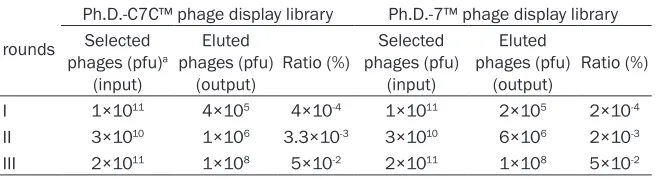
To enrich rh-midkine binding phages from the Ph.D.-C7C and Ph.D.-7 phage display libraries, three rounds of selection with the rh-midkine protein were performed. The enrichment was determined by the use of the output/input ratio of phages after each round of selection. The ratio increased approximately 10-fold after the second round of selection. After the third round of selection of the Ph.D.-C7C and Ph.D.-7 phage display libraries, the output/input ratio of phag-es increased 125-fold and 250-fold rphag-espective- respective-ly, which indicated that both linear and cyclic peptide libraries had an obvious enrichment for
the specific binding of phages to rh-midkine
[image:5.612.90.523.70.416.2](Table 1).
Estimation of binding specificities of selected phage clones
The positive clones were chosen from the sam-ples after the third round of selection with the
rh-midkine protein and the specificity was
examined by phage ELISA. 29 clones bound to the target protein and were chosen for further sequencing (Figure 1).
Analyses of sequences of positive phage clones
The ssDNA was prepared for positive phage clones and sequenced and the amino acid sequences of the midkine-binding peptides were deduced from the DNA sequences.
Analysis of the 29 clones from the
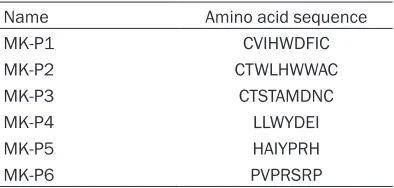
affinity-selected phage population revealed that the majority of clones fell into six groups with differ-ent sequences (Table 2). The six peptides were designated and synthesized (Table 3).
Effects of midkine-binding peptides treatment on HUVEC growth
To investigate the activity of midkine-binding peptides on the vascular endothelial cell prolif-eration, we analyzed the growth of HUVECs treated with midkine-binding peptides. From the results presented in Figure 2, the
midkine-binding peptides were observed to significantly
inhibit the proliferation of HUVECs. The inhibi-tion level ranged from 15.6 to 76.6%. Among the six peptides, MK-P3 showed the highest inhibitory activity. The percentage of the inhibi-tion was calculated as:
Inhibitory rate (%) = (OD570control-OD570treat)/ OD570control×100
Sections of xenograft tumors from the mice in all eight groups were stained for endothelial
cell specific markers to detect the number of
endothelial cells as a measure of tumor
angio-genesis. A single microvessel was defined as
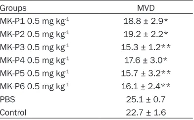
any immunohistochemistry stained endothelial cell distinguished from adjacent tumor cells and other connective tissue elements. A representa-tive result of immunostaining is shown in Figure 3. A high density of microvessels was observed in the untreated tumor sample, whereas treat-ment with the midkine-binding peptides
result-ed in a significant rresult-eduction in vascularization
(Table 5; Figure 3).
Discussion
Angiogenesis is a process of remodeling of a primitive vascular network into mature vascula-ture through sprouting, branching and differen-tial growth of blood vessels. This complex pro-cess involves endothelial cell proliferation, chemotactic migration and functional matura-tion, as well as differential recruitment of sup-porting cells. Normal or physiological angiogen-esis occurs during embryonic development, wound healing, menstruation and pregnancy. Angiogenesis is controlled and regulated by many molecules including stimulators and inhibitors. An appropriate balance between stimulators and inhibitors is pivotal for the maintenance and regulation of angiogenesis. Tumor-associated angiogenesis is critical for tumor growth and is controlled by a balance between pro- and anti-angiogenic factors. Growing evidence demonstrates the heteroge-neity of tumor angiogenesis, probably arising from the vastly different microenvironments of individual tumors [26-30]. Due to the heteroge-neous nature of tumor angiogenesis, effective anti-angiogenic therapies should be optimized and may require interference with multiple
**P < 0.01, compared with PBS control, the negative
[image:6.612.93.289.96.217.2]angiogenic pathways. A large number of natural proteins are angiogenic inhibitors and various inhibitors of angiogenesis exhibit antitumor effects [31, 32]. Selection of target proteins is very important in designing and screening anti-angiogenic peptides. Novel targets could pro-vide unique characteristics and new desirable features for further development into peptide drugs.
In this paper, we initially analyzed the effects of midkine-binding peptides on endothelial cell proliferation. The results indicated that mid-kine-binding peptides could retard HUVEC growth, suggesting that these peptides may also inhibit angiogenesis in vivo. Furthermore, we developed an in situ mouse hepatocellular
carcinoma model to determine the influence of
the midkine-binding peptides on tumor growth and angiogenesis in vivo. The results indicated
that midkine-binding peptides significantly
inhibited the mouse hepatocellular carcinoma model. Furthermore, midkine-binding peptides also suppressed tumor angiogenesis.
It is well established that tumor growth and metastasis depend on the induction of a new blood supply [33, 34]. Angiogenic activity, as determined by MVD, has been shown to corre-late with an unhealthy prognosis in a number of solid tumors [35-37]. Therefore, anti-angiogenic agents targeting either of these factors or vas-cular endothelial cells are promising therapeu-tic modalities as tumor dormancy therapies [38]. In the present study, we have addressed the potential therapeutic role of
midkine-bind-ing peptides on angiogenesis. Significant inhibi -tion of angiogenesis was achieved by the mid-kine-binding peptide MK-P3, indicating that MK-P3 represents a possible effective anti-angiogenesis agent.
Acknowledgements
This work was supported by the National Natural Science Foundation of China (No. 30772534), the Major Science and Technology Project Fund of Huzhou City (No. 2005GS01), and the Natural Science Foundation of Zhejiang (No. Y2111181 and No. LQ13H160019).
Disclosure of conflict of interest
None.
Address correspondence to: Dr. Licheng Dai, Huzhou Key Laboratory of Molecular Medicine,
Huzhou Central Hospital, Huzhou 313000, China. E-mail: [email protected]
References
[1] Folkman J. Role of angiogenesis in tumor growth and metastasis. Semin Oncol 2002; 29: 15-18.
[2] Aiello L. Keeping in touch with angiogenesis. Nat Med 2000; 6: 379-381.
[3] Rosen L. Antiangiogenic strategies and agents in clinical trials. Oncologist 2000; 5 Suppl 1: 20-27.
[4] O’Reilly MS, Holmgren L, Shing Y, Chen C, Rosenthal RA, Moses M, Lane WS, Cao Y, Sage EH, Folkman J. Angiostatin: a novel angiogen-esis inhibitor that mediates the suppression of metastases by a Lewis lung carcinoma. Cell 1994; 79: 315-28.
[5] O’Reilly MS, Boehm T, Shing Y, Fukai N, Vasios G, Lane WS, Flynn E, Birkhead JR, Olsen BR, Folkman J. Endostatin: an endogenous inhibi-tor of angiogenesis and tumor growth. Cell 1997; 88: 277-285.
[6] Tomomura M, Kadomatsu K, Matsubara S, Muramatsu T. A retinoic acid-responsive gene, MK, found in the teratocarcinoma system. J Biol Chem 1990; 265: 10765-10770.
[7] Kadomatsu K, Huang R, Suganuma T, Murata F, Muramatsu T. A retinoic acid responsive gene, MK, found in the teratocarcinoma sys-tem is expressed in a spatially and sys-temporally controlled manner during mouse embryogen-esis. J Cell Biol 1990; 110: 607-616.
[8] Kojima S, Imui T, Muramatsu H, Kimura T, Sakakiba S, Muramatsu T. Midkine is a heat and acid stable polypeptide capable of en-hancing plasminogen activator activity and neurite outgrowth extension. Biochen Biophys Res Commun 1995; 216: 574-581.
[9] Kadomatsu K, Hagihara M, Akhter S, Fan QW, Muramatsu H, Muramatsu T. Midkine induces the transformation of NIH3T3 cells. Br J Can-cer 1997; 75: 354-359.
[10] Owada K, Sanjo N, Kobayashi T, Mizusawa H, Muramatsu T, Michikawa M. Midkine inhibits caspase-dependent apoptosis via the activa-tion of mitogen-activated protein kinase and phosphatidylinositol 3-kinase in cultures neu-rons. J Neurochem 1999; 73: 2084-2092. [11] Choudhuri R, Zhang HT, Donnini S, Ziche M,
Bicknell R. An angiogenic role for the neuro-kines midkine and pleiotrophin in tumorigene-sis. Cancer Res 1997; 57: 1814-1819. [12] Miyauchi M, Shimada H, Kadomatsu K,
versus malignant lung tissues. Am J Respir Cell Mol Biol 1993; 9: 463-466.
[17] Garver RJ, Radford DM, Donis-Keller H, Wick MR, Milner PG. Midkine and pleiotrophin ex-pression in normal and malignant breast tis-sue. Cancer 1994; 74: 1584-1590.
[18] O’Brien T, Cranston D, Fuggle S, Bicknell R, Harris AL. The angiogenic factor midkine is ex-pressed in bladder cancer, and overexpression correlates with a poor outcome in patients with invasive cancers. Cancer Res 1996; 56: 2515-2518.
[19] Choudhuri R, Zhang HT, Donnini S, Ziche M, Bichnell R. An angiogenic role for neurokines midkine and pleiotrophin in tumorigenesis. Cancer Res 1997; 57: 1841-1849.
[20] Muramatsu H. Shirahama H, Yonezawa S, Maruta H, Muramatsu T. Midkine a retinoic acid-inducible growth/differentiation factor: immunochemical evidence for the function and distribution. Dev Biol 1993; 159: 392-402.
[21] Takei Y, Kadomatsu K, Matsuo S, Itoh H, Naka-zawa K, Kubota S, Muramatsu T. Antisense oli-godeoxynucleotide targeted to Midkine, a heparin-binding growth factor, suppresses tu-morigenicity of mouse rectal carcinoma cells. Cancer Res 2001; 61: 8486-8491.
[22] Dai LC, Wang X, Yao X, Min LS, Ping JL, He JF. Antisense oligonucleotides targeting midkine inhibit tumor growth in an in situ human hepa-tocellular carcinoma model. Acta Pharmacol Sin 2007; 28: 453-458.
[23] Dai LC, Wang X, Yao X, Lu YL, Ping JL, He JF. Antisense oligonucleotides targeting midkine suppresses in vivo angiogenesis. World J Gas-troenterol 2007; 13: 1208-1213.
[24] Khandare JJ, Minko T. Antibodies and peptides in cancer therapy. Crit Rev Ther Drug Carrier Syst 2006; 23: 401-435.
[25] Weidner N, Semple JP, Welch WR, Folkman J. Tumor angiogenesis and metastasis-correla-tion in invasive breast carcinoma. N Engl J Med 1991; 324: 1-8.
giogenesis and blood vessel maturation in hu-man tumors: implications for antiangiogenic tumor therapies. Cancer Res 2000; 60: 1388-1393.
[31] Ruggeri B, Singh J, Gingrich D, Angeles T, Al-bom M, Yang S, Chang H, Robinson C, Hunter K, Dobrzanski P, Jones-Bolin S, Pritchard S, Aimone L, Klein-Szanto A, Herbert JM, Bono F, Schaeffer P, Casellas P, Bourie B, Pili R, Isaacs J, Ator M, Hudkins R, Vaught J, Mallamo J, Di-onne C. CEP-7055: a novel, orally active pan inhibitor of vascular endothelial growth factor receptor tyrosine kinases with potent antian-giogenic activity and antitumor efficacy in pre-clinical models. Cancer Res 2003; 63: 5978-5991.
[32] Bergers G, Javaherian K, Lo KM, Folkman J, Hanahan D. Effects of angiogenesis inhibitors on multistage carcinogenesis in mice. Science (Wash, DC) 1999; 284: 808-812.
[33] Plate KH, Breier G, Risau W. Molecular mecha-nisms of developmental and tumor angiogen-esis. Brain Pathol 1994; 4: 207-218.
[34] Hanahan D, Folkman J. Pattern and emerging mechanisms of the angiogenic switch during tumorigenesis. Cell 1996; 86: 353-364. [35] Brawer MK, Deering RE, Brown M, Preston SD,
Bigler SA. Predictors of pathologic stage in prostatic carcinoma. The role of neovasculari-ty. Cancer 1994; 73: 678-87.
[36] Delahunt B, Bethwaite PB, Thornton A. Prog-nostic significance of microscopic vascularity for clear cell renal cell carcinoma. Br J Urol 1997; 80: 401-404.
[37] Bochner BH, Cote RJ, Weidner N, Groshen S, Chen SC, Skinner DG, Nichols PW. Angiogene-sis in bladder cancer: relationship between microvessl density and tumor prognosis. J Natl Cancer Inst 1995; 87: 1603-1612.