Original Article
HOXA10 controls proliferation, migration and invasion in
oral squamous cell carcinoma
Manoela Carrera1,2, Carolina C Bitu3, Carine Ervolino de Oliveira1, Nilva K Cervigne1,4, Edgard Graner1, Aki Manninen5, Tuula Salo1,3,6, Ricardo D Coletta1
1Department of Oral Diagnosis, School of Dentistry, University of Campinas, Piracicaba, São Paulo, Brazil; 2
De-partment of Life Sciences, Bahia State University, Bahia, Brazil; 3Department of Diagnostics and Oral Medicine,
Institute of Dentistry, and Medical Research Center, Oulu University Hospital, Oulu, Finland; 4Department of
Clinical, Faculty of Medicine of Jundiai, Jundiai, São Paulo, Brazil; 5Oulu Center for Cell-Matrix Research, Faculty
of Biochemistry and Molecular Medicine, University of Oulu, Finland; 6Institute of Dentistry, University of Helsinki,
Finland
Received January 29, 2015; Accepted March 23, 2015; Epub April 1, 2015; Published April 15, 2015
Abstract: Although HOX genes are best known for acting in the regulation of important events during embryogenesis, including proliferation, differentiation and migration, alterations in their expression patterns have been frequently
described in cancers. In previous studies we analyzed the expression profile of the members of the HOX family of homeobox genes in oral samples of normal mucosa and squamous cell carcinoma (OSCC) and identified differently
expressed genes such as HOXA10. The present study aimed to validate the increased expression of HOXA10 in OSCCs, and to investigate the effects arising from its knockdown in OSCC cells. The levels of HOXA10 mRNA were determined in human OSCC samples and cell lines by quantitative PCR, and HOXA10-mediated effects on prolifera-tion, apoptosis, adhesion, epithelial-mesenchymal transition (EMT), migration and invasion were studied in HSC-3 tongue carcinoma cells by using retrovirus-mediated RNA interference. Higher expression of HOXA10 mRNA was
observed in OSCC cell lines and in tumor tissues compared to normal controls. HOXA10 knockdown significantly re -duced the proliferation of the tumor cells which was accompaniedby increased levels of p21. HOXA10 silencing also
significantly induced the expression of EMT markers and enhanced the adhesion, migration and invasion of HSC-3 cells. No effects on cell death were observed after HOXA10 knockdown. The results of the current study confirm
the overexpression of HOXA10 in OSCCs, and further demonstrate that its expression is functionally associated with several important biological processes related to oral tumorigenesis, such as proliferation, migration and invasion.
Keywords: Oral squamous cell carcinoma, HOXA10, tumorigenesis, proliferation, p21, migration, invasion
Introduction
HOX genes are members of the superfamily of homeobox genes that encode transcription fac-tors involved in the control of cell growth and identity [1, 2], as well as in cell and cell-extracellular matrix interactions [3]. Deregu- lated expression of HOX genes has been described in leukemia, breast, cervical, ovarian and prostate cancers, among others [4-8]. Aberrant expression of HOX genes was also observed in oral squamous cell carcinomas (OSCCs) [9-19]. However, few studies have att- empted to elucidate the involvement of HOX genes in oral tumorigenesis. In previous stud-ies, we showed that most of the genes in the
HOXB network are inactive in oral normal tis-sues, but the gain of HOXB7 expression in OSCCs leads to increased tumor cell prolifera-tion [10, 14]. Increased expression of HOXA1 in OSCCs induced cell proliferation and was asso-ciated with poor prognosis [15].
proliferation, apoptosis, epithelial-mesenchy-mal transition (EMT) and treatment resistance [3, 27-29]. In OSCCs, the expression of HOXA10 was significantly higher in cancer cell lines and in primary tumors compared to controls, and the protein encoded by HOXA10 was correlated with disease stage, indicating its potential use as a prognostic marker [11]. In the present study, we further investigated the expression of HOXA10 in cell lines and in samples from nor-mal oral mucosa and OSCC. We also evaluated the effects of HOXA10 knockdown on prolifera-tion, apoptosis, adhesion, expression of EMT markers, migration and invasion of OSCC cells.
Materials and methods
Samples
Tissue samples were collected after approval of the Human Research Ethics Committee of the School of Dentistry, University of Campinas. Fresh samples of normal oral mucosa (n=12) and OSCC (n=12) were used to investigate the expression of HOXA10 using quantitative PCR (qPCR). The samples were divided into two parts: one was fixed in formalin and embedded in paraffin for hematoxylin and eosin staining, while the other was snap frozen in liquid nitro-gen. The initial diagnosis was based on clinical findings and confirmed by histopathological analysis of the specimens.
Cell cultures and generation of HOXA10 knock-down cells
Normal human gingival keratinocyte cell line (HGK) was cultured in serum-free, low calcium medium (Gibco’s Keratinocyte-SFM; Invitrogen, USA) containing specific supplements and anti-biotics [30]. The human OSCC cell lines SCC-4, SCC-9, SCC-15 and SCC-25 were obtained from American Type Culture Collection (ATCC, Ma- nassas, VA, USA), and cultured as recommend-ed in a 1:1 mixture of Dulbecco’s modifirecommend-ed Eagle’s medium and Ham’s F12 medium (DMEM/F12; Invitrogen, USA) supplemented with 10% fetal bovine serum (FBS), 400 ng/ml
hydrocortisone (Sigma-Aldrich, USA) and antibi-otics. HSC-3, a human tongue squamous cell carcinoma cell line (JCRB 0623; Osaka National Institute of Health Sciences, Japan), was cul-tured in DMEM/F-12 medium (Invitrogen, USA) supplemented with 10% FBS, 50 µg/ml ascor-bic acid (Sigma-Aldrich, USA), 400 ng/ml hydro-cortisone (Sigma-Aldrich, USA) and antibiotics. All cells were growth at 37°C in a humidified atmosphere of 5% CO2.
Transduction of HSC-3 cells with shRNA sequences was performed using the human Phoenix packaging cells (293T cells) according to the manufacturer’s instructions (OriGene Technologies, USA). After transduction, cells were selected in the presence of 1 μg/ml of puromycin (Invitrogen, USA) for 2 weeks.
qPCR
Total RNA from cell lines and fresh samples was isolated with TRIzol reagent according to the manufacturer’s protocol (Invitrogen, USA). Following DNase I treatment in order to elimi-nate genomic DNA contamination, 2 μg of total RNA per sample were used to generate cDNA using Oligo-dT primers (Invitrogen, USA) and a superscript enzyme (Superscript II RT enzyme, Invitrogen, USA). The resulting cDNAs were sub-jected to qPCR using SYBR® Green PCR master mix (Applied Biosystems, USA) in the Step- OnePlus Real Time PCR (Applied Biosystems, USA). Gene expressions were determined by the standard curve method or deltadeltaCT with normalization to the housekeeping gene PPIA (cyclophilin A). Primer sequences are described in Table 1.
Western blot
[image:2.612.91.521.83.151.2]Cells were washed with cold PBS and lysed in a protein-lysis buffer containing 20 mM Tris pH 8.0, 137 mM NaCl, 10% glycerol, 2 mM EDTA and protease inhibitors. After centrifugation, protein concentrations were measured using a protein assay according to the manufacturer’s instructions (Bio-Rad Protein Assay, Bio-Rad, USA). Eighty μg of total protein per sample were
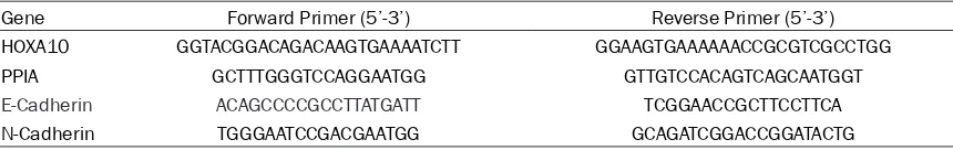
Table 1. Primer sequences used in the qPCR
Gene Forward Primer (5’-3’) Reverse Primer (5’-3’)
HOXA10 GGTACGGACAGACAAGTGAAAATCTT GGAAGTGAAAAAACCGCGTCGCCTGG
PPIA GCTTTGGGTCCAGGAATGG GTTGTCCACAGTCAGCAATGGT
E-Cadherin ACAGCCCCGCCTTATGATT TCGGAACCGCTTCCTTCA
resolved in a 10% sodium dodecyl sulphate polyacrylamide gel electrophoresis (SDS-PAGE) under reducing conditions, and transferred to nitrocellulose membranes. The membranes we- re blocked with 10% non-fat dry milk in PBS containing 0.1% Tween-20, rinsed in the same buffer, and incubated for 2 h with anti-HOXA10 (clone A20, Santa Cruz, USA), anti-p21 (clone C-19, Santa Cruz, USA) or anti-β-actin antibod-ies (Sigma-Aldrich, USA). After washing, the me- mbranes were developed using an enhanced chemiluminescent western blot kit (GE Health- care, Austria).
Bromodeoxyuridine-labeling (BrdU) index
Cells were plated in 96-well chamber slides at a density of 3,000 cells per well in complete media. After 24 h, cells were washed with PBS and cultured in serum-free media for 48 h for synchronism. Following serum starvation, cul-ture media containing 10% of FBS was added to the cells. BrdU, a thymidine analog, was added to the cultures in 1:10 dilution and kept for 2 h at 37°C in 5% CO2. After incubation, the media was removed and manufacturer’s proto-col (Cell proliferation ELISA BrdU proto-colorimetric assay kit, Roche Applied Science, Germany) was followed. Absorbance was measured at 370 nm with correction at 492 nm.
Apoptosis analysis
The apoptosis index was determined by annex-in V-FITC labelannex-ing. Succannex-inctly, cells were har-vested, washed with PBS and resuspended in the binding buffer (10 mM HEPES pH 7.4, 150 mM NaCl, 5 mM KCl, 1 mM MgCl2 and 1.8 mM CaCl2) containing annexin V-FITC (BD Biosci- ences, USA) and propidium iodide (PI, Sigma-Aldrich, USA). Samples were incubated for 20 min at room temperature, and shielded from light. Apoptosis was analyzed on a FACScalibur flow cytometer equipped with an argon laser (BD Biosciences, USA) and quantified as the number of annexin V-FITC positive and PI nega-tive cells divided by the total number of cells. A minimum of 10,000 events was analyzed in each sample.
Cell adhesion assay
A 96-well culture plate was coated with 2 μg of type I collagen or fibronectin (BD Biosciences, USA) in 100 μl of PBS for 16 h at 4°C. The wells
were washed 3 times with 200 μl of PBS and then coated with the same volume of 3% of BSA in PBS for 2 h at 37°C. Control wells were coated only with 3% BSA solution. Cells were harvested and then resuspended in DMEM supplemented with 10% FBS and 3% BSA at a final concentration of 3,000 cells in 100 μl. The wells were washed and then 100 μl of the cell suspension was added to each well. The plate was then kept for 1 h in 37°C at 5% CO2. Loose cells were washed away and adhered cells were fixed in 10% formalin for 15 min and stained with a solution of 1% toluidin blue and 1% bo- rax. Absorbance was measured at 650 nm.
Analysis of the EMT markers
The expression of the epithelial marker E- cadherin and of mesenchymal marker N-ca- dherin was carried out using western blot and qPCR analysis as described above. The mono-clonal antibodies anti-E-cadherin and anti-N-cadherin were purchased from BD Biosciences (USA) and used at concentrations of 1:2000 and 1:2500, respectively.
Migration and invasion assays
Transwell migration and invasion assays were performed in 6.5 mm inserts with 8 μm pore size (Corning, USA). For invasion assay, mem-branes were coated with 50 µl of growth factor-reduced matrigel (BD Biosciences, USA). Serum starved cells (80,000 cells/well) were plated into the upper chamber in 200 μl of serum-free DMEM. As chemoattractant, 500 μl of com-plete medium was used in the lower chamber. Experiment times varied between 24 h for migration assays and 72 h for invasion assays. Assessment of migration or invasion was per-formed by gently removing cells in the interior part of the insert with a cotton swab. Cells in the bottom of the membrane were fixed in 10% formalin for 15 min and stained with a solution of 1% toluidine blue and 1% borax. The excess dye was washed and the plate was incubated with a solution of 1% SDS for 5 min. Absorbance was measured at 650 nm.
Statistical analysis
Kruskal-Wallis test and Mann-Whitney U test were app- lied in our comparisons, and P≤0.05 was con- sidered as indicating of statistical significance.
Results
To confirm the differential expression of HOXA10 in oral cancers, we first evaluated the expression levels of HOXA10 in different cell lines and in fresh tissues. The levels of HOXA10
[image:4.612.93.524.73.273.2]mRNA were significantly higher in the OSCC cell lines SCC-4 (P<0.0001), SCC-15 (P<0.0001), SCC-25 (P=0.005) and HSC-3 (P=0.001) com-pared with the spontaneously immortalized, but not transformed epithelial cell line HGK (Figure 1A). Although higher, the expression levels of SCC-9 did not reach a statistically sig-nificant difference when compared to HGK (P=0.20). A significantly higher content of HOXA10 mRNA was observed in the fresh Figure 1. HOXA10 is overexpressed in OSCCs and OSCC-derived cell lines. Total RNA from cell lines and fresh
sam-ples were converted in cDNA and subjected to qPCR. A. HOXA10 mRNA levels were significantly higher in OSCC cell
lines compared to normal human oral keratinocyte cells (HGK), with exception of SCC-9. B. The amount of HOXA10
mRNA was significantly higher in OSCCs than in adjacent normal oral mucosa. *P=0.003.
Figure 2. RNAi-mediated silencing of HOXA10 expression in HSC-3 cells. Representative qPCR (A) and western blot
(B) analysis of HOXA10 in HSC-3-Control and HSC-3-shHOXA10, which confirmed the marked reduction of HOXA10
[image:4.612.91.522.344.537.2]tumors in comparison to normal control tissues (P=0.003, Figure 1B).
In order to comprehend the biological role of HOXA10 in the events that control tumorigene-sis, the levels of HOXA10 were specifically sil- enced using interference RNA in the HSC-3 cell line, the most invasive OSCC cell line. After stable retroviral transduction and selection of puromycin-resistant cells, both HOXA10 mRNA and protein levels were quantified by qPCR and western blot. Although the HOXA10 mRNA lev-els were only reduced by ~50% in comparison with the control cells (HSC-3-Control), the HOX10A protein levels were markedly reduced in cells transduced with specific shRNA against HOXA10 mRNA (HSC-3-shHOXA10, Figure 2). The effects of HOXA10 on proliferation and cell death (apoptosis) were investigated by using BrdU-labeling index and Annexin-V assays. As depicted in Figure 3A, no significant
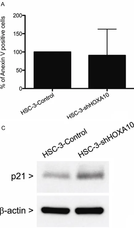
differenc-es in apoptosis were observed between the HSC-3-shHOXA10 and control cells, but the proliferation rate of the HSC-3-shHOXA10 cells was significantly reduced compared with the HSC-3-Control cells (P<0.0001, Figure 3B). As a previous study has shown that HOXA10 con-trols p21 expression leading to decrease cell cycle progression [27], we sought to evaluate p21 levels in HSC-3-shHOXA10 cells using we- stern blot analysis. Consistently, increased p21 expression was observed in HSC-3-shHOXA10 cells compared to controls (Figure 3C).
[image:5.612.92.309.73.443.2]Next, the effects of HOXA10 on cell invasion and migration were assessed in transwell chambers. Even in the presence of a large stan-dard deviation, HSC-3-shHOXA10 cells exhibit-ed significantly augmentexhibit-ed migration (P< 0.0001) and invasion (P=0.002) capacity when compared with HSC-3-Control cells (Figure 4). Furthermore, down-regulation of HOXA10 was Figure 3. Inhibition of HOXA10 alters proliferation
but not apoptosis in HSC-3 cells. A. No significant
effects on apoptosis were observed in HSC-3 cells after knock down of HOXA10 as evaluated by annexin V stain. B. BrdU incorporation assay
re-vealed a significantly reduction in proliferation of
followed by a significant increase in cell adhe-sion on surfaces coated with fibronectin (P= 0.049) or type I collagen (P=0.049). Although the adhesion of HSC-3-shHOXA10 cells was higher than HSC-3-Control in the uncoated sur-faces, the difference was not significant (P= 0.13, Figure 5). To determine whether HOX-A10 levels regulate EMT, we examined the expres-sion levels of the epithelial marker E-cadherin and of the mesenchymal marker N-cadherin in the HOXA10 silencing cells. As revealed in
Figure 6A, HSC-3-shHOXA10 had high mRNA levels of N-cadherin and reduced expression of E-cadherin. Using protein immunoblotting, we confirmed that HSC-3-shHOXA10 cells con-tained increased protein levels of N-cadherin and slight decreased levels of E-cadherin (Figure 6B).
Discussion
HOX genes encode transcription factors that control important cellular events such as cell proliferation, differentiation and death during the early phases of development [31]. Their contribution to tumorigenesis is widely accept-ed, although the exact mechanisms through which they exert their functions are not yet clear [32, 33]. The relationship between HO- XA10 and oral tumorigenesis was initially described by Hassan et al. [9] in a study that verified the expression pattern of all HOX genes in normal, dysplastic and tumor tissues. Since
then, Yamatoji and colleagues [11] detected an aberrant expression of HOXA10 in OSCCs and OSCC-derived cell lines. Similarly, our group demonstrated that HOXA10 expression was significantly higher in OSCC samples compared to both healthy oral mucosa and histologically normal mucosa adjacent to OSCC [15]. This motivated this in depth analysis on the role of HOXA10 in oral carcinogenesis. Therefore, we chose to validate our data by testing different cells lines and fresh samples from OSCC and adjacent normal oral mucosa, which confirmed our previous findings that HOXA10 mRNA levels are significantly higher in tumors when com-pared to normal oral mucosa. The most impor-tant finding of the current study was that knock-down of HOXA10 significantly reduced the proliferation of OSCC cells, while concomitantly induced cell adhesion, expression of EMT mark-ers and migration and invasion of the cells. Although several studies have verified the role of HOXA10 as a potential prognostic marker, the results showed discrepancies [1, 34-36]. Multiple evidences showed that HOXA10 is involved in the proliferation of hematopoietic stem cells and progenitor cells, leading to can-cer development through the activation of sev-eral target genes, including TGF-β2, dual-speci-ficity protein phosphatase 4 and integrin-β3 [1, 34, 36]. Up-regulation of HOX genes is associ-ated with a tumor stem-like cell phenotype of glioblastoma, and high HOXA10 protein expres-Figure 4. Migration and invasion of HSC-3 were induced after HOXA10 knock down. (A) Migration and (B) invasion
were evaluated by transwell system. Both migration and invasion were significantly increased by HOXA10-specific
[image:6.612.93.523.69.263.2]sion has been implicated with resistance to chemotherapy [35]. Furthermore, expression of HOXA10 was negatively correlated with the depth of invasion in gastric carcinomas, and the prognosis of patients with positive HOXA10 expression was significantly better than in cases where HOXA10 was absent [37]. In addi-tion, overexpression of HOXA10 was associat-ed with gastric tumors showing poor-prognostic features [38]. On the other hand, loss of HO- XA10 expression was associated with poor tumor differentiation in endometrial and ovari-an tumors, indicating that a lower expression of HOXA10 worsen patient’s prognosis [3, 39]. HO- XA10 methylation has been described in sev-eral types of cancer and was correlated with different histologic grading, as well as prognos-tic and clinicopathological characterisprognos-tics [40-43]. In breast and ovarian cancer, miR-135a directly regulated HOXA10 expression, which promoted cell migration and invasion [44, 45].
Taken together, these findings suggest that the role of HOXA10 is variable and tumor lineage dependent.
Regardless of the mechanism by which HOXA10 expression is upregulated in OSCCs, we showed in this study that HOXA10 is an important regu-lator of proliferation, adhesion, migration and invasion of OSCC cells. HOXA10 downregula-tion led to an increase in p21 expression and a reduction of cell proliferation rate. This result is in agreement with previous findings of transac-tivation of p21 by direct binding of HOXA10 onto the p21 promoter in breast cancer cells [27]. A lower proliferative capacity was also noticed in mice after bone marrow transplant with HOXA10 knockdown cells when compared to control animals [46]. As expected, in vitro
[image:7.612.91.309.72.445.2]analysis of such cells indicated a shift on cell cycle with reduced number of cells on S-phase [46]. Forced expression of HOXA10 significantly Figure 5. Effects of HOXA10 downregulation on adhesion of HSC-3 cells. Both HSC-3-Con-trol and HSC-3-shHOXA10 cells were harvest-ed and allowharvest-ed to adhere for 1 h to wells of a 96-well plate coated with type I collagen (B)
or fibronectin (C). (A) Adhesion on uncoated
plastic surface was also analyzed. HOXA10
si-lencing was able to increase significantly the
adhesion of HSC-3-shHOXA10 cells to type
reduced the invasive phenotype of breast can-cer cells as well as modulated p53 expression [28]. Recently, it was reported that HOXA10 silencing is also implicated in a lower prolifera-tive capacity of epithelial ovarian cancer cells [45]. Those findings suggested that overexpres-sion of HOXA10 promotes cell proliferation in OSCCs via downregulation of p21.
The results presented here also revealed that HOXA10 levels can modulate several biological processes related to metastasis, such as adhe-sion, EMT, migration and invasion in HSC-3 cells. Indeed, the effects of HOXA10 on some of those biological processes were already described in other cell types [36, 38, 45, 47]. For instance, HOXA10 is also implicated on controlling E-cadherin expression in endome-trial carcinoma cells through its regulatory role over Snail protein [3]. HOXA10 expression has also been associated with cell adhesion th- rough regulation of ITGB3 gene, which encodes one of the subunits of αvβ3 integrin, in endo-metrial [48] and myeloid cells [1]. HOXA10 has
also been described to play a pivotal role in controlling the TGFβ-2 expression in myeloid and pancreatic cells [36, 49]. Activation of ERK and the TGFβ2-p38 MAPK pathway may be involved in these processes [36, 49]. HOXA10 is also responsible for regulation of MMP-3 expression in pancreatic cells [49].
HOXA10 participation on apoptosis control is still uncertain. On the present study, HOXA10 knockdown was not able to influence apoptosis levels. Likely, apoptosis of human endometrial cells (HESC) [50] and acute myeloid leukemia cells [46] were also unaltered after HOXA10 silencing. Nonetheless, Tang et al. [45] recently demonstrated that HOXA10 silencing in ovarian cancer cells resulted in increased apoptosis with concomitant enhance in caspase-3 and p53 expression and reduction of Bcl-2 exp- ression.
In essence, our data demonstrates the in vitro
[image:8.612.90.521.69.437.2]involvement of HOXA10 on oral carcinogenesis. The results suggest that HOXA10 modulates
Figure 6. Effect of HOXA10 silencing on mark-ers of epithelial-mesenchymal transition. (A) qPCR and (B) western blot analysis for E-cad-herin and N-cadE-cad-herin revealed that abrogation of HOXA10 was able to induce epithelial-mes-enchymal transition. N-cadherin expression
important cellular events for the development and progression of OSCCs, and that its expres-sion may be associated with a less aggressive tumor phenotype.
Acknowledgements
This work was supported by grants from Fundação de Amparo a Pesquisa do Estado de São Paulo-FAPESP, São Paulo, Brazil; and Conselho Nacional de Desenvolvimento Científico e Tecnológico-CNPq, Brasília, Brazil.
Disclosure of conflict of interest
None.
Address correspondence to: Dr. Ricardo D Coletta, Department of Oral Diagnosis, School of Dentistry, University of Campinas, CEP 13414-018, Piracicaba, São Paulo, Brazil. E-mail: coletta@fop.unicamp.br
References
[1] Bei L, Lu YF, Bellis SL, Zhou W, Horvath E,
Eklund EA. Identification of a HOXA10 activa -tion domain necessary for transcrip-tion of the
gene encoding β3 integrin during myeloid dif -ferentiation. J Biol Chem 2007; 282: 16846-16859.
[2] Magli MC, Barba P, Celetti A, Vita GD, Cillo C, Boncinelli E. Coordinate regulation of HOX genes in human hematopoietic cells. Proc Natl Acad Sci U S A 1991; 88: 6348-6352.
[3] Yoshida H, Broaddus R, Cheng W, Xie SS, Nao-ra H. Deregulation of the HOX A10 homeobox gene in endometrial carcinoma: role in epithe-lial-mesenchymal transition. Cancer Res 2006; 66: 889-897.
[4] Calvo KR, Sykes DB, Pasillas MP, Kamps MP. Nup98-HoxA9 immortalizes myeloid progeni-tors, enforces expression of Hoxa9, Hoxa7 and
Meis1, and alters cytokine-specific responses
in a manner similar to that induced by retrovi-ral co-expression of Hoxa9 and Meis1. Onco-gene 2002; 21: 4247-4256.
[5] Naora H, Yang YQ, Montz FJ, Seidman JD,
Kur-man RJ, Roden RB. A serologically identified
tumor antigen encoded by a homeobox gene promotes growth of ovarian epithelial cells. Proc Natl Acad Sci U S A 2001; 98: 4060-4065.
[6] Hung YC, Ueda M, Terai Y, Kumagai K, Ueki K, Kanda K, Yamaguchi H, Akise D, Ueki M. Ho-meobox gene expression and mutation in cer-vical carcinoma cells. Cancer Sci 2003; 94: 437-441.
[7] López R, Garrido E, Piña P, Hidalgo A, Lazos M,
Ochoa R, Salcedo M. HOXB homeobox gene expression in cervical carcinoma. Int J Gynecol Cancer 2006; 16: 329-335.
[8] Miller GJ, Miller HL, van Bokhoven A, Lambert JR, Werahera PN, Schirripa O, Lucia MS, Nor-deen SK. Aberrant HOXC expression accompa-nies the malignant phenotype in human pros-tate. Cancer Res 2003; 63: 5879-5888. [9] Hassan NM, Hamada J, Murai T, Seino A,
Taka-hashi Y, Tada M, Zhang X, Kashiwazaki H, Yamazaki Y, Inoue N, Moriuchi T. Aberrant ex-pression of HOX genes in oral dysplasia and squamous cell carcinoma tissues. Oncol Res 2006; 16: 217-224.
[10] Destro MFSS, Bitu CC, Zecchin KG, Graner E, Lopes MA, Kowalski LP, Coletta RD. Overex-pression of HOXB7 homeobox gene in oral can-cer induces cellular proliferation and is associ-ated with poor prognosis. Int J Oncol 2010; 36: 141-149.
[11] Yamatoji M, Kasamatsu A, Yamano Y, Sakuma K, Ogoshi K, Iyoda M, Shinozuka K, Ogawara K, Takiguchi Y, Shiiba M, Tanzawa H, Uzawa K. State of homeobox A10 expression as a puta-tive prognostic marker for oral squamous cell carcinoma. Oncol Rep 2010; 23: 61-67. [12] Libório TN, Acquafreda T, Matizonkas-Antonio
LF, Silva-Valenzuela MG, Ferraz AR, Nunes FD. In situ hybridization detection of homeobox genes reveals distinct expression patterns in oral squamous cell carcinomas. Histopatholo-gy 2011; 58: 225-233.
[13] Tucci R, Campos MS, Matizonkas-Antonio LF, Durazzo M, Pinto Junior DS, Nunes FD. HOXB5 expression in oral squamous cell carcinoma. J Appl Oral Sci 2011; 19: 125-129.
[14] Bitu CC, Carrera M, Lopes MA, Kowalski LP, So-ares FA, Coletta RD. HOXB7 expression is a prognostic factor for oral squamous cell carci-noma. Histopathology 2012; 60: 662-665. [15] Bitu CC, Destro MF, Carrera M, da Silva SD,
Graner E, Kowalski LP, Soares FA, Coletta RD. HOXA1 is overexpressed in oral squamous cell carcinomas and its expression is correlated with poor prognosis. BMC Cancer 2012; 12: 146.
[16] Moon SM, Ahn MY, Kwon SM, Kim SA, Ahn SG, Yoon JH. Homeobox C5 expression is associat-ed with the progression of 4-nitroquinoline 1-oxide-induced rat tongue carcinogenesis. J Oral Pathol Med 2012; 41: 470-476.
[17] Rodini CO, Xavier FC, Paiva KB, De Souza Setúbal Destro MF, Moyses RA, Michaluarte P, Carvalho MB, Fukuyama EE; Head And Neck Genome Project Gencapo, Tajara EH, Okamoto OK, Nunes FD. Homeobox gene expression
profile indicates HOXA5 as a candidate prog -nostic marker in oral squamous cell carcino-ma. Int J Oncol 2012; 40: 1180-1188. [18] Hakami F, Darda L, Stafford P, Woll P, Lambert
[19] Libório-Kimura TN, Jung HM, Chan EK.
miR-494 represses HOXA10 expression and inhib-its cell proliferation in oral cancer. Oral Oncol 2015; 51: 151-157.
[20] Thorsteinsdottir U, Sauvageau G, Hough MR, Dragowska W, Lansdorp PM, Lawrence HJ, Largman C, Humphries RK. Overexpression of HOXA10 in murine hematopoietic cells per-turbs both myeloid and lymphoid differentia-tion and leads to acute myeloid leukemia. Mol Cell Biol 1997; 17: 495-505.
[21] Ma XJ, Salunga R, Tuggle JT, Gaudet J, Enright E, McQuary P, Payette T, Pistone M, Stecker K, Zhang BM, Zhou YX, Varnholt H, Smith B, Gadd
M, Chatfield E, Kessler J, Baer TM, Erlander MG, Sgroi DC. Gene expression profiles of hu -man breast cancer progression. Proc Natl Acad Sci U S A 2003; 100: 5974-5979.
[22] Miao J, Wang Z, Provencher H, Muir B, Dahiya S, Carney E, Leong CO, Sgroi DC, Orsulic S. HOXB13 promotes ovarian cancer progression. Proc Natl Acad Sci U S A 2007; 104: 17093-17098.
[23] Plowright L, Harrington KJ, Pandha HS, Morgan R. HOX transcription factors are potential ther-apeutic targets in non-small-cell lung cancer (targeting HOX genes in lung cancer). Br J Can-cer 2009; 100: 470-475.
[24] Shah N, Sukumar S. The Hox genes and their roles in oncogenesis. Nat Rev Cancer 2010; 10: 361-371.
[25] Li B, Cao X, Weng C, Wu Y, Fang X, Zhang X, Liu G. HoxA10 induces proliferation in human prostate carcinoma PC-3 cell line. Cell Bio-chem Biophys 2014; 70: 1363-1368.
[26] Zhang L, Wan Y, Jiang Y, Ma J, Liu J, Tang W, Wang X, Cheng W. Upregulation HOXA10 ho-meobox gene in endometrial cancer: role in cell cycle regulation. Med Oncol 2014; 31: 52. [27] Bromleigh VC, Freedman LP. p21 is a transcrip-tional target of HOXA10 in differentiating my-elomonocytic cells. Genes Dev 2000; 14: 2581-2586.
[28] Chu MC, Selam FB, Taylor HS. HOXA10 regu-lates p53 expression and matrigel invasion in human breast cancer cells. Cancer Biol Ther 2004; 3: 568-572.
[29] Kim JW, Kim JY, Kim JE, Kim SK, Chung HT, Park CK. HOXA10 is associated with temozolo-mide resistance through regulation of the ho-mologous recombinant DNA repair pathway in glioblastoma cell lines. Genes Cancer 2014; 5: 165-174.
[30] Salo T, Lyons JG, Rahemtulla F, Birkedal-Han-sen H, Larjava H. Transforming growth factor-βl up-regulates type IV collagenase expression in cultured human keratinocyte. J Biol Chem 1991; 266: 11436-11441.
[31] Ford HL. Homeobox genes: a link between de-velopment, cell cycle, and cancer Cell Biol Int 1998; 22: 397-400.
[32] Abate-Shen C. Deregulated homeobox gene expression in cancer: cause or consequence? Nat Rev Cancer 2002; 2: 777-785.
[33] Samuel S, Naora H. Homeobox gene expres-sion in cancer: insights from developmental regulation and deregulation. Eur J Cancer 2005; 41: 2428-2437.
[34] Wang H, Lu Y, Huang W, Papoutsakis ET, Fuhrken P, Eklund EA. HOXA10 activates tran-scription of the gene encoding mitogen-acti-vated protein kinase phosphatase 2 (Mkp2) in myeloid cells. J Biol Chem 2007; 282: 16164-16176.
[35] Murat A, Migliavacca E, Gorlia T, Lambiv WL, Shay T, Hamou MF, de Tribolet N, Regli L, Wick W, Kouwenhoven MC, Hainfellner JA, Heppner FL, Dietrich PY, Zimmer Y, Cairncross JG, Jan-zer RC, Domany E, Delorenzi M, Stupp R, Hegi ME. Stem cell-related “self-renewal” signature and high epidermal growth factor receptor ex-pression associated with resistance to con-comitant chemoradiotherapy in glioblastoma. J Clin Oncol 2008; 26: 3015-3024.
[36] Shah CA, Wang H, Bei L, Platanias LC, Eklund EA. HoxA10 regulates transcription of the gene encoding transforming growth factor beta2 (TGFbeta2) in myeloid cells. J Biol Chem 2011; 286: 3161-3176.
[37] Sentani K, Oue N, Naito Y, Sakamoto N, Anami K, Oo HZ, Uraoka N, Aoyagi K, Sasaki H, Yasui W. Upregulation of HOXA10 in gastric cancer with the intestinal mucin phenotype: reduction during tumor progression and favorable prog-nosis. Carcinogenesis 2012; 33: 1081-1088. [38] Lim JY, Yoon SO, Seol SY, Hong SW, Kim JW,
Choi SH, Lee JS, Cho JY. Overexpression of miR-196b and HOXA10 characterize a poor-prognosis gastric cancer subtype. World J Gas-troenterol 2013; 19: 7078-7088.
[39] Andikyan V, Taylor HS. WT1 represses HOX gene expression in the regulation of gynaeco-logic tumour histogynaeco-logic type. J Cell Mol Med 2009; 13: 4522-4531.
[40] Chen H, Chung S, Sukumar S. HOXA5-induced apoptosis in breast cancer cells is mediated by caspases 2 and 8. Mol Cell Biol 2004; 24: 924-935.
[41] Fiegl H, Windbichler G, Mueller-Holzner E, Goe-bel G, Lechner M, Jacobs IJ, Widschwendter M. HOXA11 DNA methylation-a novel prognostic biomarker in ovarian cancer. Int J Cancer 2008; 123: 725-729.
[42] Park SY, Kwon HJ, Lee HE, Ryu HS, Kim SW, Kim JH, Kim IA, Jung N, Cho NY, Kang GH. Pro-moter CpG island hypermethylation during breast cancer progression. Virchows Arch 2011; 458: 73-84.
Go-hlke H, Gardin G, Merlo DF, Mantovani V, Ro-mani M. Quantitative methylation analysis of HOXA3, 7, 9, and 10 genes in glioma: associa-tion with tumor WHO grade and clinical out-come. J Cancer Res Clin Oncol 2012; 138: 35-47.
[44] Chen Y, Zhang J, Wang H, Zhao J, Xu C, Du Y, Luo X, Zheng F, Liu R, Zhang H, Ma D. miRNA-135a promotes breast cancer cell migration and invasion by targeting HOXA10. BMC Can-cer 2012; 12: 111.
[45] Tang W, Jiang Y, Mu X, Xu L, Cheng W, Wang X. MiR-135a functions as a tumor suppressor in epithelial ovarian cancer and regulates HOXA10 expression. Cell Signal 2014; 26: 1420-1426.
[46] Orlovsky K, Kalinkovich A, Rozovskaia T, She-zen E, Itkin T, Alder H, Ozer HG, Carramusa L, Avigdor A, Volinia S, Buchberg A, Mazo A, Kollet O, Largman C, Croce CM, Nakamura T, Lapidot T, Canaani E. Down-regulation of homeobox genes MEIS1 and HOXA in MLL-rearranged acute leukemia impairs engraftment and re-duces proliferation. Proc Natl Acad Sci U S A 2011; 108: 7956-7961.
[47] Bei L, Shah C, Wang H, Huang W, Roy R, Eklund EA. β-Catenin activates the HOXA10 and CDX4 genes in myeloid progenitor cells. J Biol Chem 2012; 287: 39589-39601.
[48] Daftary GS, Troy PJ, Bagot CN, Young SL, Taylor HS. Direct regulation of beta3-integrin subunit gene expression by HOXA10 in endometrial cells. Mol Endocrinol 2002; 16: 571-579. [49] Cui XP, Qin CK, Zhang ZH, Su ZX, Liu X, Wang
SK, Tian XS. HOXA10 promotes cell invasion
and MMP-3 expression via TGFβ2-mediated
activation of the p38 MAPK pathway in pancre-atic cancer cells. Dig Dis Sci 2014; 59: 1442-1451.