Original Article
Expression of miR-9 in the serum of patients with acute
ischemic stroke and its effect on neuronal damage
Yufeng Xue1, Min Li1, Donghong Liu2, Qibing Zhu3, Huijun Chen3
Departments of 1Neurology, 2Laboratory, 3Pharmacy, Tai Zhou First People’s Hospital, China
Received August 29, 2018; Accepted October 9, 2018; Epub December 1, 2018; Published December 15, 2018
Abstract: Background: This research was aimed to measure the expression of miR-9 in serum of acute ischemic stroke (AIS) patients and explore the role of miR-9 on OGD-induced neuronal damage. Methods: In the present study, we measured the expression of miR-9 in serum of 65 AIS patients by real-time quantitative PCR (RT-qPCR) and the effect of miR-9 on oxygen-glucose deprivation (OGD)-induced neuronal injury was detected by CCK-8 in vitro. Western blot was used to measure the expression of protein. Results: We found that the serum level of miR-9 in 65 AIS patients was significantly higher than that in control group (no-AIS), and was positively correlated with NIHSS score (r=0.627, P<0.001), infarct volume ((r=0.576, P<0.001), serum IL-8 (r=0.376, P=0.002), TNF-α (r=0.418, P<0.001), IL-6 (r=0.545, P<0.001), and IL-1β (r=0.592, P<0.001). miR-9 expression levels were upregulated in cultured neurons with OGD treatment. The downregulation of miR-9 significantly alleviated OGD-induced neuronal injury. Dual-luciferase reporter assay demonstrated that SIRT1 was a target gene of miR-9, and miR-9 negatively regulated SIRT 1 expression and positively regulated p65 expression. Conclusions: All in all, our data showed that downregulation of miR-9 protected neurons against OGD/R-induced injury by the SIRT1-mediated NF-kB pathway.
Keywords:miR-9, acute ischemic stroke, neuronal, inflammation
Introduction
Acute ischemic stroke (AIS) is an infarction of brain tissue caused by occlusion of cerebral arteries, accompanied by the injury of neurons, astrocytes, and oligodendrocytes. It is the most important central nervous system vascular event leading to lethality and disability in mod-ern society. Unlike other organs in the human body, although the brain accounts for only 2 percent of human body weight, adult brain tis-sue oxygen consumption is 20 percent of the total systemic oxygen consumption. It can rely on little substance to provide energy, while the ability to store energy is poor. This results in poor tolerance to ischemic injury. Once isch-emic injury occurs, the brain tissue will be dev-astated and cause great harm to the human body [1]. Therefore, it is very important to explore the pathologicprocess of cerebral ischemia.
MicroRNAs are endogenous non-coding RNAs with a length of about 20 base pairs, which can
bind to the 3’UTR region of the downstream tar-get gene to regulate downstream gene expres-sion, thereby regulating the expression level of the corresponding protein and playing a corre-sponding biological role. It has been confirmed that miRNAs are widely present in humans and play a crucial regulatory role in the alteration of self-expression levels in a variety of diseases [2, 3]. At the same time, a variety of miRNAs were found in brain tissue [4, 5] and are involved in the regulation of the pathophysiological pro-cesses of central nervous system diseases [6, 7]. In recent years, with the development of bio-chip technology, more miRNAs and their down-stream regulatory genes have been found to be involved in the development and progression of neurological diseases [6], especially ischemic stroke [7].
and various malignant tumor diseases [13]. Ji et al. [14] found that the brain-specific miR-9 was increased in the serum exosomes of acute ischemic stroke patients. However, the expres-sion of miR-9 in serum of AIS patients and the role of miR-9 remains on OGD-induced neuro-nal damage remain unknown. In the present study, we found that miR-9 expression was upregulated in the serum of AIS patients, and downregulation of miR-9 protected neurons against OGD/R-induced injury via SIRT1-mediated NF-kB pathway in vitro.
Materials and methods
Patients and blood sample
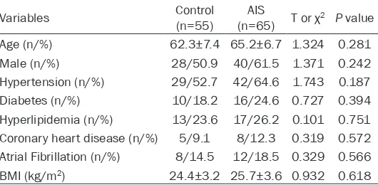
65 AIS patients and 55 controls (no-AIS) in Taizhou First People’s Hospital were selected for this research. AIS patients were diagnosed by brain magnetic resonance imaging or com-puted tomography. Two neurologists who were deputy director and above performed NIHSS scores on AIS patients within 24 hours of cere-bral ischemia. Non-stroke controls were recruit-ed from those who underwent an annual mrecruit-edi- medi-cal examination at our hospital. Clinimedi-cal data are shown in Table 1. For the whole blood sam-ples, they were centrifuged at 1,000×g for 10 min to collect serum, and Elisa kit was used to measure the content of IL-8 (H-EL-IL-8, ZYsci- ence, USA), TNF-α (50R-E.1693H, BIOVALUE, AUS), IL-6 (H-EL-IL-6, ZYscience, USA), and IL-1β (50R-E.1095H, BIOVALUE, AUS).
All subjects (or their guardians) included in this study consented the research protocol and signed an informed consent form. The ethics committee of Taizhou First People’s Hospital approved this research protocol.
with a pasteurized tube, and the cell suspen-sion was prepared and allowed to stand for 3 minutes, and the supernatant cell suspension was transferred to another centrifuge tube (the above steps were repeated 3 times). The super-natant was discarded by centrifugation (800 rpm for 5 min), and the cells were resuspended in DMEM medium (12491-15, ThermoFisher, CA, USA) to which was added 10% of fetal bovine serum (10100-147, ThermoFisher, CA, USA), and cultured (37°C, 5% CO2) in the cell culture incubator. After 4-8 hours of culture, the whole medium was changed to Neurobasal medium (21103-049, ThermoFisher, CA, USA), and then changed every half for 2 d. The neu-rons were cultured for 9-12 d for a later experiment.
Immunofluorescence staining with MAP2 was done to identify neuronal cells, and primary antibody was anti-MAP2 antibody (ab5392, 1:500, Abcam, UK), and secondary antibody was Goat Anti-Rabbit IgG H&L (Alexa Fluor®
488) (ab150077, 1:500, Abcam, UK). Hoechst (H3570, 1:5000, Abcam, UK) was used to stain the nucleus.
Oxygen glucose deprivation
1×10⁷ neuronal cells were seeded into a 100
mm dish, and the culture medium was replaced with HEPES medium (15630106, ThermoFisher, CA, USA). Then the cells were placed in an oxy-gen-free incubator for 0, 0.5, 1.0, 1.5 and 2.0 hours.
Cell transfection
miR-9-NC, miR-9-mimic, and miR-9-inhibitor were synthesized by Shenggong Bioengineering Co., Ltd. (Shanghai, China), and were directly
[image:2.612.93.358.85.215.2]transferred into cells by Lipofectamine™ 2000 transfection reagent (11668019, Invitrogen, CA, USA). For wild type or mutation mRNA 3’-UTR of SIRT1, they were first connected to pisCHECK2 (Promega, WI, USA) and then trans-fected into cells as miRNA.
Real-time quantitative PCR
Real-time quantitative PCR (RT-qPCR) was used to measure the expression of miRNA and mRNA. For serum samples, total RNA was
iso-AG-3’; GAPDH-F: 5’-TGGCCTTCCGTGTTCCTAC-3’; GAPDH-R: 5’-GAGTTGCTGTTGAAGTCGCA-3’; U6- F: 5’-CTCGCTTCGGCAGCACA-3’; U6-R: 5’-AA- CGCTTCACGAATTTGCGT-3’.
Western blot
[image:3.612.92.519.70.208.2]Cell lysates were separated by SDS-page and then transferred to PVDF membrane. Primary antibody was selected as follows: anti-SIRT1 (ab110304, 1:1000), anti-p65 (ab16502, 1: 1000), or anti-GAPDH (ab9484, 1:3000). Se-
Figure 1. The expression of serum miR-9 in different groups. (A) Comparison of levels of serum miR-9 between AIS patients and non-stroke controls; (B, C) Correlations between the levels of serum miR-9 and the NIHSS score (B) or infarction volume (C) in 65 AIS patients.
Figure 2. Correlation between serum level of miR-9 and TNF-α, IL-6, IL-8, and IL-10 in 65 AIS patients.
[image:3.612.90.377.269.536.2]condary antibody was selected as follows: goat anti-mouse (ab6789, 1:3000) or goat anti-rab-bit (ab150077, 1:3000). Primary antibody was incubated overnight at 4°C and secondary anti-body was incubated for 1 hour at room temper-ature. All antibodies are purchased from Abcam unless otherwise stated.
CCK-8 assay
The cell culture medium was removed in the 96-well plates, and we added 10 μl of CCK-8 (40203ES60, Yeasen, Shanghai, China) to each well, and placed the 96-well plate back to the cell culture incubator at 37°C in the dark for 2 hours. Absorbance values of different treat-ment groups were read at 450 nm using a microplate reader (Bio-Rad 680, USA), and cell viability was calculated based on absorbance values.
Statistical analysis
The expression of miR-9, mRNA, protein and other serum indexes were represented as (Mean ± standard deviation), and Student’s t test was used to compare differences between
the two groups. Pearson’s method was used to analyze the correlation between two types of clinical data. P<0.05 was considered signifi-cant in all the analyses.
Results
miR-9 was highly expressed in the serum of patients with AIS
The serum level of miR-9 in AIS group (n=135) and control group (n=45) were measured by RT-qPCR, and as shown in Figure 1A, the serum level of miR-9 in AIS group was significantly higher than that in control group. NIHSS score and infarct volume were used to assess the condition of patients with AIS, and their rela-tionship with serum miR-9 levels was analyzed by Pearson correlation. As shown in Figure 1B
and 1C, the level of serum miR-9 in 65 AIS patients was positively correlated with NIHSS score (r=0.627, P<0.001) and infarct volume (r=0.576, P<0.001).
[image:4.612.93.525.73.319.2]Based on the above results, it appears that serum miR-9 levels were related to the develop-ment of AIS.
Correlation between serum miR-9 and inflam-matory factors in AIS patients
The most important pathophysiologic mecha-nism of ischemic stroke is inflammation, and previous studies had shown that inflammatory factors were significantly elevated in ischemic stroke patients, and these inflammatory fac-tors function and influence each other, which determines the transformation of ischemic stroke [15, 16]. In this study, we analyzed the correlation between serum inflammatory fac-tors and miR-9 in AIS patients, and we found that the level of serum miR-9 in AIS patients had a positive correlation with serum IL-8 (r=0.376, P=0.002), TNF-α (r=0.418, P<0.001), IL-6 (r=0.545, P<0.001), and IL-1β (r=0.592, P<0.001). As shown in Figure 2. This suggested miR-9 might participate in the development of AIS by mediating the inflammatory response. miR-9 and SIRT1 expression in primary neuro-nal cells after OGD
Neonatal rat neuronal cells were isolated and identified, and as shown in Figure 3A, MAP2-stained neurons were in a green fluorescent
state, and their cell bodies and synapses grew well, forming a dense network of cells that could be used in subsequent experiments. CCK-8 kit was used to measure cell viability, and we defined normal cultured (OGD for 0 hours) neuronal cell activity as 100%. As shown in Figure 3B-D, the proportion of apoptotic cells in neurons increased, miR-9 expression was upregulated, and SIRT1 expression was down-regulated with longer OGD time.
miR-9 regulated OGD-induced neuronal apop-tosis by targeting SIRT1
miR-9 is a non-coding RNA that must play a bio-logical role by regulating the expression of the encoded protein RNA. We predicted the target genes of miR-9 through bioinformatics, and found that there was a complementary sequence to miR-9 at the 3’-UTR end of SIRT1 mRNA (Figure 4A). To confirm that miR-9 regu-lated SIRT 1 expression by binding to SIRT1 3’-UTR, we validated the luciferase gene report-er system (Figure 4B), and found that transfec-tion of miR-9-mimic significantly decreased WT type 3’-UTR luciferase activity (P<0.001) in neuronal cells, but did not work in MUT.
[image:5.612.93.521.71.312.2]protein in neurons (Figure 4E). This suggests that miR-9 activates the NF-kB pathway by tar-geting SIRT1.
Discussion
In this study, we measured the expression of miR-9 in the serum of AIS patients, and found that the level of serum miR-9 in 65 AIS patients was not only significantly higher than that in control group, but also was positively correlated with NIHSS score and infarct volume. miR-9 is one of the most widely studied miRNAs, and previous studies confirmed that miR-9 was abnormally expressed in a variety of malignant solid tumor tissues, as it was up-regulated in glioma [17], laryngeal squamous cell carcinoma [18], breast cancer [19, 20], and down-regulat-ed in colorectal cancer [21], colon cancer [22], and gastric cancer [23]. In recent years, researchers had found and confirmed that miR-9 was involved in the development and dif-ferentiation of nerve cells and the maintenance of traits, and played an indispensable role in nerve cells. Krichevsky et al. [24] indicated that inhibition of miR-124, miR-128 and miR-9 in neural stem cells could reduce the differentia-tion of neural stem cells into glial cells or neu-rons, and inhibition of miR-9 and miR-124 could maintain neural stem cell dryness by inhibiting phosphorylation of STAT3. In addition, overex-pressed miR-9 could induce neuronal differen-tiation by inducing Hes1 protein expression to induce nerve cells to exit the cell cycle [25]. Uncontrolled inflammatory response caused by central nervous system ischemia was one of the main causes of secondary injury of cerebral ischemia [26]. Cerebral ischemia can lead to increased expression of a variety of inflamma-tory cytokines, which increase the vulnerability of neurons, leading to the destruction of the blood-brain barrier, causing inflammation to be
after cerebral ischemia, and the main neuro-toxic effects included IL-1β, TNF-α-mediated neuroedema formation, promotion of gliosis, increased Ca2+ in neurons, and released white
blood cells [29]. All in all, inflammation that is abnormally increased after cerebral ischemia is one of the main causes of neuronal damage. Since serum miR-9 was positively correlated with inflammatory factors, miR-9 might partici-pate in the development of AIS by mediating the inflammatory response.
To further investigate the role of miR-9 in the development of ischemic stroke, we estab-lished a neuronal OGD model, and found that miR-9 expression was gradually upregulated and SIRT1 (silent information regulator 1) expression was gradually downregulated with the prolongation of OGD time. Dual-luciferase reporter assay demonstrated that SIRT1 was a target gene of miR-9. SIRT1 is a histone deacet-ylase that is widely expressed in human cells, and carries out important biological functions by deacetylating multiple transcription factors, such as p53 [30], UCP2 [31], P300 [32] and NF-kB [33]. p65 is an important component of NF-kB, which only functions after it is acetylat-ed. In inflammatory responses, SIRT1 deacety-lates p65 thus inhibiting the transcription of TNF-α, IL-6 and other inflammatory genes downstream of NF-kB [33]. p65 is an important protein in the TLR/NF-kB signaling pathway, and its phosphorylation-mediated transloca-tion (from cytoplasm to nucleus) is an impor-tant marker of the activation of NF-kB signaling. In this study, we found that miR-9 negatively regulated SIRT1 expression and positively regu-lated p65 expression, which meant miR-9 posi-tively regulated the NF-kB pathway.
epi-genetic inactivation of miR-9 family microRNAs in chronic lymphocytic leukemia-implications on constitutive activation of NFκB pathway. Gu et al. [35] found that miR-9 regulated the devel-opment of knee osteoarthritis through the Nf-kappa B1 pathway in chondrocytes. More importantly was that miR-9-mimic could increase OGD-induced neuronal apoptosis, and miR-9-inhabitor could decrease OGD-induced neuronal apoptosis. Combined with the above clinical data analysis, we it appears that upreg-ulated miR-9 enhanced inflammation via the SIRT1-mediated NF-kB pathway, and then regu-lated neuronal damage.
Conclusion
The level of serum miR-9 in AIS patients was upregulated and had a positive correlation with NIHSS score, infarct volume, and serum inflam-matory factor. Moreover, miR-9 could activate the NF-kB signaling pathway by targeting inhibi-tion of SIRT1 expression, thereby enhancing OGD-induced neuronal damage in vitro.
Acknowledgements
This research was supported by the fund of Taizhou City Project Foundation of Taizhou First People’s Hospital ([2015]43-15yw05).
Disclosure of conflict of interest
None.
Address correspondence to: Min Li, Department of Neurology, Tai Zhou First People’s Hospital, 218, Hengjie Road, Huangyan District, Taizhou 318020, Zhejiang Province, China. Tel: +86-576-84230161; E-mail: limin0516218@163.com
References
[1] Hu X, De Silva TM, Chen J, Faraci FM. Cerebral vascular disease and neurovascular injury in ischemic stroke. Circ Res 2017; 120: 449-471. [2] Bang C, Batkai S, Dangwal S, Gupta SK, Foin-quinos A, Holzmann A, Just A, Remke J, Zim-mer K, Zeug A. Cardiac fibroblast-derived mi-croRNA passenger strand-enriched exosomes mediate cardiomyocyte hypertrophy. J Clin In-vest 2014; 124: 2136-2146.
[3] Thomson DW, Dinger ME. Endogenous microR-NA sponges: evidence and controversy. Nat Rev Genet 2016; 17: 272.
[4] Schratt GM, Tuebing F, Nigh EA, Kane CG, Sa-batini ME, Kiebler M, Greenberg ME. A
brain-specific microRNA regulates dendritic spine development. Nature 2006; 439: 283-289. [5] Gillet V, Hunting DJ, Takser L. Turing revisited:
decoding the microRNA messages in brain ex-tracellular vesicles for early detection of neuro-developmental disorders. Curr Environ Health Rep 2016; 3: 188-201.
[6] Cao XY, Lu JM, Zhao ZQ, Li MC, Lu T, An XS, Xue LJ. MicroRNA biomarkers of Parkinson’s dis-ease in serum exosome-like microvesicles. Neurosci Lett 2017; 644: 94-99.
[7] Rink C, Khanna S. MicroRNA in ischemic stroke etiology and pathology. Physiol Genomics 2011; 43: 521-8.
[8] Chang KH, Wu YR, Chen CM. Down-regulation of miR-9* in the peripheral leukocytes of Hun-tington’s disease patients. Orphanet J Rare Dis 2017; 12.
[9] Wang K, Long B, Zhou J, Li PF. miR-9 and NFATc3 regulate myocardin in cardiac hypertro-phy. J Biol Chem 2010; 285: 11903-11912. [10] Han R, Kan Q, Sun Y, Wang S, Zhang G, Peng T,
Jia Y. MiR-9 promotes the neural differentia-tion of mouse bone marrow mesenchymal stem cells via targeting zinc finger protein 521. Neurosci Lett 2012; 515: 147-152.
[11] Nowak JS, Choudhury NR, de Lima Alves F, Rappsilber J, Michlewski G. Lin28a regulates neuronal differentiation and controls miR-9 production. Nat Commun 2014; 5: 3687. [12] Marion C, Shauna K, Laure BC. miR-9: a
versa-tile regulator of neurogenesis. Front Cell Neu-rosci 2013; 7: 220.
[13] Sromek M, Glogowski M, Chechlinska M, Ku-linczak M, Szafron L, Zakrzewska K, Owczarek J, Wisniewski P, Wlodarczyk R, Talarek L. Changes in plasma miR-9, miR-16, miR-205 and miR-486 levels after non-small cell lung cancer resection. Cell Oncol (Dordr) 2017; 40: 1-8.
[14] Ji Q, Ji Y, Peng J, Zhou X, Chen X, Zhao H, Xu T, Chen L, Xu Y. Increased brain-specific MiR-9 and MiR-124 in the serum exosomes of acute ischemic stroke patients. PLoS One 2016; 11: e0163645.
[15] Oto J, Suzue A, Inui D, Fukuta Y, Hosotsubo K, Torii M, Nagahiro S, Nishimura M. Plasma pro-inflammatory and anti-pro-inflammatory cytokine and catecholamine concentrations as predic-tors of neurological outcome in acute stroke patients. J Anesth 2008; 22: 207-212. [16] Zhang G, Xia F, Zhang Y, Zhang X, Cao Y, Wang
L, Liu X, Zhao G, Shi M. Ginsenoside Rd is effi-cacious against acute ischemic stroke by sup-pressing microglial proteasome-mediated in-flammation. Mol Neurobiol 2016; 53: 2529-2540.
HW, Wang HC, Shen CY. Abstract 2775: prog-nostic significance of triple miRNAs, miR-9, miR-221, and miR-200c in breast cancer. Can-cer Res 2015; 75: 2775-2775.
[21] Bandres E, Agirre X, Bitarte N, Ramirez N, Za-rate R, Romangomez J, Prosper F, Garciafoncil-las J. Epigenetic regulation of microRNA ex-pression in colorectal cancer. Int J Cancer 2010; 125: 2737-2743.
[22] Lujambio A, Calin GA, Villanueva A, Ropero S, Sánchez-Céspedes M, Blanco D, Montuenga LM, Rossi S, Nicoloso MS, Faller WJ. A microR-NA DmicroR-NA methylation signature for human can-cer metastasis. Proc Natl Acad Sci U S A 2008; 105: 13556-13561.
[23] Luo H, Zhang H, Zhang Z, Zhang X, Ning B, Guo J, Nie N, Liu B, Wu X. Down-regulated miR-9 and miR-433 in human gastric carcinoma. J Exp Clin Cancer Res 2009; 28: 82.
[24] Krichevsky AM, Sonntag KC, Isacson O, Kosik KS. Specific microRNAs modulate embryonic stem cell-derived neurogenesis. Stem Cells 2010; 24: 857-864.
[25] Tan SL, Ohtsuka T, González A, Kageyama R. MicroRNA9 regulates neural stem cell differ-entiation by controlling Hes1 expression dy-namics in the developing brain. Genes Cells 2012; 17: 952-961.
[26] Iadecola C, Alexander M. Cerebral ischemia and inflammation. Curr Opin Neurol 2001; 14: 89-94.
[30] Lai M, Du G, Shi R, Yao J, Yang G, Wei Y, Zhang D, Xu Z, Zhang R, Li Y. miR-34a inhibits migra-tion and invasion by regulating the SIRT1/p53 pathway in human SW480 cells. Mol Med Rep 2015; 11: 3301-3307.
[31] Li X, Xie Z, Lin M, Huang R, Liang Z, Huang W, Jiang W. Renalase protects the cardiomyocytes of sprague-dawley rats against ischemia and reperfusion injury by reducing myocardial cell necrosis and apoptosis. Kidney Blood Press Res 2015; 40: 215.
[32] Kim EK, Choi EJ. Compromised MAPK signal-ing in human diseases: an update. Arch Toxicol 2015; 89: 867-882.
[33] Wagner SA, Satpathy S, Beli P, Choudhary C. SPATA2 links CYLD to the TNF-α receptor sig-naling complex and modulates the receptor signaling outcomes. EMBO J 2016; 35: 1868-1884.
[34] Wang LQ, Kwong YL, Chi SBK, Wong KF, Wong KY, Ferracin M, Calin GA, Chim CS. Epigenetic inactivation of miR-9 family microRNAs in chronic lymphocytic leukemia--implications on constitutive activation of NFκB pathway. Mol Cancer 2013; 12: 173.