Original Article
Down-regulation of anti-apoptosis protein
livin promotes HMR1275 (flavopiridol)-induced
apoptosis of endometrial carcinoma cell line ishikawa
Fenqin Zhao1, Zhihui Xie1, Yongxiu Yang2, Nana Li1, Xinyi Wang1
1Gansu College of Traditional Chinese Medicine, Lanzhou, PR China; 2The Department of Obstetrics and Gynecology, Lanzhou First Hospital of University, Lanzhou, PR China
Received October 19, 2016; Accepted December 3, 2016; Epub February 1, 2017; Published February 15, 2017
Abstract: HMR1275 (flavopiridol) is one of the commonly used drugs for anti-cancer treatment, and plays a pivotal role in breast cancer and lung cancer. The treatment of endometrial carcinoma is always delayed due to misdiagno
-sis for gynecological inflammation. In addition, it remains unclear how HMR1275 (flavopiridol) attenuates lesion of endometrial carcinoma. Our study was focused on the exploration of molecular mechanism underlying anti-cancer effect of HMR1275 (flavopiridol) via Livin. Endometrial carcinoma cell line Ishikawa was treated with HMR1275 (1 μmol/L). MTT assay and flow cytometry examination were performed to assess the cell growth, proliferation and apoptosis of Ishikawa cells. Western blot was performed to examine the protein expression of Livin. siRNA was used for HMR1275 (1 μmol/L) treated Ishikawa cells to inhibit the expression of Livin, and apoptosis was also examined by MTT assay and flow cytometry. HMR1275 (1 μmol/L) treatment significantly reduced the growth rate of ishikawa cells, resulted in plasma membrane translocation of phosphatidylserine and activation of caspase. Protein expres
-sion of Livin was remarkably decreased after HMR1275 (1 μmol/L) treatment. Moreover, down-regulation of Livin further enhanced apoptosis of ishikawa cells after HMR1275 (1 μmol/L) treatment. Down-regulation of anti-apop
-tosis protein Livin promotes HMR1275 (flavopiridol)-induced apop-tosis, and HMR1275 was possible to alleviate cancer via decreasing the expression of Livin.
Keywords: HMR1275 (flavopiridol), livin, endometrial carcinoma, apoptosis, ishikawa
Introduction
Endometrial carcinoma is a kind of epithelial cancer [1]. Although mortality of endometrial carcinoma varies in different regions, global analysis data showed that its mortality ranked
the third among common gynecologic
malig-nancy [2]. Mortality of endometrial carcinoma
gradually increases yearly in China, and is ran-
ked second among genital malignancy [3].
Surgery is the main treatment for endometrial
carcinoma [4]. For endometrial carcinoma pati-
ents at early stage, surgery can be used for
many purposes, including operation-pathologic
staging, judgement of disease extent, progno
-sis estimation, uterectomy, removal of meta
-static lesion and choice of adjuvant therapy. Routine protocols of surgery were performed as follows: peritoneal lavage, epifacial panhys
-terectomy, bilateral salpingo-oophorectomy, pe- lvic lymph node dissection and para-ortic lym-
phadenectomy. Although most of early-stage
patients could be cured by standard surgery,
efficacy of current therapies is not promising for late-stage patients or patients with high recurrence risk according to operation-patho
-logic staging. Thus such high-risk patients
al-ways need adjuvant therapy. In addition, endo-metrial carcinoma patients are always compli-cated with multiple diseases, including hyper-tension, diabetes, obesity and other cardiovas-cular or cerebrovascardiovas-cular diseases. Accordingly,
combined therapy should be individualized for
late-stage patients [3].
Chemotherapy drugs were developed for can -cer therapy, such as platinum drugs, taxol and adriamycins. Flavonoids are novel anti-cancer
flavonoids, HMR1275 (flavopiridol) was proved to have promising efficacy in breast cancer and
lung cancer. However, the anti-cancer
mecha-nisms of HMR1275 remain unclear [5].
Apoptosis is a programmed cell death regulat-ed by genes to maintain homeostasis. Previous
studies demonstrated apoptosis was
feasi-ble to alleviate tumor lesions. Anti-apoptosis
proteins are potential targets for cancer treat
-ment, and some have been used for drug
de-sign, including P53, CrmA, IAPs, FLIPs and Bcl-2. Recent studies demonstrated that Livin
was a protein kinase influencing apoptosis [3] and programmed necrosis [6]. Inhibition of Livin promoted apoptosis of cancer cells, which provided a potential mechanism for HMR1275 (flavopiridol).
Our study is focused on the exploration of mo-lecular mechanism underlying anti-cancer ef-fect of HMR1275 (flavopiridol), and Livin could be regulated by HMR1275 (flavopiridol).
Materials and methods
Reagents and cells
MTT assays, lipidosome and Livin antibody
were purchased from Sigma (USA). Reagents for examination of apoptosis were purchased from Beyotime (China), including tetramethyl -rhodamineethylester (TMRE), FITC-Annexin-V
and caspase Kit. DMEM and fetal bovine serum were purchased from Dingguo (China). siRNA for inhibition of Livin expression was designed
and synthesized by GenePharma (China), and
sequences are as follows: 5’-TTCCGGGGGATT-GAATTT-3’ and 5’-ATTCCAATAGAATAATTT-3’.
Cell culture
Ishikawa cell line was purchased from ATCC
and stored at -80°C. Cell resuscitation was
performed with routine protocol. Resuspended Ishikawa cells into DMEM (5% fetal bovine serum). Add 1 μmol/L HMR1275 (flavopiridol)
into experimental group, and the same amount
of DMEM into control group. Ishikawa cells were treated for 24 hours.
Lipidosome transfection
Transfer Ishikawa cells into 24-well plate. Dilute Escort™ Transfection Reagent and siRNA at
room temperature. Add lipidosome into 24-well
plate. Detailed protocols were performed as fol -lows: Add 400 μL ddH2O into reaction liquid
with Ishikawa cells. Maintain reaction at vibra
-tion for 10 seconds to dissolve flocculent pre -cipitation. Store reaction liquid at -20°C. Trans-
fect reaction liquid with Ishikawa cells at an
appropriate mixing ratio (lipidosome: DNA = 1:1-1:2). Store reaction liquid at room
tempera-ture for 10 to 15 minutes. Discard cultempera-ture medi -um in 24-well plate, and wash gently with PBS
or serum-free medium. Add reaction liquid into
24-well plate, and maintain culture for 1 hour.
Discard culture medium in 24-well plate, add
complete medium and maintain culture for 24
to 48 hours [7]. MTT assays
MTT assays were performed to examine the survival and growth of Ishikawa cells with rou
-tine protocols [8]. Transfer Ishikawa cells into
24-well plate. Add MTT reagents to terminate
culture. Data of absorbance value (560 nm) was analyzed for establishing the growth chart.
Flow cytometry test
As reported previously, TMRE (cytochemical
stains for membrane potential) and FITC-Annexin-V (cytochemical stains for phosphati
-dylserine) were used for flow cytometry test
[9, 10].
Detailed protocols of TMRE stain were per
-formed as follows: Resuspend appropriate
amount of Ishikawa cells with 0.5 ml cell cul
-ture fluid. Add 0.5 ml TMRE treatment fluid, and incubate at 37°C for 20 minutes. Prepare
TMRE buffer solution: mix TMRE treatment fluid with ddH2O (1:4) and store in ice-bath.
Centrifuge (600×g) at 4°C for 3 to 4 minutes after incubation. Discard supernatant.
Wash cells twice with TMRE buffer solution, details were as follows: Resuspend cells with 1 ml TMRE buffer solution. Centrifuge (600 g) at 4°C for 3 to 4 minutes after incubation.
Discard supernatant. Add appropriate amount
of TMRE buffer solution and analyze with flow
cytometry. Western blot
Western blot are performed according to previ
Results
HMR1275 (flavopiridol) inhibited growth of
Ishikawa cells
As showed in Figure 1, compared with control
group (DMSO treated), the cell growth of Ishi-kawa cells in HMR1275 group was significantly
decreased.
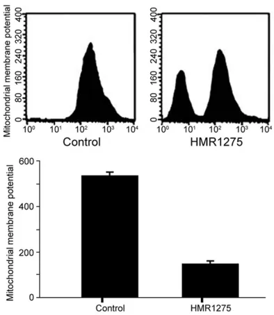
HMR1275 (flavopiridol) induced decrease of
membrane potential
HMR1275 (flavopiridol) treatment significantly
decreased the mitochondrial membrane po-
[image:3.612.93.288.69.217.2]tential of Ishikawa cells, suggesting HMR1275 possibly regulated apoptosis of Ishikawa cells
(Figure 2).
HMR1275 (flavopiridol) enhanced apoptosis of
Ishikawa cells
HMR1275 (flavopiridol) treatment significantly increased the percentage of phosphatidylser -ine translocation, suggesting HMR1275 indeed
enhanced apoptosis of Ishikawa cells (Figure 3).
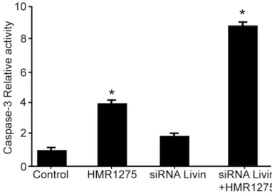
HMR1275 (flavopiridol) activated caspase-3 in
Ishikawa cells
As showed in Figure 4, compared with control
group (DMSO treated), activation of caspase-3 in Ishikawa cells was enhanced in HMR1275 group, confirming HMR1275 increased the apo-ptosis of Ishikawa cells.
HMR1275 (flavopiridol) down-regulated
ex-pression of livin in ishikawa cells
As showed in Figure 5, compared with control
group (DMSO treated), the expression of Livin in Ishikawa cells was decreased in HMR1275
group, suggesting HMR1275 regulated apopto-sis via Livin.
Down-regulation of livin promoted HMR1275 (flavopiridol)-induced apoptosis
siRNA indeed decreased the expression of Livin, while inhibition of Livin alone did not increase the apoptosis. Moreover, inhibition of Livin promoted apoptosis of HMR1275-trea-ted Ishikawa cells (Figure 6), suggesting
down-regulation of Livin promoted HMR1275
(flavo-piridol)-induced apoptosis.
Examination of Caspase-3 activation
As reported previously, examination of Cas-pase-3 activation was performed according to routine protocols [12]. Data of absorbance va-lue (560 nm) was analyzed for different groups.
Statistical analysis
SPSS 12.0 software was used for data
pro-cessing. Measurement data were represented as mean ± standard deviation (SD). One-Way
ANOVA was performed for analysis of the statis
-tical significance. P value < 0.05 was
[image:3.612.90.287.266.493.2]consid-ered to be statistically significant.
Figure 1. Analysis of cell growth in two groups. *P <
0.05 versus control group.
demonstrated that down-regulation of Livin en-hanced HMR1275 (flavopiridol)-induced apop
-tosis of endometrial carcinoma cell Line Ishi-kawa, which was consistent with previous
re-port [6].
We explored the effect of HMR1275 (flavopiri
-dol) on growth and survival of Ishikawa cells, and found that HMR1275 (flavopiridol) indeed inhibited the growth of Ishikawa cells. Furt-hermore, HMR1275 (flavopiridol) enhanced the apoptosis of Ishikawa cells, verified by signifi
-cant decrease of mitochondria membrane po-tential and increase of Caspase-3 activity. All
above were consistent with previous results
[13, 14]. What’s more, although previous study reported HMR1275 (flavopiridol) also resulted in apoptosis of breast cancer cells, Ishikawa
cells seemed to be more vulnerable at the
same concentration of HMR1275 (1 μmol/L), verified by higher levels of apoptosis than
breast cancer cells. This suggested HMR1275
(flavopiridol) was more effective in the treat
-ment of endometrial carcinoma, which could be explained by different chemo-sensitivities
among cells [15-17].
We further explored the mechanisms underly
-ing anti-cancer effect of HMR1275 (flavopiri
-dol). Activation of caspase-3 induced by HMR-1275 (flavopiridol) suggested HMRHMR-1275 (flavo
-piridol) enhanced apoptosis of Ishikawa cells
via mitochondrial signaling pathway, which is consistent with previous studies [18-20]. As
a novel biomarker in mitochondrial [4], Livin was possibly regulated by multiple kinds of
microRNAs. Our study demonstrated that HMR- 1275 enhanced apoptosis via down-regulat-
ing Livin, verified by siRNA experiment. This suggested inhibition of Livin increased the sen
-sitivity of Ishikawa cells treated with HMR1275 (flavopiridol).
Discussion
HMR1275 (flavopiridol) has been proved as
[image:4.612.93.284.72.235.2]an important anti-cancer drug [1, 2]. Our study
Figure 3. Analysis of percentage of phosphatidylser -ine translocation in two groups.
[image:4.612.324.520.73.183.2]Figure 4. Analysis of activation of caspase-3 in Ishikawa cells. *P < 0.05 versus control group.
Figure 5. Protein bands of Livin and β-actin in two
[image:4.612.91.286.288.442.2]groups.
[image:4.612.91.282.495.633.2][5] Kikuchi M, Kuroki S, Kayama M, Sakaguchi S, Lee KK and Yonehara S. Protease activity of procaspase-8 is essential for cell survival by
inhibiting both apoptotic and nonapoptotic cell death dependent on receptor-interacting
protein kinase 1 (RIP1) and RIP3. J Biol Chem
2012; 287: 41165-41173.
[6] Xu J, Huang Y, Li Y, Pu L, Xia F, Jiang C, Liu H
and Jiang Z. [Small interfering RNA-mediated RIP1 knockdown enhances L-OHP sensitivity of
human oral squamous carcinoma cells]. Nan Fang Yi Ke Da Xue Xue Bao 2013; 33: 1004-1007.
[7] Henrich CJ, Brooks AD, Erickson KL, Thomas CL, Bokesch HR, Tewary P, Thompson CR, Pom
-pei RJ, Gustafson KR, McMahon JB and Sayers
TJ. Withanolide E sensitizes renal carcinoma cells to TRAIL-induced apoptosis by increasing cFLIP degradation. Cell Death Dis 2015; 6: e1666.
[8] Ashkenazi A. Targeting the extrinsic apoptotic pathway in cancer: lessons learned and future
directions. J Clin Invest 2015; 125: 487-489. [9] Singh K, Poteryakhina A, Zheltukhin A, Bhate
-lia K, Prajapati P, Sripada L, Tomar D, Singh R,
Singh AK and Chumakov PM. NLRX1 acts as
tumor suppressor by regulating TNF-alpha in-duced apoptosis and metabolism in cancer cells. Biochim Biophys Acta 2015; 1853: 1073-1086.
[10] Kim JS, Oh D, Yim MJ, Park JJ, Kang KR, Cho IA,
Moon SM, Oh JS, You JS, Kim CS, Kim DK, Lee SY, Lee GJ, Im HJ and Kim SG. Berberine in-duces FasL-related apoptosis through p38 ac-tivation in KB human oral cancer cells. Oncol Rep 2015; 33: 1775-1782.
[11] Chou YC, Chang MY, Wang MJ, Harnod T, Hung CH, Lee HT, Shen CC and Chung JG. PEITC
induces apoptosis of human brain glioblasto -ma GBM8401 cells through the extrinsic-and intrinsic-signaling pathways. Neurochem Int 2015; 81: 32-40.
[12] Sun L, Fan H, Yang L, Shi L and Liu Y. Tyrosol
prevents ischemia/reperfusion-induced cardi
-ac injury in H9c2 cells: involvement of ROS,
Hsp70, JNK and ERK, and apoptosis. Mole-cules 2015; 20: 3758-3775.
[13] Rogalska A and Marczak A. Epothilone B in -duces human ovarian cancer OV-90 cell apop-tosis via external pathway. Environ Toxicol Pharmacol 2015; 39: 700-712.
[14] Yang HJ, Wang M, Wang L, Cheng BF, Lin XY
and Feng ZW. NF-kappaB regulates caspase-4
expression and sensitizes neuroblastoma cells to Fas-induced apoptosis. PLoS One 2015; 10: e0117953.
[15] Liu YK, Chen KH, Leu YL, Way TD, Wang LW,
Chen YJ and Liu YM. Ethanol extracts of cin
-namomum kanehirai hayata leaves induce
apoptosis in human hepatoma cell through Four aspects should be focused in the future
studies. 1) Clinical specimens should be
col-lected for examination of Livin expression. 2) Analogue of HMR1275 (flavopiridol) will be studied for better understanding of the phar -macological action. 3) Mice models with Livin
gene knock-out should be established to con
-firm our findings [21]. 4) The relationship bet-ween Livin and HMR1275 (flavopiridol) should be confirmed in vivo.
In conclusion, down-regulation of anti-apopto
-sis protein Livin promotes HMR1275
(flavopiri-dol)-induced apoptosis via increasing the drug sensitivity, and HMR1275 was possible to
alle-viate cancer via decreasing the expression of
Livin.
Acknowledgements
This work was supported by Gansu province natural science fund project (NO. 148RJZ072).
Disclosure of conflict of interest
None.
Address correspondence to: Dr. Fenqin Zhao, Gansu
College of Traditional Chinese Medicine, 35 Dingxi
Road, Lanzhou 730000, PR China. Tel:
+86-931-8765321; Fax: +86-931-+86-931-8765321; E-mail: fenqin -zhaoasd@163.com; Dr. Yongxiu Yang, Department
of Obstetrics and Gynecology, Lanzhou First Hospital of University, 1 Donggang Road, Lanzhou 730000,
Gansu, PR China. Tel: 931-8765321; Fax: +86-931-8765321; E-mail: yongxiuyangqwe@163.com
References
[1] Vandamme M, Pauwels W and Bleecker JD. A case of delayed oxaliplatin-induced pseudo-obstruction: an atypical presentation of oxali -platin neurotoxicity. Acta Clin Belg 2015; 70: 207-210.
[2] Aroldi F, Prochilo T, Bertocchi P and Zaniboni A. Oxaliplatin-induced hypersensitivity reaction: underlying mechanisms and management. J Chemother 2015; 27: 63-66.
[3] Yao Z, Zhang P, Guo H, Shi J, Liu S, Liu Y and Zheng D. RIP1 modulates death receptor me-diated apoptosis and autophagy in macro-phages. Mol Oncol 2015; 9: 806-817.
[4] Park EJ, Min KJ, Lee TJ, Yoo YH, Kim YS and
[19] Chen G, Cheng X, Zhao M, Lin S, Lu J, Kang J and Yu X. RIP1-dependent bid cleavage medi-ates TNFalpha-induced but
caspase-3-inde-pendent cell death in L929 fibroblastoma
cells. Apoptosis 2015; 20: 92-109.
[20] Zhang W, Zhao L, Liu J, Du J, Wang Z, Ruan C and Dai K. Cisplatin induces platelet apoptosis through the ERK signaling pathway. Thromb Res 2012; 130: 81-91.
[21] Zhang W, Liu J, Sun R, Zhao L, Du J, Ruan C and Dai K. Calpain activator dibucaine induces platelet apoptosis. Int J Mol Sci 2011; 12: 2125-2137.
caspase-3 cascade. Onco Targets Ther 2015; 8: 99-109.
[16] Chen TL, Zhu GL, He XL, Wang JA, Wang Y and Qi GA. Short-term pretreatment with
atorvas-tatin attenuates left ventricular dysfunction, reduces infarct size and apoptosis in acute myocardial infarction rats. Int J Clin Exp Med
2014; 7: 4799-4808.
[17] Lutz A, Sanwald J, Thomas M, Feuer R, Sawod-ny O, Ederer M, Borner C, Humar M and
Mer-fort I. Interleukin-1beta enhances FasL-in -duced caspase-3/-7 activity without increasing apoptosis in primary mouse hepatocytes. PLoS One 2014; 9: e115603.
[18] Guo H, Omoto S, Harris PA, Finger JN, Bertin J,
Gough PJ, Kaiser WJ and Mocarski ES. Herpes