Int J Clin Exp Pathol 2015;8(1):636-642
www.ijcep.com /ISSN:1936-2625/IJCEP0004286
Original Article
Detection of MGMT promoter methylation in
glioblastoma using pyrosequencing
Hao Xie, Raymond Tubbs, Bin Yang
Robert J. Tomsick Pathology & Laboratory Medicine Institute, Cleveland Clinic, Cleveland, OH
Received November 30, 2014; Accepted December 29, 2014; Epub January 1, 2015; Published January 15, 2015
Abstract: Recent clinical trials on patients with glioblastoma revealed that O6-Methylguanine-DNA methyltransferase
(MGMT) methylation status significantly predicts patient’s response to alkylating agents. In this study, we sought to develop and validate a quantitative MGMT methylation assay using pyrosequencing on glioblastoma.We quantified promoter methylation of MGMT using pyrosequencing on paraffin-embedded fine needle aspiration biopsy tissues from 43 glioblastoma.Using a 10% cutoff, MGMT methylation was identified in 37% cases of glioblastoma and 0% of the non-neoplastic epileptic tissue. Methylation of any individual CpG island in MGMT promoter ranged between 33% and 95%, with a mean of 65%. By a serial dilution of genomic DNA of a homogenously methylated cancer cell line with an unmethylated cell line, the analytical sensitivity is at 5% for pyrosequencing to detect MGMT methyla-tion. The minimal amount of genomic DNA required is 100 ng (approximately 3,000 cells) in small fine needle biopsy specimens. Compared with methylation-specific PCR, pyrosequencing is comparably sensitive, relatively specific, and also provides quantitative information for each CpG methylation.
Keywords: MGMT, methylation, glioblastoma, epigenetics, pyrosequencing
Introduction
Glioblastoma multiforme (GBM), or high grade glioblastoma, is the most common malignant brain tumor in adults. It has a very poor progno-sis with a median overall survival of only 12-15 months. Current treatments of GBM include surgical resection, radiotherapy, and
adminis-tration of alkylating agents such as temozolo
-mide [1]. Alkylating agents inhibit DNA replica -tion and induce tumor cell apoptosis by
produc-ing cross-links between adjacent DNA strands. The most common site for alkylation is the O6 position of guanine [2]. MGMT as a DNA repair
enzyme specifically removes promutagenic alkyl groups from the O6 position of guanine in DNA. Repair of O6-alkylguanine adducts in
tumor DNA reduces the cytotoxicity of alkylat -ing chemotherapeutic agents and thus confers tumor chemoresistance [3].
Both preclinical and clinical evidence showed that the expression of MGMT is mainly regulat-ed at the epigenetic level by promoter methyla-tion [4]. Under normal circumstances, the CpG sites spanning a large portion of the promoter
some tumor cells, however, the cytosines at certain CpG sites are methylated. This prevents transcriptional factors from binding and eventu-ally leads to the reduced expression of MGMT
protein, which sensitizes the cancer cells to alkylating agents.
Many studies have established a significant
correlation between methylation of MGMT and a positive clinical outcome for patients with
glioblastoma treated with alkylating agents such as temozolomide, carmustine, and procar
-bazine with or without radiotherapy [2, 5-7]. Furthermore, significant differences were dem -onstrated among varying levels of methylation, with higher MGMT methylation level associated with longer overall survival [8]. These studies suggested that MGMT methylation can predict
glioblastoma chemosensitivity to alkylating
agents and help determine the patient
popula-tion who likely benefit from such treatment.
Several methods have been used to identify MGMT methylation status [3, 9-12].
Methylation-specific PCR (MSP) with its high sensitivity is
increased false-positivity and its qualitative nature. Its mediocre compatibility with
forma-lin-fixed paraffin-embedded (FFPE) tissue spec -imens and requirement of high quality DNA lim-ited its application in the clinical setting [11]. Pyrosequencing is a highly reproducible and
quantitative method for confirming methylation
status. It has been shown to be the best approach for assessing MGMT methylation sta-tus in GBM patients and correlating with clinical outcomes [3]. However, no studies have been done to validate this method in the clinical set-ting for its compatibility with FFPE tissue speci-mens, analytical quality, and clinical feasibility. In this study, we aimed to validate a quantita-tive MGMT methylation assay using pyrose-quencing on FFPE biopsy tissue of glioblastoma and determine its analytical sensitivity and pre-cision as well as clinical feasibility for
patholog-ic diagnosis and predpatholog-iction of glioblastoma chemosensitivity.
Materials and methods
A total of 53 cases of brain tissue for quantita-tive analysis of MGMT promoter methylation status were selected for the study. These tis-sue specimens came from 10 patients with epi-lepsy as non-neoplastic controls, 43 patients with resected GBM or stereotaxic biopsy for GBM. All specimens were FFPE tissue trans-ported and stored at room temperature. No special preparation was needed.
The genomic DNA was extracted from 10 µm tissue sections of FFPE tissue samples using
the Gentra Puregene tissue kit (Qiagen Inc.,
Valencia, CA). DNA was further cleaned and
[image:2.612.90.524.76.444.2]purified by running through the QIAamp
MGMT methylation pyrosequencing
MiniElute column (Qiagen Inc., Valencia, CA) according to the manufacture’s manual. The
DNA concentration, protein to nucleic acid ratio, and DNA to RNA ratio for purity were assessed by spectrophotometer (NanoDrop products, Wilmington, DE). Approximately 100
to 200 ng total DNA was subjected to bisulfite conversion using EZ DNA Methylation Gold kit
(Zymo Research, Orange, CA).
A total of 10-20 ng bisulfite-treated DNA was carried on for PCR using the PyroMark PCR kit (Qiagen Inc., Valencia, CA). The PCR control was
reaction mixture with water in place of DNA. The PCR conditions for MGMT gene were 95°C for 15 minutes; 45 cycles of 95°C for 20
sec-onds, 53°C for 20 secsec-onds, and 72°C for 20 seconds; 72°C for 5 minutes, and then on hold at 4°C. The PCR products were immobilized to
beads and strand separation. We then conduct-ed pyrosequencing methylation assay using the
sequencing primer provided in the PyroMark CpG MGMT kit on the PyroMark Q96ID pyrose
-quencer (Qiagen Inc., Valencia, CA). The Pyro-mark CpG MGMT kit detected the level of meth -ylation on 5 CpG sites located in exon 1 of
MGMT gene. A cytosine not followed by a gua-nine served as an internal control for
comple-tion of bisulfite conversion. The percent meth
-ylation (% of C’s present) was reported for each
CpG site. A sample with methylation above 10% of any single CpG island or the average of all 5 CpG islands was interpreted as positive. A sam-ple below 10% methylation was interpreted as negative.
Two cell lines were used in analytical sensitivity tests. SW40, a colon cancer cell line with a mean MGMT methylation of 95% was used as
the positive control; MRC5, a fibroblast cell line
with 0% MGMT methylation was used as the negative control. We diluted SW40 DNA with MRC5 DNA from a ratio of 1:10 to 1:1000. The most diluted concentration with detectable methylation was recorded as the analytical sensitivity, which was compared with the stan-dard MSP method. In addition, we tested 50, 100, 150, and 200 ng DNA obtained from FFPE tissues in MGMT methylation pyrosequencing for the minimal requirement of DNA volume. To
measure the biopsy tissue size needed for
[image:3.612.92.524.71.363.2]reviewed 50 consecutive GBM biopsy speci-mens in 2010 at our institution and
document-ed the size of these specimens. We chose 16
biopsy specimens that represent the biopsy
size below 30 percentile for testing. Genomic
DNA was extracted from 8 sections of 15 µm
thick FFPE blocks. The concentration of DNA
was measured in duplicate in each case. Statistical analysis for this study was descrip-tive in nature. Thus we did not perform any
sample size calculation or power estimation. We summarized categorical data as frequency
counts and percentages, and continuous mea-sures as means, standard deviations (s.d.), medians, and ranges. All the statistical analy-ses were performed using VassarStats (Richard Lowry, PhD, Vassar College, NY).
Results
Optimizing the DNA extraction and purification procedure for pyrosequencing assay
We optimized the experimental conditions for
MGMT methylation pyrosequencing assay. After extraction, genomic DNA was further
puri-fied to generate highly puripuri-fied DNA for pyrose -quencing. The amount of DNA sample loss was less than 15%. We used EZ DNA methylation
gold kit for bisulfite conversion. This method
converts greater than 95% of unmethylated
cytosines in the genome which satisfies the
requirement of PyroMark Q96ID pyrosequencer
for consistent results.
MGMT methylation in GBM and epilepsy brain tissues
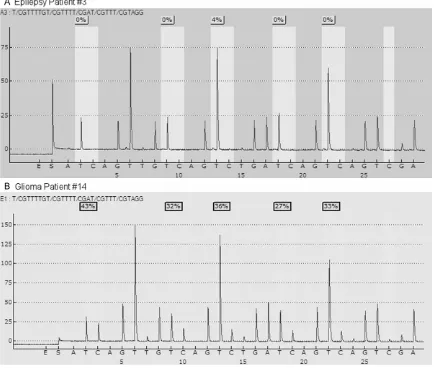
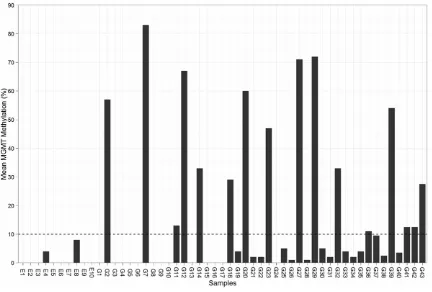
We determined the level of methylation on 5 CpG sites located in exon 1 of MGMT gene from the brain tissues of both patients with epilepsy as non-neoplastic control and those with glio-blastoma. Typical examples of pyrograms are shown in Figure 1. Shaded boxes encompass T/C pairs with methylation of 5 CpG sites mea-sured quantitatively. In a typical tissue speci-men from an epilepsy patient, most of the CpG sites are unmethylated with occasional sites bearing less than 10% methylation. In contrast, a typical tumor specimen from a glioma patient often has more than 25% methylation in all the CpG sites. As shown in Figure 2, we measured MGMT gene methylation in 10 non-neoplastic epilepsy patients. None of the epilepsy brain tissues harbors > 10% MGMT methylation. We only observed low level of MGMT methylation in two cases (4% and 8%, respectively). In con-trast, among the glioblastoma tissues from 43 patients, the range of methylation percentage of any CpG island in MGMT gene is between 33% and 95%, with a mean of 65%. We
observed > 10% MGMT methylation in 37%
[image:4.612.93.525.78.293.2]MGMT methylation pyrosequencing
ylation is the best cutoff to distinguish MGMT methylation positive cases from negative cases.
Comparing the analytical sensitivity between pyrosequencing and MSP assays
We determined the analytical sensitivity of this methylation test by pyrosequencing using a series of DNA dilutional tests. By a serial dilu-tion of a methylated cancer cell line with an unmethylated normal cell line, pyrosequencing can detect 1 tumor cell harboring MGMT ylation out of 80-100 cells without MGMT meth-ylation. For clinical application, in comparison with MSP, pyrosequencing is comparably sensi-tive with less false-posisensi-tive cases and also pro-vide quantitative methylation value of each CpG island.
Minimal DNA amount and minimal tissue vol-ume needed for pyrosequencing assay
Our measurement of minimal DNA requirement revealed that the minimal amount of genomic DNA required for successful detection of MGMT methylation by pyrosequencing is 100 ng (approximately 3,000 cells). Our efforts to
determine the tissue size that is needed for
MGMT methylation pyrosequencing test dem-onstrated that most of the clinical GBM biopsy
specimens range from 0.1 to 3.0 cm in size
with a median of 1.0 cm. We chose the cases
with biopsy size less than 1.0 cm. to test the
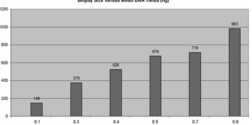
minimal tissue needed for obtaining enough genomic for conducting pyrosequencing-based MGMT methylation assay. As shown in Figure 3, the mean DNA yield increased in line with the
increase of biopsy tissue size. Four of 15 µm thick tissue sections should yield approximate -ly > 300 ng genomic DNA, which corresponds
to 0.3 cm biopsy size with less than 15%. We
could use 150 ng DNA samples for each run
and fulfill the clinical requirement of duplicate tests. After testing protocol has been finalized, we run a precision test with a known MGMT
methylation case along with positive and nega-tive controls to polish up the procedure. Results of duplicated tests were consistent and com-patible with expected results.
Discussion
Since the establishment of MGMT methylation as a favorable predictor of positive clinical
out-come for patients treated for glioblastoma [2], several methods have been developed to deter-mine its methylation status including the widely used MSP method in the research setting [9, 11]. However, various limitations of these meth-ods such as qualitative result report, low
speci-ficity, and high tissue quality requirement hin
-dered their further utilization in the clinical set -ting [3, 11]. This fact was clearly demonstrated
by Karayan-Tapon and coworkers [3] during
their assessment of prognostic value of MGMT
status in glioblastoma patients with five differ -ent methods. It turned out that pyrosequencing is the best approach to stratify patients treated
with temozolomide into different prognostic
groups based on their measured MGMT meth-ylation status.
The reliability and reproducibility of most of the previously developed methods require high-quality DNA. These methods such as MSP often
require fresh frozen specimen [7]. However, in
most clinical settings, FFPE specimens are needed for easy transport and storage. It has been reported that MSP can only provide reli-able MGMT methylation status on two thirds of
the FFPE tumor specimens [7]. To solve this problem, we further purified the genomic DNA extract from FFPE tissue samples using Qiagen
MiniElute column. After elution with 100 µl AE
buffer, we were able to generate highly purified
genomic DNA for the following steps with less than 15% DNA loss.
Bisulfite conversion of cytosine is a crucial step
toward success. Complete conversion of unmethylated cytosine to uracil is the basis of
methylation quantification. Incomplete conver -sion leads to overestimation of MGMT gene methylation. It also severely compromises the robustness of methods for clinical samples often with low DNA quantity. In our study, we
compared two bisulfite treatment methods: EZ DNA methylation gold kit and Qiagen DNA methylation kit. The former method converts
greater than 95% of unmethylated cytosines in
the genome which satisfies the requirement of PyroMark Q96ID pyrosequencer for consistent
results. However, the latter method used in some of the previous studies converts only about 90% of unmethylated cytosines which
does not fulfill the requirement of
pyrosequ-encer.
level of methylation (8%) in the control group. No formal analysis of receiver operating char-acteristics was conducted due to small sample
size. However, both the 10% cutoff and the
prevalence of MGMT hypermethylation in the tumor tissues of glioblastoma patients thereof were consistent with the previous reports [4, 11].
The analytical sensitivity of our method for MGMT methylation detection is from 1:80 to
1:100, which is approximately five times less
than MSP method (1:500). Although MSP is more sensitive than pyrosequencing, it often generates approximately 10% false positive results [10]. This concern with false-positivity using MSP was mentioned in the literature due to its “super-sensitivity” and mispriming (PCR bias), which can be further aggravated by high cycle number and nested primers used for decreased sample quality of PPFE specimen [13]. Since most testing samples will be GBM lesional materials by FNAB and are less diluted with normal brain tissue, sensitivity of 1:80-1:100 should be good enough for clinical usage. Further analysis in this study demonstrated approximately 95% of cases matched between pyrosequencing and MSP assays. On the other hand, this non-inferior sensitivity of
pyrose-quencing is accompanied by its higher specific -ity with quantitative value of both average and individual CpG island methylation status. In contrast, MSP is not quantitative and cannot tell methylation status of individual CpG island which may be more predictive to clinical out-comes as previously reported [11].
In our study, 100 ng DNA is adequate to repeat-edly detect MGMT methylation by pyrosequenc-ing, which is much less than the
recommenda-tion of Qiagen kit using > 300 ng genomic DNA for research purposes. The biopsy size require -ment (0.3 cm) of our method is a lot smaller
than the current median biopsy size of 1.0 cm
(range: 0.1-3.0 cm). The DNA yielded from even
the smallest GBM biopsy size (0.1 cm) at our
institution in 2010 was enough for duplicated assays. Thereby our tissue requirement is
rea-sonable clinically and is significantly less
demanding than the currently commercially
available kits of MSP method, which requires >
1.0 cm tissue with minimal 500 ng input for their assays. Last, we created both positive and negative report templates compatible with CAP MP reporting guidelines.
In summary, we described the first validated
pyrosequencing-based MGMT methylation test on clinical FFPE biopsy tissue from 33 patients with glioblastoma and 10 patients with epilep-sy as control. We demonstrated that pyrose-quencing detection of MGMT methylation has an analytical sensitivity, feasible DNA and biop-sy tissue requirement suitable for routine clini-cal application. In addition to pathologic diag-nosis, MGMT methylation assay shall provide a
useful molecular biomarker for prediction of
chemosensitivity in patients with glioblastoma multiforme.
Disclosure of conflict of interest
None.
Address correspondence to: Dr. Bin Yang, Robert J. Tomsick Pathology & Laboratory Medicine Institute, Cleveland Clinic, L25, 9500 Euclid Avenue, Cleve- land, OH 44195. Tel: 0306; Fax: 216-445-6967; E-mail: yangb@ccf.org
References
[1] Wen PY, Kesari S. Malignant gliomas in adults. N Engl J Med 2008; 359: 492-507.
[2] Esteller M, Garcia-Foncillas J, Andion E, Goodman SN, Hidalgo OF, Vanaclocha V, Baylin SB, Herman JG. Inactivation of the DNA-repair gene MGMT and the clinical response of glio-mas to alkylating agents. N Engl J Med 2000; 343: 1350-4.
[3] Karayan-Tapon L, Quillien V, Guilhot J, Wager M, Fromont G, Saikali S, Etcheverry A, Hamlat A, Loussouarn D, Campion L, Campone M, Vallette FM, Gratas-Rabbia-Ré C. Prognostic value of O6-methylguanine-DNA methyltrans-ferase status in glioblastoma patients, as-sessed by five different methods. J Neurooncol 2010; 97: 311-22.
[4] Esteller M, Herman JG. Generating mutations but providing chemosensitivity: the role of O6-methylguanine DNA methyltransferase in hu-man cancer. Oncogene 2004; 23: 1-8. [5] Paz MF, Yaya-Tur R, Rojas-Marcos I, Reynes G,
Pollan M, Aguirre-Cruz L, García-Lopez JL, Piquer J, Safont MJ, Balaña C, Sanchez-Cespedes M, García-Villanueva M, Arribas L, Esteller M. CpG island hypermethylation of the DNA repair enzyme methyltransferase predicts response to temozolomide in primary gliomas. Clin Cancer Res 2004; 10: 4933-8.
MGMT methylation pyrosequencing
[7] Hegi ME, Diserens AC, Gorlia T. MGMT gene si-lencing and benefit from temozolomide in glio -blastoma. N Engl J Med 2005; 352: 997-1003. [8] Dunn J, BaboriE A, Alam F, Alam F, Joyce K,
Moxham M, Sibson R, Crooks D, Husband D, Shenoy A, Brodbelt A, Wong H, Liloglou T, Haylock B, Walker C. Extent of MGMT promoter methylation correlates with outcome in glio-blastomas given temozolomide and radiother -apy. Brit J Cancer 2009; 101: 124-31.
[9] Herman JG, Graff JR, Myöhänen S, Nelkin BD, Baylin SB. Methylation-specific PCR: a novel PCR assay for methylation status of CpG is-lands. Proc Nati Acad Sci U S A 1996; 93: 9821-6.
[10] Shaw RJ, Akufo-Tetteh EK, Risk JM, Field JK, Liloglou T. Methylation enrichment pyrose-quencing: combining the specificity of MSP with validation by pyrosequencing. Nucleic Acids Res 2006; 34: e78.
[11] Mikeska T, Bock C, El-Maarri O, Hübner A, Ehrentraut D, Schramm J, Felsberg J, Kahl P, Büttner R, Pietsch T, Waha A. Optimization of quantitative MGMT promoter methylation anal-ysis using pyrosequencing and combined bi-sulfite restriction analysis. The J Mol Diagn 2007; 9: 368-81.
[12] Jha P, Suri V, Jain A, Sharma MC, Pathak P, Jha P, Srivastava A, Suri A, Gupta D, Chosdol K, Chattopadhyay P, Sarkar C. O6-methylguanine DNA methyltransferase gene promoter methyl-ation status in gliomas and its correlmethyl-ation with other molecular alterations: first Indian report with review of challenges for use in customized treatment. Neurosurgery 2010; 67: 1681-91. [13] Derks S, Lentjes MHFM, Hellebrekers DMEI.